Digital Poster
Radial Acquisition & Analysis Techniques
ISMRM & ISMRT Annual Meeting & Exhibition • 03-08 June 2023 • Toronto, ON, Canada

Digital Poster
Radial Acquisition & Analysis Techniques
| Computer # | |||
|---|---|---|---|
4791.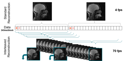 |
21 |
Real-time MRI with Simultaneous Multi-slice and Compressed
Sensing
Isaac Watson1,2,3,
Martin Trefzer1,
David Mitchell2,
Angelika Sebald2,
and Aneurin Kennerley3,4
1School of Physics, Engineering and Technology, University of York, York, United Kingdom, 2York Cross-disciplinary Centre for Systems Analysis, University of York, York, United Kingdom, 3York Neuroimaging Centre, University of York, York, United Kingdom, 4Institute of Sport, Manchester Metropolitan University, Manchester, United Kingdom Keywords: Data Acquisition, Data Acquisition Real time (rt) MRI is currently predominantly a single slice acquisition technique. We show that, by combining compressed sensing, simultaneous multi-slice acquisition and interleaved data ordering with a highly undersampled radial trajectory, multi-slice rtMRI images with a high temporal resolution can be acquired. This offers new opportunities, for example, the non-invasive monitoring of oral and maxillo facial dynamics.
|
|
4792.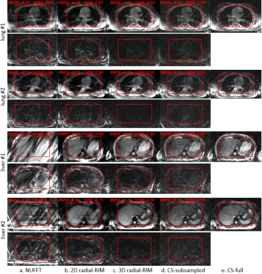 |
22 |
Radial-RIM: accelerated radial 4D MRI using the recurrent
inference machine
Chaoping Zhang1,
Matthan W.A. Caan2,3,4,
Robin Navest1,
Jonas Teuwen1,5,6,
and Jan-Jakob Sonke1
1Netherlands Cancer Institute, Amsterdam, Netherlands, 2Amsterdam UMC location University of Amsterdam, Amsterdam, Netherlands, 3Amsterdam Neuroscience, Amsterdam, Netherlands, 4Oslo University Hospital, Oslo, Norway, 5University of Amsterdam, Amsterdam, Netherlands, 6Radboud University Medical Center, Nijmegen, Netherlands Keywords: Image Reconstruction, Image Reconstruction, radial, radiotherapy, liver, lung, respiratory motion To accelerate the acquisition and reconstruction of the respiratory correlated (4D) radial MRI for image guided radiotherapy guidance, we propose a 3D radial-RIM network by adapting the forward model for Cartesian k-space in the regular RIM to radial k-space. Experiments show that the radial-RIM reconstructs images with better quality than the XD-GRASP compressed sensing for 3-4 times accelerated 4D radial free-breathing lung/liver acquisitions, with reconstruction time per slice reduced from 2 minutes to 1.9 seconds. Signal correlation exploited by the radial-RIM over the extra motion-state dimension contributes substantially to the image quality. |
|
4793.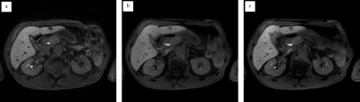 |
23 |
Free-Breathing Gadoxetic Acid-Enhanced Hepatobiliary Phase
Imaging Using Stack-of-Stars Radial Sampling and Compressed
SENSE
Tetsuro Kaga1,
Yoshifumi Noda1,
Nobuyuki Kawai1,
Kimihiro Kajita2,
Yu Ueda3,
Masatoshi Honda3,
Fuminori Hyodo4,
Hiroki Kato1,
and Masayuki Matsuo1
1Department of radiology, Gifu university, Gifu, Japan, 2Gifu university hospital, Gifu, Japan, 3Philips Japan, Tokyo, Japan, 4Institute for Advanced Study, Gifu university, Gifu, Japan Keywords: Data Acquisition, Liver, Free breathing, Radial sampling The gadoxetic acid-enhanced hepatobiliary phase imaging is an effective sequence for detecting hepatic lesions; however, degraded image quality is often observed due to poor breath holding. Free-breathing sequence using radial stack-of-stars acquisition (3D VANE) has been introduced and can provide diagnosable image quality. Recently, Compressed SENSE (CS), that is one of the acceleration techniques, has been applicable in 3D VANE. In this study, we evaluated the feasibility of 3D VANE with CS in hepatobiliary phase imaging. Our results showed that 3D VANE with CS at section thickness of 2 mm had almost comparable image quality as conventional breath-holding cartesian sampling. |
|
4794.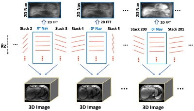 |
24 |
Navistar: Golden-Angle Stack-of-Stars Sampling with Embedded 2D
Navigators for Highly-Accelerated 4D Dynamic MRI
Ding Xia1,
Sera Saju1,
Kai Tobias Block2,
and Li Feng1
1BioMedical Engineering and Imaging Institute (BMEII), Icahn School of Medicine at Mount Sinai, New York, NY, United States, 2Center for Advanced Imaging Innovation and Research (CAI2R), NYU Grossman School of Medicine, New York, NY, United States Keywords: Data Acquisition, Motion Correction This work proposes a new radial sampling scheme called Navistar for highly-accelerated 4D dynamic MRI. Navistar periodically inserts 2D navigators into a golden-angle radial stack-of-stars acquisition. The embedded 2D navigators provide spatially-resolved information that can be used for different purposes, such as improved temporal-basis estimation for image reconstruction or respiratory-motion estimation. In our experiments, we have shown that Navistar sampling can be combined with low-rank subspace-constrained image reconstruction for highly-accelerated free-breathing 4D DCE-MRI of the liver with sub-second temporal resolution, which helps reduce intraframe respiratory blurring and eliminates the need for explicit respiratory-motion compensation. |
|
4795.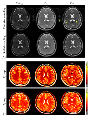 |
25 |
Full-brain multi-pathway relaxometry using a golden-angle radial
scheme and sparsely-sampled radial spokes
Li-Ya Shao1,
Zih-Ci Wang2,
Bruno Madore3,
and Cheng-Chieh Cheng2
1Department of Electrical Engineering, National Sun Yat-sen University, Kaohsiung, Taiwan, 2Department of Computer Science and Engineering, National Sun Yat-sen University, Kaohsiung, Taiwan, 3Department of Radiology, Brigham and Women's Hospital, Harvard Medical School, Boston, MA, United States Keywords: Data Acquisition, Relaxometry Quantitative MRI is emerging as a powerful diagnostic tool for neuroimaging applications. A motion-robust, golden-angle radial acquisition version of the ‘triple-echo steady-state’ (TESS) relaxometry method was implemented here, to mitigate the artifacts induced by the flowing motion of the cerebrospinal fluid (CSF). To improve the scan efficiency, a probability density function-based sparse sampling scheme was introduced in each radial spoke. Close agreement was obtained between reference and TESS scans for both T1 and T2 values, using a multi-compartment phantom. In vivo whole-brain results were further obtained (3D T1 and T2 maps, 1-mm isotropic resolution, whole-brain coverage, 7.5-minute scan). |
|
4796.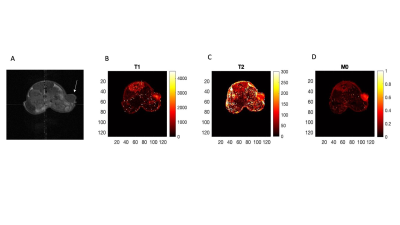 |
26 |
Radial Magnetic Resonance Fingerprinting for quantification of
T1 in metastatic breast tumor.
Chetan B. Dhakan1,
Jorge De La Cerda1,
William Schuler1,
Christina J. MacAskill2,
Chris A. Flask2,
Mark D Pagel1,
and Gary V. Martinez3
1Cancer Systems Imaging, MD Anderson Cancer Center, Houston, TX, United States, 2Department of Radiology, Case Western Reserve University, Cleveland, OH, United States, 3Imaging Physics, MD Anderson Cancer Center, Houston, TX, United States Keywords: MR Fingerprinting/Synthetic MR, Cancer Quantitative Magnetic Resonance Imaging (MRI) involves pixel-wise mapping of longitudinal relaxation time T1, transverse relaxation time T2 and proton density M0 and other relevant parameters at each location in the tissue to be characterized. The goal of this study is to investigate potential benefits of Magnetic Resonance Fingerprinting in quantifying T1 relaxation times in metastatic breast tumor using radial acquisition in tumor with high temporal and spatial resolution. |
|
4797.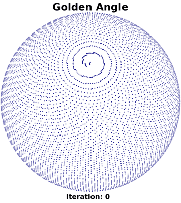 |
27 |
Electric Potential Energy Optimized 3D Radial Trajectories
Christopher Huynh1,
Datta Singh Goolaub1,
and Christopher K Macgowan1,2
1Translational Medicine, Hospital for Sick Children, Toronto, ON, Canada, 2Medical Biophysics, University of Toronto, Toronto, ON, Canada Keywords: Data Acquisition, Artifacts, Optimized 3D Trajectory The commonly used golden angle trajectory for 3D radial dynamic MRI suffers from spiral-shaped sample clustering around the z-axis, often resulting in image streaking artifact. We develop a method that corrects for this for center-out 3D radial trajectories. The electric potential energy of the trajectory is minimized through the use of repulsive forces, producing a spherically uniform distribution while maintaining the quasirandom quality of the golden angle trajectory. ELECTRic potential energy Optimized (ELECTRO) trajectories are relatively inexpensive to obtain and can produce images with lower MSE compared to the golden angle counterpart. |
|
4798.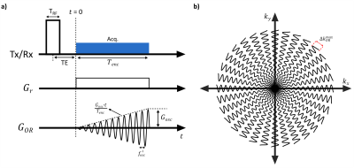 |
28 |
Ultra-fast Radial MRI with Silent Oscillating Gradients
Serhat Ilbey1,
Sebastian Littin1,
Feng Jia1,
Niklas Wehkamp1,
Philipp Amrein1,
Maxim Zaitsev1,
Michael Bock1,
and Ali Caglar Özen1
1Division of Medical Physics, Department of Radiology, University Medical Center Freiburg, Freiburg, Germany Keywords: New Trajectories & Spatial Encoding Methods, New Trajectories & Spatial Encoding Methods The sampling efficiency of radial center-out MRI was increased by using additional oscillating readout gradients, which lead to an accelerated image acquisition. The idea of radial sampling along an oscillating spoke was demonstrated using a clinical MRI system with ultra-fast UTE MRI. |
|
4799.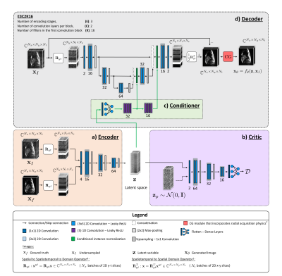 |
29 |
A Physics-informed Conditional Wasserstein Autoencoder to
Quantify Uncertainties in Accelerated 2D Dynamic Radial MRI
Sherine Brahma1,
Tobias Schaeffter1,2,3,
Christoph Kolbitsch1,3,
and Andreas Kofler1
1Physikalisch-Technische Bundesanstalt (PTB), Braunschweig and Berlin, Germany, 2Department of Biomedical Engineering, Technical University of Berlin, Berlin, Germany, 3School of Imaging Sciences and Biomedical Engineering, King’s College London, London, United Kingdom Keywords: Image Reconstruction, Cardiovascular, Radial Acquisition Uncertainty quantification (UQ) can provide important information about deep learning algorithms and help interpret the obtained results. UQ for multi-coil dynamic MRI is challenging due to the large scale of the problem and scarce training data. We approach these issues by learning distributions in a lower dimensional latent space using a conditional Wasserstein autoencoder while utilizing the MR data acquisition model and by exploiting spatio-temporal correlations of the cine MR images. Our results indicate excellent image quality accompanied with uncertainty maps that correlate well with estimation errors. |
|
4800. |
30 |
T1 Relaxation-Enhanced Steady-State (T1RESS) Imaging with Radial
Sampling for Robust Brain Examination at 3T and 0.55T
Ruoxun Zi1,
Kai Tobias Block1,
Ioannis Koktzoglou2,
Mahesh Keerthivasan3,
Thomas Benkert4,
Jakob Asslaender1,
Daniel K Sodickson1,
and Robert R Edelman2
1The Bernard and Irene Schwartz Center for Biomedical Imaging, Department of Radiology, New York University Grossman School of Medicine, New York, NY, United States, 2Radiology, Northshore University HealthSystem, Evanston, IL, United States, 3Siemens Medical Solutions, New York, NY, United States, 4Application Development, Siemens Healthcare GmbH, Erlangen, Germany Keywords: Data Acquisition, Brain A new class of sequences, named T1 relaxation-enhanced steady-state (T1RESS), has been recently proposed and showed improved lesion conspicuity in contrast-enhanced exams. T1RESS sequences combine steady-state acquisition, either balanced or unbalanced with weak gradient spoiling, with periodic contrast-modifying preparation pulses to create T1 weighting of the contrast. Here, we present radial versions of T1RESS sequences using the stack-of-stars trajectory, which offer improved motion robustness and enable dynamic imaging through use of advanced reconstructions such as GRASP. Volunteer scans acquired at 3T are shown for both sequences. Moreover, the balanced version is demonstrated at 0.55T to highlight its SNR efficiency. |
|
4801.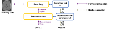 |
31 |
Stochastic optimization of 3D non-Cartesian sampling trajectory
(SNOPY)
Guanhua Wang1,
Jon-Fredrik Nielsen1,
Jeffrey A. Fessler2,
and Douglas C. Noll1
1Biomedical Engineering, University of Michigan, Ann Arbor, MI, United States, 2EECS, University of Michigan, Ann Arbor, MI, United States Keywords: New Trajectories & Spatial Encoding Methods, Machine Learning/Artificial Intelligence Efficient k-space trajectories are crucial for accelerated MRI. SNOPY proposes a generalized gradient-based method for optimizing 3D non-Cartesian sampling patterns. The algorithm can simultaneously tune multiple properties of sampling patterns, including image quality, hardware constraints (maximum slew rate and gradient strength), reduced peripheral nerve stimulation (PNS), and parameter-weighted contrast. The proposed method applies to various scenarios, such as optimizing gradient waveforms or optimizing rotation angles of radial/spiral trajectories. We adopted several computational strategies to address this non-convex and large-scale problem. Various simulated and in-vivo experiments demonstrated the effectiveness of SNOPY. |
|
4802.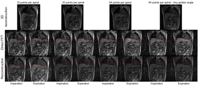 |
32 |
Cartesian Spiral acquisitions for radiotherapy on an MR-Linac
Bastien Lecoeur1,2,
Prashant Nair1,
Rosie Goodburn1,
Tom Bruijnen3,
Uwe Oelfke1,
Wayne Luk2,
and Andreas Wetscherek1
1Joint Department of Physics, The Institue of Cancer Research, Sutton, United Kingdom, 2Department of Computing, Imperial College London, London, United Kingdom, 3Department of Radiotherapy, University Medical Center Utrecht, Utrecht, Netherlands Keywords: Image Reconstruction, Radiotherapy
Respiratory-resolved 4D-MRIs could measure the extent of
respiratory motion for online MR-guided radiotherapy on
MR-Linacs. Cartesian spiral (CASPR) trajectories are an
alternative to radial sampling for self-gated 4D-MRIs,
because they could avoid prohibitively long image
reconstruction times related to the non-uniform Fourier
Transform. We studied the effect of different sampling
parameters on image quality for CASPR-based sequences on an
MR-Linac and reconstructed abdominal 4D-MRI, we found that
increasing the number of points per spiral arm improved the
reconstructed image quality, creating a trade-off between
spatial and temporal resolutions. |
|
4803.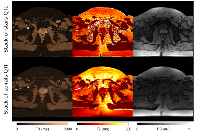 |
33 |
Radial vs. Spiral – A Comparison of Stack-of-stars and
Stack-of-spirals Spatial Encoding Schemes in Multiparametric
Body MRI with QTI
Carolin M Pirkl1,
Rolf F Schulte1,
Pablo García-Polo2,
Matteo Cencini3,
Sebastian Endt1,4,5,
Laura Biagi3,
Michela Tosetti3,
and Marion I Menzel1,4,5
1GE Healthcare, Munich, Germany, 2GE Healthcare, Madrid, Spain, 3IRCCS Stella Maris, Pisa, Italy, 4Technische Hochschule Ingolstadt, Ingolstadt, Germany, 5Technical University of Munich, Munich, Germany Keywords: MR Fingerprinting/Synthetic MR, Data Acquisition Highly accelerated multiparametric MRI techniques are more and more demonstrating their diagnostic potential. To achieve short scan times, these techniques rely on non-Cartesian (under-) sampling. Initially developed for MRI applications in the brain, these sequences are now also applied in body MRI. In this work, we present a phantom and in vivo study to demonstrate and evaluate the strengths and pitfalls of the two most common non-Cartesian – spiral and radial – sampling schemes for the application of rapid, free-breathing 3D T1, T2 and proton density (PD) mapping with quantitative transient-state imaging (QTI) in the prostate gland. |
|
4804.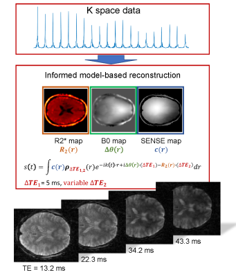 |
34 |
Single-shot 2D Radial Echo Planar Imaging using a KWIC Filter
and Model-based Reconstruction Approach
Christoph Rettenmeier1,
Zidan Yu2,
and V. Andrew Stenger3
1Medicine, University of Hawaii, Honolulu, HI, United States, 2Univeristy of Hawaii, Honolulu, HI, United States, 3University of Hawaii, Honolulu, HI, United States Keywords: Data Acquisition, Brain High quality brain images at spatial resolutions of 2x2x3 mm3 and 3.3x3.3x3 mm3 were obtained using single-shot 2D rEPI. The approach is based on an R2*, B0 and coil sensitivity informed model-based reconstruction in combination with k-space weighted image contrast (KWIC) filtering. Signal loss due to phase inconsistencies are starkly reduced by the linear phase model and R2* contrast was modifiable by adjusting the target echo time in the model of the reconstruction and KWIC filter. Bold activation maps were generated from fMRI datasets at 2x2 mm2 resolution showing activation in the visual cortex. |
|
4805.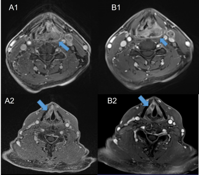 |
35 |
Values of golden-angle radial-VIBE in DCE-MRI in laryngeal and
hypopharyngeal squamous cell carcinoma: comparison with
conventional VIBE
Yan Wen1,
Liling Long1,
Chenhui Li1,
Huiting Zhang2,
and Xiaodong Zhong3
1Department of Radiology, The First Affiliated Hospital of Guangxi Medical University, Nanning, China, 2MR Scientific Marketing, Siemens Healthineers Ltd., Wuhan, China, 3MR R&D Collaborations, Siemens Medical Solutions, Los Angeles, CA, United States Keywords: Image Reconstruction, Head & Neck/ENT This study aimed to investigate the value of a dynamic contrast-enhanced (DCE)-MRI with golden-angle radial volumetric‑interpolated breath‑hold examination (radial-VIBE) research application sequence by comparing with conventional VIBE in patients with laryngeal and hypopharyngeal squamous cell carcinoma (SCC) under free breathing conditions. Our results showed that compared with conventional contrast-enhanced VIBE images with single phase, the morphological radial-VIBE images of multitemporal synthesis had significantly higher image quality based on subjective (edge, artifact, and confidence) and objective (signal-to-noise ratio, contrast, and contrast-to-noise ratio) assessments. Therefore, radial-VIBE can be used in DCE‑MRI examination in laryngeal and hypopharyngeal SCC in the free breathing state. |
|
4806.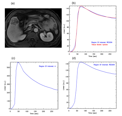 |
36 |
Evaluation of perfusion on Golden-Angle Radial Sparse Parallel
(GRASP) MRI in discriminating benign and malignant liver tumors
Yu Wang1,
Xiaohui Duan1,
Mengzhu Wang2,
Lingjie Yang1,
Zhuoheng Yan1,
and Huijun Hu1
1Department of Radiology, Sun Yat-Sen Memorial Hospital, Sun Yat-Sen University, Guangzhou, China, 2MR Scientific Marketing, Siemens Healthineers Ltd., Guangzhou, China Keywords: Data Analysis, Cancer This study investigated the utility of quantitative parameters of golden-angle radial sparse parallel (GRASP) dynamic contrast enhanced magnetic resonance imaging (DCE-MRI) for evaluation of perfusion and differentiation of benign and malignant liver tumors. The results showed the quantitative parameters in GRASP DCE-MRI can effectively evaluate the perfusion characteristics and differentiation in liver tumors with good diagnostic performance. This indicated that the GRASP quantitative parameters may be useful to evaluate and predict the pathological stage of liver lesions. |
|
4807.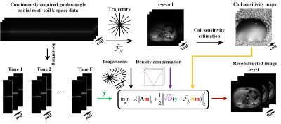 |
37 |
A Convergence Analysis for Projected Fast Iterative
Soft-thresholding Algorithm under Radial Sampling MRI
Biao Qu1,
Zuwen Zhang2,
Yewei Chen2,
Chen Qian2,
Taishan Kang3,
Jianzhong Lin3,
Lihua Chen4,
Zhigang Wu5,
Jiazheng Wang5,
Gaofeng Zheng1,
and Xiaobo Qu2
1Department of Instrumental and Electrical Engineering, Xiamen University, Xiamen, China, 2Department of Electronic Science, Biomedical Intelligent Cloud R&D Center, Fujian Provincial Key Laboratory of Plasma and Magnetic Resonance, Xiamen University, Xiamen, China, 3Department of Radiology, Zhongshan Hospital of Xiamen University, School of Medicine, Xiamen University,, Xiamen, China, 4Department of Radiology, Chongqing University Cancer Hospital, School of Medicine, Chongqing University, Chongqing, China, 5Philips, Beijing, China Keywords: Image Reconstruction, Image Reconstruction Radial sampling is a fast magnetic resonance imaging technique. The projected fast iterative soft-thresholding algorithm (pFISTA) has shown the advantage to solved tight frame sparse reconstruction model for removing undersampling image artifacts. However, the convergence of this algorithm under radial sampling has not been clearly set up. In this work, the authors derived a theoretical convergence condition for this algorithm and an optimal step size was further suggested to allow the fastest convergence. Verifications were made in vivo data of static brain imaging and dynamic contrast-enhanced (DCE) liver imaging, demonstrating that the recommended parameter allowed fast convergence in radial MRI. |
|
4808.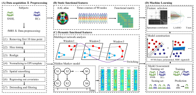 |
38 |
Machine learning for detecting sensorineural hearing loss
utilizing functional imaging with a combination of static and
dynamic brain features
Xiao-Min Xu1 and
Yu-Chen Chen1
1Radiology, Nanjing First Hospital, Nanjing, China Keywords: Head & Neck/ENT, fMRI (resting state) Alterations of static and dynamic brain function have been found in sensorineural hearing loss (SNHL). The combination of data-driven machine learning based classifiers and multiple imaging features can identify SNHL and healthy controls automatically. The spearman rank correlation with radial basis functional kernel support vector machine (SVM) and sigmoid SVM provides promising neural biomarkers for clinical classifier of SNHL. |
|
4809.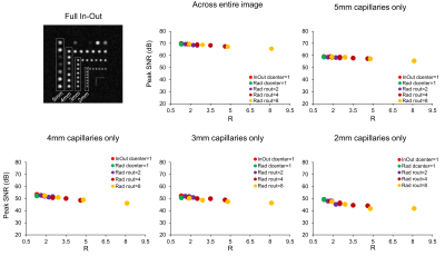 |
39 |
Reducing scan-time for 3D imaging with undersampled
cartesian-radial phase encoding on a point-of-care 46 mT Halbach
MRI scanner
Chloé Najac1,
Kirsten Koolstra2,
Tom O’Reilly1,
and Andrew Webb1
1C.J. Gorter MRI Center, Department of Radiology, Leiden University Medical Center, Leiden, Netherlands, 2Philips, Best, Netherlands Keywords: Low-Field MRI, New Trajectories & Spatial Encoding Methods Low-field MRI systems (with B0<0.1T) for point-of-care applications are becoming increasingly widespread. Imaging at low-field remains challenging due to the low intrinsic SNR. We evaluated speeding up 3D imaging using radial-based Cartesian undersampled phase-encodings (PEs). In phantoms, we tested different undersampling schemes and compared them to full in-out Cartesian PE in terms of peak SNR (PSNR) to take account of spatial resolution and image SNR. Results suggests that a radial-based Cartesian PE trajectory with an overall acceleration factor of two can be implemented while preserving image quality (PSNR~69dB with R=2 vs. ~70dB with R=1.3 in a discrete spatial resolution phantom). |
|
4810.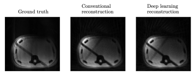 |
40 |
Fast Deep Learning Reconstruction of Interventional MRI Data
with Radial, Undersampling k-Space Trajectories
Johanna Topalis1,
Balthasar Schachtner1,
Andreas Mittermeier1,
Philipp Wesp1,
Tobias Weber1,2,
Anna Theresa Stüber1,2,
Max Seidensticker1,
Jens Ricke1,
Katia Parodi3,
Michael Ingrisch1,
and Olaf Dietrich1
1Department of Radiology, University Hospital, LMU Munich, Munich, Germany, 2Department of Statistics, LMU Munich, Munich, Germany, 3Department of Medical Physics, Faculty of Physics, LMU Munich, Munich, Germany Keywords: Image Reconstruction, Machine Learning/Artificial Intelligence, Radial Acquisition; Undersampled; MR-Guided Interventions To reduce the data acquisition time and increase frame rates for image guidance during percutaneous needle interventions in the liver, k-space data can be acquired with a radial, undersampling acquisition scheme. The purpose of this work was to optimize a deep learning model for the fast reconstruction of k-space data in this context. The proposed deep learning model reconstructed artificial data with a better image quality compared to conventional reconstruction. Successful reconstruction of interventional phantom data suggests its potential for application during percutaneous needle interventions in the liver. |
|
 Back to Meeting Home
 Back to the Program-at-a-Glance
The International Society for Magnetic Resonance in Medicine is accredited by the Accreditation Council for Continuing Medical Education to provide continuing medical education for physicians.
 Back to Meeting Home
 Back to the Program-at-a-Glance
 View Presentation Video