Digital Poster
Quantitative Imaging, AI & Miscellaneous
ISMRM & ISMRT Annual Meeting & Exhibition • 03-08 June 2023 • Toronto, ON, Canada

Digital Poster
Quantitative Imaging, AI & Miscellaneous
| Computer # | |||
|---|---|---|---|
2411.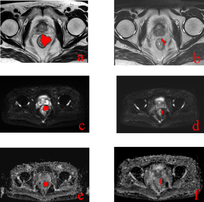 |
61 | MRI Radiomics Signature to Predict Lymph Node Metastasis after Neoadjuvant Chemoradiation Therapy in locally advanced Rectal Cancer
Zhu Fang1, Hong Pu1, Xiaoli Chen2, Yi Yuan1, Feng Zhang1, and Hang Li1
1Department of Radiology, Sichuan Academy of Medical Sciences and Sichuan Provincial People's Hospital, Chengdu, China, 2Department of Radiology, Affiliated Cancer Hospital of Medical School, University of Electronic Science and Technology of China, Sichuan Cancer Hospital, Chengdu, China Keywords: Radiomics, Radiomics To develop and validate clinical-radiomics models for predicting lymph node metastasis following neoadjuvant chemoradiation therapy in locally advanced rectal cancer .83 patients were retrospectively enrolled.pre-,post- and delta radiomics signatures of T2WI and ADC images were constructed by support vector machine model.These models were applied to predict LNM and 5-year disease-free survival.The clinical-deltaADC radiomics combined model presented good performance for predicting post-CRT LNM in the training cohort (AUC=0.895) and validation cohort (AUC=0.900). In ypT0-T2 stage, this model could predict 5-year RFS. Clinical-deltaADC radiomics combined model has good performance to predict LNM after nCRT and helped identify patients with poor prognosis. |
|
2412.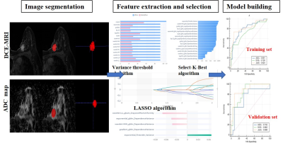 |
62 | The value of whole volume radiomics machine learning model based on multi-parameter MRI in predicting triple negative breast cancer
Tingting Xu1, Xueli Zhang1, Ting Hua1, Guangyu Tang1, lin Zhang1, and Xiance Zhao2
1Radiology, Shanghai Tenth People’s Hospital, School of Medicine, Tongji University, Shanghai, China, 2Philips Healthcare, Shanghai, China Keywords: Radiomics, Machine Learning/Artificial Intelligence, Triple-negative breast cancer. DCE-MRI. ADC maps 43 TNBCs and 84 Non-TNBCs were allocated in this retrospective study.The lesions were manually segmented with ITK-SNAP software then whole volume radiomics features were extracted with Radcloud radiomics platform based on DCE-MRI and ADC maps, respectively. Three prediction models were constructed by using support vector machine (SVM) classifier, including Model A (based on the selected features of ADC maps), Model B (based on the selected features of DCE-MRI), and Model C (based on the selected features of both combined). The radiomics features model combining DCE-MRI and ADC maps can improve the diagnostic performance of predicting TNBC. |
|
2413.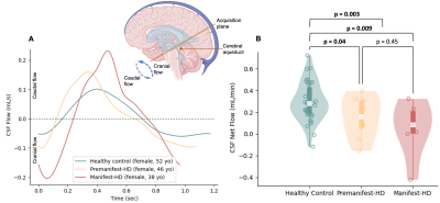 |
63 | MRI biomarkers of neurofluid production and egress provide support for aberrations in the neurofluid circuit in Huntington’s disease
Kilian Hett1, Colin D. McKnight2, Jarrod Eisma1, Adreanna B. Hernandez1, Melanie Leguizamon1, Jason Elenberger1, Ciaran M. Considine1, Daniel O. Claassen1, and Manus J. Donahue1
1Department of Neurology, Vanderbilt University Medical Center, Nashville, TN, United States, 2Department of Radiology, Vanderbilt University Medical Center, Nashville, TN, United States Keywords: Data Analysis, Neurodegeneration The overall goal of this work is to apply novel MRI methods, sensitive to CSF production and clearance, to test fundamental hypotheses regarding altered neurofluid circulation in patients with Huntington’s disease (HD). In participants with HD and healthy age- and sex-matched controls, we applied multi-modal MRI to investigate deviations of neuroimaging biomarkers of the neurofluid circuit at different locations: choroid plexus, cerebral aqueduct, and PSD. Analyses show a significant increase in ChP volume and PSD volume, and a decrease in ChP perfusion and CSF net flow, in HD participants relative to healthy controls. |
|
2414.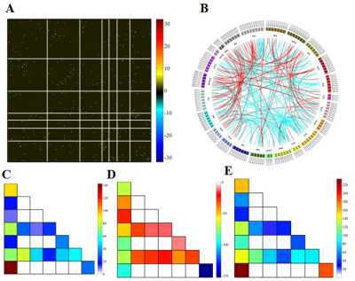 |
64 | Associated high-order resting-state functional connectivity network for diagnosis of schizophrenia
Dafa Shi1, Haoran Zhang1, Guangsong Wang1, and Ke Ren1
1Department of Radiology, Xiang’an Hospital of Xiamen Uneversity,School of Medicine, Xiamen University, Xiamen, China Keywords: Data Analysis, Brain Connectivity, high-order functional connectivity Schizophrenia (SZ) is one of the most prevalent mental disorders; however, its accurate diagnosis is difficult in clinical practice. Currently, the underlying mechanism of SZ remains poorly understood. The associated higher-order functional connectivity (HOFC) which constructed based on the conventional FC is promising for understanding pathological changes of brain connectome. In our study,we found the model constructed with associated HOFC outperformed the model constructed with conventional FC. SZ-related brain regions were widely distributed in frontal, parietal, insula, occipital, subcortical, and limbic lobes, which are the core brain areas of the subcortical, fronto-parietal, sensorimotor, limbic, and default mode networks. |
|
2415.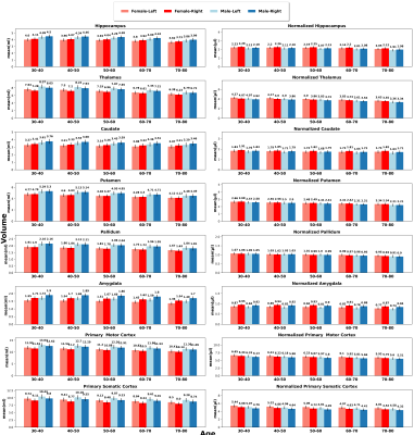 |
65 | Volumetric analysis of Brain Regions: A cross-sectional study on a large healthy dataset
Nasrin Akbari1, Saurabh Garg1, Thanh-Duc Nguyen1, Saqib Basar1, Mostafa Fatehi2, Yosef Chodakiewitz3, Rajpaul Attariwala1,3, Sean London3, and Sam Hashemi1,3
1Voxelwise Imaging Technology Inc, Vancouver, BC, Canada, 2Vancouver General Hospital, Vancouver, BC, Canada, 3Prenuvo, Vancouver, BC, Canada Keywords: Data Analysis, Brain, A cross-sectional study, Brain quantification, Brain volume analysis Cross-sectional neuroimaging studies have indicated hemispheric asymmetry, gender differences and a decline in brain volume with advancing age. However, these studies often have a small sample size (≤ 500) and unbalanced groupings, resulting in contradictory findings that might not be reproducible. Precise estimates on a large cohort will facilitate the assessment and quantification of brain volume, resulting in improved diagnostics and establishing statistical norms. In this study we analyze 6739 individuals to draw normative conclusions about typical patterns of age-related brain changes. We provide whole-brain and regional volume aging-curves for males and females for raw and head-size adjusted data. |
|
2416.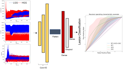 |
66 | Classification of low- and high-grade gliomas through multimodal temporal MRI and PET data
Marianna Inglese1,2, Matteo Ferrante1, Tommaso Boccato1, Shah Islam3, Matthew Williams4,5, Adam D Waldman6, Kevin O'Neill7, Eric O Aboagye3, and Nicola Toschi1,8
1Biomedicine and Prevention, University of Rome Tor Vergata, Rome, Italy, 2Surgery and Cancer, Imperial College London, London, United Kingdom, 3Department of Surgery and Cancer, Imperial College London, London, United Kingdom, 4Computational Oncology Group - Department of Surgery and Cancer, Imperial College London, London, United Kingdom, 5Institute for Global Health Innovation, Imperial College London, London, United Kingdom, 6Centre for Clinical Brain Sciences, University of Edinburgh, Edimburgh, United Kingdom, 7Imperial College Healthcare NHS Trust, London, United Kingdom, 8Department of Radiology, Athinoula A. Martinos Center for Biomedical Imaging, Boston, NY, United States Keywords: Data Processing, Modelling, Deep Learning, convolutional filters Stratifying human brain gliomas using imaging techniques is extremely challenging. Valuable insight into the characterization and classification of gliomas can be provided by integrating two imaging modalities, i.e. 18F-FPIA PET and MRI. This study introduces a new approach for glioma stratification based on the extraction of temporal features from tissue time activity curves (TACs) extracted from dynamic PET/MRI data. We exploit tissue-specific biochemical properties embedded in the TACs through deep learning and achieve good discrimination results while foregoing pharmacokinetic fitting and hence invasive measurement of the AIF. |
|
2417.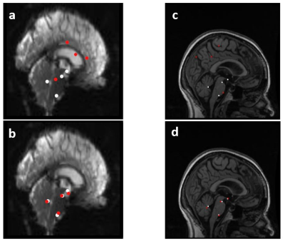 |
67 | Implementation of a convolutional neural network for brainstem landmark detection and co-registration
Owen Bleddyn Woodward1, Ian Driver1, Michael Germuska1, and Richard Wise2
1CUBRIC, Cardiff University, Cardiff, United Kingdom, 2Department of Neuroscience, Imaging and Clinical Science, 'G. D'Annunzio University' of Chieti-Pescara, Chieti, Italy Keywords: Data Processing, Machine Learning/Artificial Intelligence, Co-registration Accurate brainstem co-registration is important when analysing brainstem functional MRI data. We trained a convolutional neural network (CNN) to detect a set of brainstem landmarks and to define a brainstem region-of-interest and used these to co-register the brainstem between functional and anatomical space using previously developed landmark-based and automated brainstem co-registration (LBC and ABC) methods. The use of CNNs to supply these features to LBC and ABC produced results that compared well to conventional methods. Similar CNNs could be applied to other brain regions and such methods may be useful to automate the analysis of large functional datasets. |
|
2418.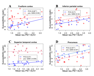 |
68 | DECOMPOSE-QSM improves the tracking of iron-related neurodegenerative pathology
Maruf Ahmed1, Jingjia Chen1, Yann Cobigo2, Suzanne Baker3, and Chunlei Liu1,4
1EECS, University of California, Berkeley, Berkeley, CA, United States, 2University of California, San Francisco, San Francisco, CA, United States, 3Lawrence Berkeley National Laboratory, Berkeley, CA, United States, 4Helen Wills Neuroscience Institute, University of California, Berkeley, Berkeley, CA, United States Keywords: Data Processing, Quantitative Susceptibility mapping DECOMPOSE-QSM method is used to extract a sub-voxel level paramagnetic component susceptibility (PCS) in Alzheimer’s disease (AD) related neurodegeneration. Correlation between both PCS and thresholded QSM values with tau PET SUVr are studied. PCS shows stronger association strength with tau PET measures compared to susceptibility measured using only the thresholded positive voxels (positive QSM) in the QSM map in known regions related to tau pathological progression. By separating sub-voxel paramagnetic and diamagnetic sources, DECOMPOSE-QSM provides a more specific and sensitive measure of iron-related pathology. |
|
2419.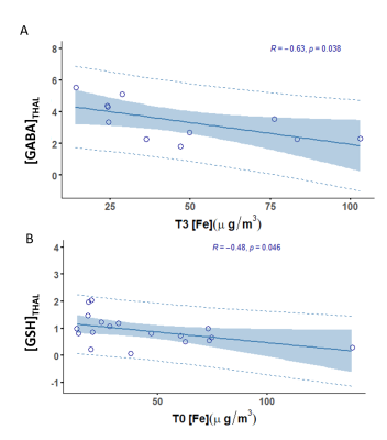 |
69 | Exposure to Welding Fumes: Evaluating the metabolite-metal relationship
Gianna Nossa1, Humberto Monsivais2, Chang Guen Lee2, Jae Hong Park2, and Ulrike Dydak2,3
1Purdue University, West Lafayette, IN, United States, 2School of Health Science, Purdue University, West Lafayette, IN, United States, 3Department of Radiology and Imaging Sciences, Indiana University School of Medicine, Indianapolis, IN, United States Keywords: Data Analysis, Toxicity Chronic exposure to Mn and Fe, through inhalation of welding fumes, is known to cause neurotoxicity. In this MRS study, we aim to determine if toenail Mn and Fe levels can be predictive of neurotoxicity identified through changes in GABA, GSH, and/or Glu concentrations. Significant GABA and GSH correlations with Fe indicate that toenail Fe concentrations can reflect decreased GABA and GSH levels in the welders’ brain. This emphasizes that Fe should not be ignored and might contribute to oxidative stress in Mn neurotoxicity. |
|
2420.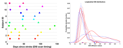 |
70 | Longitudinal Modeling of Stroke using Stochastic Distances and NODDI Diffusion Model
Anuja Sharma1 and Edward DiBella1
1Radiology and Imaging Sciences, University of Utah, Salt Lake City, UT, United States Keywords: Data Analysis, Machine Learning/Artificial Intelligence, Stroke diffusion modeling Limited prior work has explored longitudinal modeling of human brain stroke using advanced diffusion techniques. We aim to address this gap by analyzing longitudinal stroke data from diffusion spectrum imaging for modeling and predicting clinical markers of stroke recovery. Our proposed data analysis method uses a mixed-effect model which exploits stochastic distances from these images for improved regression model statistics and handling of imbalanced, inconsistent longitudinal data. We also demonstrate that this aids in differentiating between population-level and patient-level effects and the corresponding key contributing predictors at each of these levels. |
|
2421.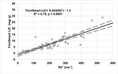 |
71 | Comparison of Single-Slice Liver R2* Estimation with FerriSmart for Assessment of Hepatic Iron overload
Juan Pablo Esparza1, Cara Morin2,3, Chris Goode3, Zachary Abramson3, and Aaryani Tipirneni-Sajja1,3
1Biomedical Engineering, The University of Memphis, Memphis, TN, United States, 2Radiology, Cincinnati Children's Hospital Medical Center, Cincinnati, OH, United States, 3Diagnostic Imaging, St. Jude Children's Research Hospital, Memphis, TN, United States Keywords: Data Analysis, Liver FerriScan is considered the gold standard to quantify liver iron content (LIC) by MRI. Recently, FerriSmart, an AI—based algorithm was developed to permit faster postprocessing of R2-based LIC. To our knowledge, FerriSmart results have not been compared to R2*-LIC methods. In this work, R2*, R2*-LIC, and FerriSmart LIC were compared to evaluate their agreement. |
|
2422.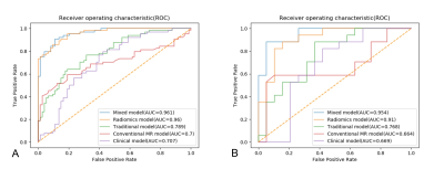 |
72 | Magnetic resonance imaging radiomics to differentiate ovarian sex cord-stromal tumors and primary epithelial ovarian cancers
Cheng Meiying1, Tan Shifang1, Ren Tian2, Zhu Zitao3, Wang Kaiyu4, Zhang Lingjie1, Meng Lingsong1, Yang Xuhong5, Yang Zhexuan1, and Zhao Xin1 1Department of Radiology, The Third Affiliated Hospital of Zhengzhou University, Zhengzhou, China, 2Department of Information, The Third Affiliated Hospital of Zhengzhou University, Zhengzhou, China, 3Wuhan University, Wuhan, China, 4MR Research China, GE Healthcare, Beijing, China, 5Huiying Medical Technology, Beijing, China Keywords: Machine Learning/Artificial Intelligence, Urogenital Ovarian sex cord-stromal tumors (SCSTs) are rare nonepithelial neoplasms that usually are benign or at early stages, but sometimes they are confused with malignant tumors such as epithelial ovarian cancers (EOCs). We constructed five models including clinical model, conventional MR model, traditional model, radiomics model and mixed model based on logistic regression classifier to distinguish SCSTs and EOCs. The performance of each model was evaluated. The radiomics approach showed excellent prediction results, and the mixed model stood out among all the models. |
|
2423.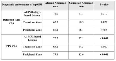 |
73 | Investigating Racial Disparities for the Performance of mpMRI in Diagnosis of Prostate Cancer
Fatemeh Zabihollahy1, Qi Miao2, Ida Sonni1, Harrison Kim3, Robert E Reiter4, Steven S Raman1, and Kyunghyun Sung1
1Department of Radiological Sciences, David Geffen School of Medicine at University of California, Los Angeles, Los Angeles, CA, United States, 2The First Affiliated Hospital of China Medical University, Shenyang City, China, 3Department of Radiology, University of Alabama at Birmingham, Birmingham, AL, United States, 4Department of Urology, David Geffen School of Medicine at University of California, Los Angeles, Los Angeles, CA, United States Keywords: Data Analysis, Multimodal Race and ethnicity strongly impact the risk of prostate cancer (PCa). In this study, the differences in the performance of mpMRI for PCa diagnosis are investigated between African American (AA) and Caucasian American (CA) men. After matching commonly known clinical risk factors, the diagnostic performances were compared between two racial groups regarding cancer prevalence, detection rates, and positive predictive values. Our result demonstrated that there are significant differences between AA and CA men in detecting clinically significant PCa when stratified by different prostate zones. |
|
2424. |
74 | Evaluation of renal function in chronic kidney disease using histogram analysis based on diffusion multi-models
Guimian Zhong1, Mengzhu Wang2, Qijia Han1, and Zhiming Xiang1
1Department of Radiology, Guangzhou Panyu Center Hospital, Guangzhou, China, 2Siemens Healthineers Ltd, Guangzhou, China Keywords: Radiomics, Diffusion/other diffusion imaging techniques, Histogram analysis;magnetic resonance imaging;Renal function This study established and validated a predictive model based on histogram features of four diffusion models to identify and evaluate early renal impairment in CDK. The results suggested that the model based on the ADC and MK could distinguish the normal and mild CDK well, and could accurately and noninvasively evaluate and predict early CDK renal dysfunction. |
|
2425. |
75 | Novel subject-specific method of visualising group differences from multiple DTI metrics without averaging
Eryn E Kwon1,2,3, Maryam Tayebi1,3, Joshua McGeown1,2, Matthew McDonald1,2, Patrick McHugh1,4, Paul Condron1,2, Leigh Potter1,5, Davidson Taylor1,6, Jerome Maller7, Miao Qiao8, Alan Wang1,2,3, Poul Nielsen3,8, Justin Fernandez1,3,8, Miriam Scadeng1,2, Samantha Holdsworth1,2, and Vickie Shim1,3
1Mātai Medical Research Institute, Tairāwhiti/Gisborne, New Zealand, 2Faculty of Medical and Health Sciences & Centre for Brain Research, University of Auckland, Auckland, New Zealand, 3Auckland Bioengineering Institute, University of Auckland, Auckland, New Zealand, 4Tūranga Health, Tūranganui-a-kiwa, Tairāwhiti/Gisborne, New Zealand, 5Ngāti Porou, Ngāti Kahungunu, Rongomaiwahine, Rongowhakaata, Tairāwhiti/Gisborne, New Zealand, 6Ngai Tāmanuhiri, Rongowhakaata, Ngāti Porou, Tairāwhiti/Gisborne, New Zealand, 7General Electric Healthcare, Victoria, Australia, 8Department of Engineering Science, Faculty of Engineering, University of Auckland, Auckland, New Zealand Keywords: Data Analysis, Data Analysis, DTI, diffusion MRI, PCA, Mesh-fitting, subject-specific model, data aggregation Averaging is commonly used for data reduction/aggregation to summarise such high-dimensional data, resulting in information loss. However, individual variability makes group-wise comparisons difficult without data reduction/aggregation. To address these issues, we developed a novel technique that integrates diffusion tensor (DTI) metrics along the whole volumes of the fibre bundle using a mesh-fitting technique. Using the right Corticospinal Tract (rCST) as an example, we demonstrate the utility of the method in detecting differences in DTI metrics of contact-sports and non-contact-sports athletes. |
|
2426.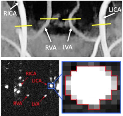 |
76 | Radiomics Applied to Phase Contrast MRI Images Successfully Distinguishes Healthy Subjects and Multiple Sclerosis Patients
Eros Montin1,2, Marco Muccio1, Yulin Ge1, and Riccardo Lattanzi1,2,3
1Center for Advanced Imaging Innovation and Research (CAI2R) Department of Radiology, Radiology Department, New York University Grossman School of Medicine, New York, New York, USA, New York, NY, United States, 2Bernard and Irene Schwartz Center for Biomedical Imaging, Department of Radiology, New York University Grossman School of Medicine, New York, New York, USA, New York, NY, United States, 3Vilcek Institute of Graduate Biomedical Sciences, New York University Grossman School of Medicine, New York, New York, USA, New York, NY, United States Keywords: Radiomics, Multiple Sclerosis In this study, we applied imaged-based radiomic techniques to phase contrast (PC) MRI images to distinguish the blood flow through the neck-feeding arteries of healthy controls (HC) from MS patients. By applying a simple machine learning model, k nearest neighbor, we found that first order features of the arteries’ regions of interest (ROI), drawn on phase images, reported the best accuracy (0.80) in labeling MS patients and HCs. PC-MRI is a fast and reliable imaging technique that, in conjunction with radiomics, offers great clinical potential to further quantify the diagnosis of MS, currently relying on qualitative approaches. |
|
2427.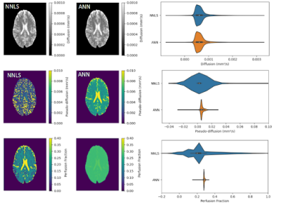 |
77 | Artificial Neural Network for Intravoxel Incoherent Motion Fitting: an Application in Glioma
Lucas Murilo da Costa1, André Monteiro Paschoal2, and Renata Leoni1
1University of São Paulo, Ribeirão Preto, Brazil, 2Instituto de Radiologia e Departamento de Radiologia e Oncologia, Faculdade de Medicina, Universidade de Sao Paulo, São Paulo, Brazil Keywords: Data Processing, Diffusion/other diffusion imaging techniques We proposed an Artificial Neural Network (ANN) to fit the IVIM images and estimate diffusion, pseudo-diffusion, and perfusion fraction values. The ANN was first trained with simulated data and tested with images of 40 healthy controls and one patient with glioma. ANN provided parameter values similar to those obtained with a standard fitting method, but D* maps with less noise. The ANN fitting process was less time-consuming and was shown to be a possible tool for replacing a standard fitting algorithm. |
|
2428. |
78 | Post mortem magnetic resonance imaging simulation system
Hiroyuki Kabasawa1, Masatoshi Kojima2, Daisuke Yajima3, and Yohsuke Makino4
1Department of Radiological Sciences, Internationa University of Health and Walfare, Narita, Japan, 2Department of Legal Medicine, Chiba University, Chiba, Japan, 3Department of Forensic Medicine, International University of Health and Welfare, Narita, Japan, 4Department of Forensic Medicine, The University of Tokyo, Tokyo, Japan Keywords: Software Tools, Software Tools, post mortem imaging A MR Image simulation system for post mortem subject scanning is developed and validated. The system has MR console like user interface. It has body temperature input as well as tissue fixation condition input, so that post mortem MR imaging can be simulated for various scanning condition. The system was validated for T1 weighted image and FLAIR for low body temperature condition. The result showed the system could simulate the actual acquired image contrast change associated with body temperature change. The proposed system can be used to predict MR contrast change associated with body temperature as well as fixation condition. |
|
2429.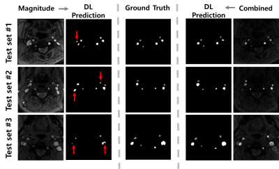 |
79 | Automatic Quantification of Total Cerebral Blood Flow from Phase-Contrast MRI Using Deep Learning
Jinwon Kim1, Hyebin Lee2, Jinhee Jang2, and Hyunyeol Lee1
1School of Electronic and Electrical Engineering, Kyungpook National University, Daegu, Korea, Republic of, 2Department of Radiology, Seoul St. Mary’s Hospital, College of Medicine, The Catholic University of Korea, Seoul, Korea, Republic of Keywords: Machine Learning/Artificial Intelligence, Quantitative Imaging In this work, we aim to develop a deep learning (DL)-based processing pipeline that enables rapid and correct segmentation of brain-feeding arteries in neck phase-contrast (PC) MR images, thereby achieving accurate quantification of total cerebral blood flow (tCBF) in an automated manner. To this end, we implemented a U-Net architecture where magnitude/phase-combined PC images are provided for network training. The results suggest that the present, automated method yields accurate measurements of tCBF in comparison to ground truth values obtained from manual vessel segmentation. |
|
2430.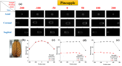 |
80 | Adverse effect of non-linearity in gradient on MRI and their impact on localization and morphology of testing sample
Rajesh Harsh1, URVASHI SINGH1, and Dharmesh Verma1
1Medical Systems Division, S.A.M.E.E.R. Mumbai, Mumbai, India Keywords: Data Analysis, Artifacts Study demonstrate the analysis of surface morphology and sample localization with variation from iso-center in the presence of non-linearity in gradient. In these experiments, we have analyzed that gradient non-linearity plays a crucial role in surface morphology and localization of samples. For the end position 200 mm and -200 mm, size of the object appears small. Additionally, the results suggest that when a large sample size is taken into account, gradient non-linearity exhibits greater negative impacts. |
|
 Back to Meeting Home
 Back to the Program-at-a-Glance
The International Society for Magnetic Resonance in Medicine is accredited by the Accreditation Council for Continuing Medical Education to provide continuing medical education for physicians.
 Back to Meeting Home
 Back to the Program-at-a-Glance
 View Presentation Video