Digital Poster
AI Application in Body Imaging I
ISMRM & ISMRT Annual Meeting & Exhibition • 03-08 June 2023 • Toronto, ON, Canada

Digital Poster
AI Application in Body Imaging I
| Computer # | |||
|---|---|---|---|
1467.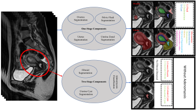 |
1 | A Deep Learning-Based Tool for Analyzing the Female Reproductive System in MR images
Javad Khaghani1, Saqib Basar1, Yosef Chodakiewitz2, Sean London2, Rajpaul Attariwal1, and Sam Hashemi1
1Voxelwise Imaging Technology Inc., Vancouver, BC, Canada, 2Prenuvo, Vancouver, BC, Canada Keywords: Uterus, Data Analysis MRI is a powerful imaging technique for examining the anatomy of the female reproductive system. However, due to cost-related concerns, ease of access, acquisition time, and necessity of expert reviewers for MR images, ultrasound is the primary modality of choice. To mitigate some of these concerns, we developed an AI-driven tool comprising seven neural networks that segments the regions of interest for the whole uterus, uterine zones, ovaries, and further identifies common benign gynecological conditions. We evaluated our package on a large representative population of 2955 sagittal T2-weighted pelvic scans to obtain normative aging-curves for various regions of interest. |
|
 |
1468.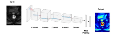 |
2 | Deep-learning based rectal tumor segmentation on multi-parametric MRI using pre-treatment and near-end-treatment data
Yang Zhang1,2, Liming Shi3, Xiaonan Sun3, Min-Ying Su2, Taoran Cui1, Ning J. Yue1, and Ke Nie1
1Department of Radiation Oncology, Rutgers-Cancer Institute of New Jersey, New Brunswick, NJ, United States, 2Department of Radiological Sciences, University of California, Irvine, CA, United States, 3Department of Radiation Oncology, Department of Radiation Oncology, Sir Run-run Shaw Hospital, Zhejiang University School of Medicine, Hangzhou, China Keywords: Cancer, Machine Learning/Artificial Intelligence In this study, the convolutional neural network (CNN) was implemented to segment the rectal cancer in 185 patients, with tumor ROI outlined by a radiologist on multiparametric magnetic resonance images (mp-MRI). The Dice similarity coefficient (DSC) and mean symmetrical surface distance (MSD) values were used to evaluate the results of the proposed algorithm. The mean DSC and MSD were 0.88 and 2.2 cm respectively for pre-treatment MR images. The transfer-learning model using paired pre-treatment and near-end-treatment images could also lead to acceptable result for boost volume segmentation with the mean DSC of 0.83 and MSD of 2.7 cm. Our work showed the deep-learning with combined image sequences was promising for automatic tumor localization and segmentation of locally advanced rectal cancer. After applying transfer learning with the prior information obtained from the pre-treatment images, the proposed method can be further used to identify boost volume for radiation dose escalation which has been demonstrated beneficial in improving pathological complete response rate. |
|
1469.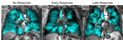 |
3 | Hyperpolarized 129Xe MRI ventilation textures predict short- and long-term response to Anti-IL-5Rα Biologic Therapy in Eosinophilic Asthma
Marrissa J McIntosh1,2, Maksym Sharma1,2, Harkiran K Kooner1,2, Hana Serajeddini2,3, Anurag Bhalla3, Cory Yamashita3, and Grace Parraga1,2,3,4
1Department of Medical Biophysics, Western University, London, ON, Canada, 2Robarts Research Institute, London, ON, Canada, 3Division of Respirology, Department of Medicine, Western University, London, ON, Canada, 4School of Biomedical Engineering, Western University, London, ON, Canada Keywords: Lung, Hyperpolarized MR (Gas), asthma, biologic therapy It was previously shown that 129Xe MR ventilation images contain embedded texture features which help predict (before treatment was initiated) those severe asthma patients who were more likely to experience early response to anti-IL-5Rα biologic therapy. Thus, we postulated that such texture features would also help identify patients with an enduring, late response. Here we identified specific 129Xe MRI features that predicted both early and late response to anti-IL-5 therapy, which were superior to clinical measurements. These promising results suggest that 129Xe MRI texture features sensitively predict patients with response to biologic therapy interventions. |
1470.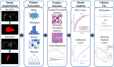 |
4 | Delta-Radiomic Based on Dynamic Contrast Enhanced MRI Predicts axillary Response after neoadjuvant chemotherapy in breast cancer patients
Shasha Liu1, Siyao DU1, Si Gao1, Lizhi Xie2, Yuee Teng3, Feng Jin4, and Lina Zhang1
1Department of Radiology, The First Hospital of China Medical University, shenyang, China, 2MR Research, GE Healthcare, Beijing, China, 3Departments of Medical Oncology and Thoracic Surgery, The First Hospital of China Medical University, shenyang, China, 4Department of Breast Surgery, The First Hospital of China Medical University, shenyang, China Keywords: Breast, Radiomics, DCE-MRI This prospective study with multiple follow-up time points investigated the early predictive value of the delta-radiomic model of axillary lymph node (ALN) using dynamic contrast-enhanced (DCE) MRI for axillary pathological complete response (pCR) in breast cancer patients after neoadjuvant chemotherapy (NAC). The results indicated that the delta-radiomic model based on early changes of ALN features performed better among all radiomic models. Moreover, when combined with clinical features, the combined model achieved the best diagnostic performance of any model we tested. The delta-radiomic + clinical model may be a promising method for ALN pCR prediction in the initial phase of NAC. |
|
1471.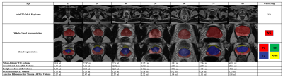 |
5 | Deep Learning Strategy to Quantify Whole Prostate and Zonal Volumes, Trends in Aging and Detection of Benign Prostatic Hyperplasia
Javad Khaghani1, Lucas Porto2, Saurabh Garg1, Saqib Basar1, Yosef Chodakiewitz3, Sean London3, Rajpaul Attariwal1, and Sam Hashemi1
1Voxelwise Imaging Technology Inc., Vancouver, BC, Canada, 2Voxelwise Imaging Technology Inc., San Francisco, CA, United States, 3Prenuvo, Vancouver, BC, Canada Keywords: Prostate, Data Analysis, Artificial Intelligence/ Machine Learning AI-assisted prostate whole-gland and zonal volume quantification enable quantitative reproducibility and enhance read-time efficiency. Our whole-gland AI-segmentation solution enhanced measurement accuracy by 23.59%, compared with traditional volume estimates. Zonal solution enabled us to generate population normative aging-curves and we used a shallow classifier to identify patients with BPH. Our findings show the transitional zone grows 2.05 ml and 3.58 ml per decade for the entire population and patients with BPH, respectively, while peripheral zone grows 0.70 ml per decade. Further, the transitional and peripheral zones grow 2.43 and 1.4 times their size over lifetime. |
|
1472.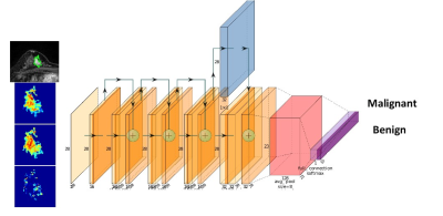 |
6 | Integration of Slice-Based Diagnostic Probabilities Predicted by 2D Deep Learning Using ResNet50 to Yield Lesion-Based Diagnosis in Breast MRI
Yang Zhang1,2, Jiejie Zhou3, Zhongwei Chen3, You-Fan Zhao3, Meihao Wang3, Yan-Lin Liu2, Jeon-Hor Chen2, Ke Nie1, and Min-Ying Su2
1Department of Radiation Oncology, Rutgers-Cancer Institute of New Jersey, New Brunswick, NJ, United States, 2Department of Radiological Sciences, University of California, Irvine, CA, United States, 3Department of Radiology, First Affiliated Hospital of Wenzhou Medical University, Wenzhou, China Keywords: Cancer, Machine Learning/Artificial Intelligence A deep learning model using 2D ResNet50 CNN was trained to differentiate a dataset of 103 malignant vs 73 benign breast lesions, then tested in a testing dataset of 53 malignant and 31 benign cases. The 2D slice-based results were used to calculate a probability for each lesion, by using 5 methods: (1) slice-based average, (2) tumor area weighted average, (3) tumor perimeter weighted average, (4) using the probability of the largest tumor slice, (5) using the highest probability among all slices. The results showed using the highest probability to convert from slice-based to lesion-based diagnosis had the best performance. |
|
1473.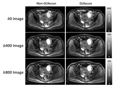 |
7 | Deep Learning Reconstruction to Pelvis Multi-Shot DWI Improved Image Quality with Less Image Distortion: A Preliminary Study
Elaine Yuen Phin Lee1, Chia-Wei Li2, Patricia Lan3, Xinzeng Wang4, Arnaud Guidon5, and Chien-Yuan Lin2
1Department of Diagnostic Radiology, Queen Mary Hospital, The University of Hong Kong, Hong Kong SAR, China, 2GE Healthcare, Taipei, Taiwan, 3GE Healthcare, Menlo Park, CA, United States, 4GE Healthcare, Houston, TX, United States, 5GE Healthcare, Boston, MA, United States Keywords: Pelvis, Machine Learning/Artificial Intelligence, Deep learning reconstruction, Multi-shot DWI This study introduced the deep learning reconstruction (DLRecon) to multi-shot DWI (MUSE-DWI) in the pelvis and aimed to investigate the changes in SNR and image quality with MUSE-DWI DLRecon. Compared with the MUSE-DWI non-DLRecon, the MUSE-DWI DLRecon showed higher SNR with stable ADC quantification. With that, DLRecon could reduce image distortion using higher shots MUSE-DWI with comparable SNR and scan time to clinically used 2-shot MUSE-DWI. This preliminary result showed the potential power of DLRecon in the pelvis allowing higher shots MUSE-DWI to be integrated in clinical practice to reduce image distortion. |
|
1474. |
8 | Predictive Model for Breast Lesion Assessment Using Restriction Spectrum Imaging
Stephane Loubrie1, Ana Rodriguez-Soto1, Tyler Seibert1,2,3, Michael Hahn1, Vandana Dialani4,5, Catherine Wei6, Zahra Karimi4, Joshua Kuperman1, Anders Dale1,7, Etta Pisano4,8, Jingjing Zou9, and Rebecca Rakow-Penner1,3
1Radiology, University of California, San Diego, San Diego, CA, United States, 2Radiation medicine, University of California, San Diego, San Diego, CA, United States, 3Bioengineering, University of California, San Diego, San Diego, CA, United States, 4Radiology, Beth Israel Hospital, Boston, MA, United States, 5Harvard Medical School, Boston, MA, United States, 6Commonwealth Radiology Associates, Boston, MA, United States, 7Neurosciences, University of California, San Diego, San Diego, CA, United States, 8American College of Radiology, Reston, VA, United States, 9Biostatistics, Herbert Wertheim School of Public Health and Human Longevity Science, University of California, San Diego, San Diego, CA, United States Keywords: Breast, Diffusion/other diffusion imaging techniques Diffusion weighted imaging (DWI) holds great potential in improving specificity of findings detected on contrast enhanced breast MRI. Restriction spectrum imaging (RSI), an advanced diffusion imaging model, has potential in discriminating between malignant and fibroglandular breast tissue. In this abstract, we evaluate RSI’s performance, combined with a random forest model, in differentiating lesions requiring biopsies from lesions that do not. The model showed significant preliminary results. |
|
1475.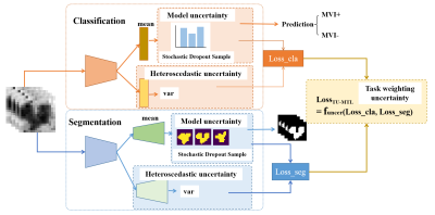 |
9 | Multi-task deep learning with triplet uncertainty for segmentation and microvascular invasion prediction of hepatocellular carcinoma
Yanyan Xie1, shangxuan Li1, Baoer Liu2, Yikai Xu2, and Wu Zhou1
1School of Medical Information Engineering, Guangzhou University of Chinese Medicine, Guangzhou, China, 2Department of Medical Imaging Center, Nanfang Hospital, Southern Medical University, Guangzhou, China Keywords: Liver, Machine Learning/Artificial Intelligence Multi-task learning has been widely used for jointly tumor segmentation and classification. Uncertainty estimation of the subtask weight coefficient in multi-task learning has been investigated. However, due to the presence of noise in medical image, data uncertainty will affect the performance of multi-task learning. In addition, model uncertainty has not been conducted for multi-task learning. In this work, we propose a triplet-uncertainty in multi-task deep learning network (TU-MTL), simultaneously considering the uncertainty estimation of subtask weight coefficient, data uncertainty estimation and model uncertainty estimation. Experimental results of clinical hepatocellular carcinoma (HCC) demonstrate the effectiveness of the proposed method. |
|
1476.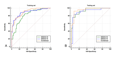 |
10 | Improved differentiation of BI-RADS 4 breast lesions based on ultrafast dynamic contrast-enhanced MRI radiomics and artificial neural network
Lingsong Meng1, Xin Zhao1, Jinxia Guo2, Lin Lu1, Meiying Cheng1, Qingna Xing1, Honglei Shang1, Yan Chen1, Penghua Zhang1, and Xiaoan Zhang1
1The Third Affiliated Hospital of Zhengzhou University, Zhengzhou, China, 2General Electric (GE) Healthcare, MR Research China, Beijing, Beijing, China Keywords: Breast, Radiomics Improving the assessment of Breast Imaging Reporting and Data System (BI-RADS) 4 lesions can avoid unnecessary biopsies. As an emerging field, radiomics has been successfully explored as a means to aid decision-making for the diagnosis and risk stratification of several kinds of cancers1-4. In this study, we combined radiomics features extracted from ultrafast dynamic contrast-enhanced magnetic resonance imaging (DCE-MRI) (using the Differential sub-sampling with cartesian ordering (DISCO) technique) with an artificial neural network (ANN) to improve diagnostic performance in assessing BI-RADS 4 lesions and evaluate the potential to avoid unnecessary biopsies. |
|
1477.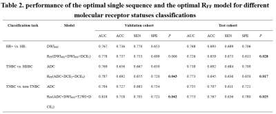 |
11 | Assessment of Breast Cancer Molecular Subtypes with a mMRI-based Feature Fusion Radiomics Model: Mimicking Radiologists’ Diagnostic Approach
Wanli Zhang1,2, Fangrong Liang1,2, Jiamin Li1,2, Yongzhou Xu3, Aaron Zhang3, Xinqing Jiang1,2, Xin Zhen4, and Ruimeng Yang1,2
1Department of Radiology, The Second Affifiliated Hospital, School of Medicine, South China University of Technology, Guangzhou, China, 2Department of Radiology, Guangzhou First People’s Hospital, Guangzhou, China, 3Philips Healthcare, Guangzhou, China, 4School of Biomedical Engineering, Southern Medical University, Guangzhou, China Keywords: Breast, Radiomics, breast cancer, molecular receptor status, feature fusion Since breast cancer is a highly heterogeneous tumor, prognosis and treatment response differ significantly according to different molecular subtypes. Based on multiparametric magnetic resonance imaging (MRI), we developed a feature fusion radiomics (RFF) model and investigated its performance in identifying the molecular receptor status of breast cancer. Mimicking the diagnostic approach of the radiologists by integrating image information from different MR sequences, the RFF model outperformed any single MRI-based radiomics model, demonstrating its potential for molecular subtypes classification of breast tumors. |
|
1478.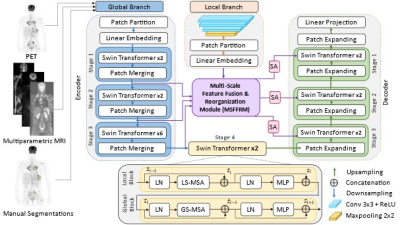 |
12 | Automated Segmentation of Metastatic Lymph Nodes in FDG PET/MRI for Lymphoma Cancer Patients Using Multi-Scale Swin Transformers
Anum Masood1,2, Sølvi Knapstad2, Håkon Johansen3, Trine Husby3, Live Eikenes 1, Mattijs Elschot 1,3, and Pål Erik Goa2,3
1Department of Circulation and Medical Imaging, NTNU, Trondheim, Norway, 2Department of Physics, NTNU, Trondheim, Norway, 3Department of Radiology and Nuclear Medicine, St. Olavs Hospital, Trondheim University Hospital, Trondheim, Norway Keywords: Machine Learning/Artificial Intelligence, PET/MR, Automated Segmentation, Deep Learning, Transformers Automated segmentation of tumors and designing a computer-assisted diagnosis system requires co-learning of imaging features from the complementary PET/MRI. We aim to develop an automated method for the segmentation of cancer-affected lymph nodes on Positron Emission Tomography/Magnetic Resonance Imaging (PET/MRI) using a modified Swin Transformer model (ST) integrated with a novel Multi-Scale Feature Fusion & Reorganization Module (MSFFRM). Our results with Hodgkin lymphoma (HL) and Non-Hodgkin lymphoma subtype Diffuse Large B-Cell Lymphoma (DLBCL) datasets show that our proposed model ST-MSFFRM achieved better performance in lesion segmentation in comparison to other state-of-the-art segmentation methods. |
|
1479.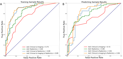 |
13 | Multi-parametric radiomics of MRI for preoperative assessment of microvascular invasion and prognosis of hepatocellular carcinoma
Lili Wang1, Junqiang Lei1, Junfeng Li2, Shunlin Guo2, Gang Wang2, and Rui Wang3
1Radiology, First Hospital of Lanzhou University, Lanzhou, China, 2First Hospital of Lanzhou University, Lanzhou, China, 3First Clinical Medical School of Lanzhou University, Lanzhou, Chile Keywords: Liver, Radiomics To preoperatively predict MVI in HCC patients, this study developed and validated an MVI nomogram prediction model that reveals the features derived from tumor and peritumor tissue of different sequences in Gd-EOB-DTPA dynamic contrast-enhanced MR and combines the clinical and radiological signatures. The nomogram included AFP, capsule appearance, arterial peritumoral enhancement, RVI, and radiomics score, which were independent risk factors for MVI. Finally, this nomogram model achieved satisfactory performance in predicting MVI in both the training and validation cohorts. Moreover, the RFS of the nomogram model was similar to the histopathology outcome. |
|
1480. |
14 | Non-invasively and Pre-Operatively Evaluate the Micro-Vascular Invasion with Susceptibility-Weighted Imaging based Radiomics Strategy
Zhijun Geng1, Yunfei Zhang2, Yongming Dai2, and Chuanmiao Xie1
1Sun Yat-sen University Cancer Center, Guangzhou, China, 2MR Collaboration, Central Research Institute, United Imaging Healthcare, Shanghai, China Keywords: Liver, Radiomics, Micro-Vascular Invasion MVI has widely been considered as an important prognostic biomarker of hepatocellular carcinoma (HCC). This study aims to non-invasively identify MVI in HCC with susceptibility-weighted-imaging (SWI)-derived radiomics. The results indicated that SWI-derived radiomics are valuable for noninvasively and accurately identifying the MVI status of HCC. Furthermore, the integration of radiomics and clinical factors yielded a predictive nomogram with satisfying diagnostic performance and potential clinical benefits. |
|
1481.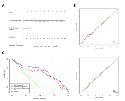 |
15 | Development and validation of an MRI-based radiomics nomogram for evaluation of chronic outcome in drug-induced liver injury
Zhehan Shen1, Ruokun Li1, and Fuhua Yan1
1Radiology, Ruijin Hospital Affiliated to Shanghai Jiao Tong University, Shanghai, China Keywords: Liver, Radiomics, drug-induced liver injury Drug-induced liver injury (DILI) occurs in a small fraction of individuals exposed to drugs, herbs or dietary supplements. DILI is challenging because of the lack of biomarkers to predict chronic outcomes. In our study, we developed a radiomics model with excellent performance for prediction of chronic outcome based on MRI and clinical data. Decision curve analysis confirmed the clinical utility of the radiomics model. This model may be used to stratify DILI patients at high risk of chronic outcome. |
|
1482.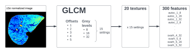 |
16 | Additional value of T2 mapping and texture analysis for the improvement to MR elastography based liver fibrosis Machine Learing classification
Diana Bencikova1, Sarah Poetter-Lang1, Marcus Raudner1, Martin Krššák2, Siegfried Trattnig1,3, and Ahmed Ba-Ssalamah1
1Department of Biomedical Imaging and Image-Guided Therapy, Medical University Vienna, Vienna, Austria, 2Division of Endocrinology and Metabolism, Department of Medicine III, Medical University Vienna, Vienna, Austria, 3Christian Doppler Laboratory for Clinical Molecular Imaging, Vienna, Austria Keywords: Liver, Machine Learning/Artificial Intelligence, Elastography, MR Value, Radiomics, Relaxometry MR elastography is currently the most accurate non-invasive diagnostic method for liver fibrosis. However, other pathologic processes co-existing with liver fibrosis influence the stiffness measurement and including other MR-based measures might improve fibrosis assessment. In this work we suggest to add texture analysis features calculated on liver T2 maps together with T2 values from fast radial turbo-spin-echo sequence into a machine-learning classification model and to compare the performance of the model after adding selected parameters. Our results show that including both, texture analysis and T2 values significantly improves the classification performance of the model. |
|
1483.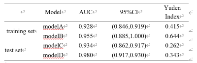 |
17 | Multiparametric MRI–based Radiomics Analysis of Endometrial Carcinoma for Preoperative Risk stratification
Bai min1
1Radiology, Liaocheng People's Hospital, Liao'cheng, China Keywords: Uterus, Radiomics Preoperative risk stratification of endometrial cancer (EC) is one of the important prognostic factors and is crucial for clinical treatment planning. We aimed to evaluate the predictive value of radiomics features based on multiparameter magnetic resonance imaging (MP-MRI) for preoperative risk stratification in patients with EC. The clinical and radiomic combined predictive models has a better performance than the model based on clinical characteristics. In the training set and the test set, the IDI and he reclassification improvement index of combined predictive models is higher than that of clinical model. |
|
1484.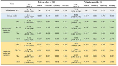 |
18 | Multi-parametric MRI-based Peritumoral Radiomics for Stage IIA and IIB Classification of Cervical Cancer:A Multicenter Study
Jin Fang1, Xiao Zhang2, Xiaoyun Liang2, Feng Huang2, and Shuixing Zhang1
1The First Affiliated Hospital of Jinan University, Guangzhou, China, 2Neusoft Medical Systems Co, Shanghai, China Keywords: Uterus, Radiomics Appropriate management and treatment decisions for cervical cancer depend on accurate staging, but the differentiation between IIA and IIB by imaging assessment remains difficult. This retrospective study investigated the performance of intratumoral and peritumoral multi-parameter MRI texture information in differentiating IIA and IIB using non-invasive radiomics analysis, which was also compared with clinical factors and imaging assessment by radiologists. Among all the comparisons, peritumoral-based radiomics models outperformed the radiologists and performed the best. This study offers a viable approach in non-invasively and accurately differentiating IIA and IIB for cervical cancer based on peritumoral texture information. |
|
1485.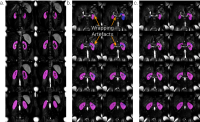 |
19 | Automated Segmentation of Multi-Vendor Kidney Images using an Iteratively Trained Convolutional Neural Network
Alexander J Daniel1, Eleanor F Cox1, Rachael A Evans2, Louise V Wain2, Christopher Brightling2, Elizabeth Tunnicliffe3,4, Stefan Neubauer3,4, Betty Raman3,4, and Susan T Francis1
1Sir Peter Mansfield Imaging Centre, University of Nottingham, Nottingham, United Kingdom, 2Institute for Lung Health, Leicester NIHR Biomedical Research Centre, University of Leicester, Leicester, United Kingdom, 3Division of Cardiovascular Medicine, Radcliffe Department of Medicine, Oxford University, Oxford, United Kingdom, 4Oxford University Hospitals NHS Foundation Trust, Oxford University, Oxford, United Kingdom Keywords: Kidney, Segmentation Measures of total kidney volume (TKV) help to evaluate disease progression, and masks to define the kidney are important for the automatic assessment of multiparametric images collected in the same data space. For accurate measures in multicentre studies an automated method which is vendor agnostic and robust against image artefacts is needed. Here a single-vendor convolutional neural network is retrained and shown to be accurate on two vendors of scanner and robust against image artefacts associated with wrapping in the phase-encode direction. |
|
1486.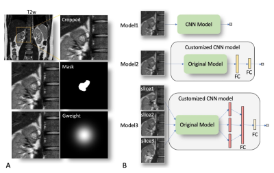 |
20 | Influential factors in deep learning classification of clear cell renal carcinoma cancer
Junyu Guo1, Keith Hulsey1, Yin Xi1, and Ivan Pedrosa1
1Radiology, UT Southwestern Medical Center, Dallas, TX, United States Keywords: Kidney, Machine Learning/Artificial Intelligence Deep learning has been successful in predicting tumor malignancy. Clear cell renal carcinoma (ccRCC) diagnosis may help in decision making between active surveillance and definitive intervention. In this study, we investigate the effects of different factors on the performance of deep learning classification of ccRCC using T2w images. We demonstrate that the performance of 15 different CNN models varied substantially. The choice of CNN models, the cropped image size, and the type of inputs greatly altered the performance of ccRCC classification. We achieved the best AUC value of 0.81, which is close to the reported performance of radiologists. |
|
 Back to Meeting Home
 Back to the Program-at-a-Glance
The International Society for Magnetic Resonance in Medicine is accredited by the Accreditation Council for Continuing Medical Education to provide continuing medical education for physicians.
 Back to Meeting Home
 Back to the Program-at-a-Glance
 View Presentation Video