Digital Poster
AI Application in Body Imaging II
ISMRM & ISMRT Annual Meeting & Exhibition • 03-08 June 2023 • Toronto, ON, Canada

Digital Poster
AI Application in Body Imaging II
| Computer # | |||
|---|---|---|---|
1644.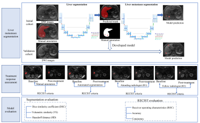 |
1 | Automatic Segmentation of Liver Metastases Based on a Deep Learning: Assessment of Tumor Treatment Response According to the RECIST 1.1 Criteria
xiang liu1 and xiaoying wang1
1peking university first hospital, Beijing, China Keywords: Liver, Cancer This retrospective study aims to develop an automated algorithm for segmentation of liver metastases based on a deep learning method and assess its efficacy for treatment response assessment according to the RECIST 1.1 criteria. One hundred and sixteen treated patients with clinically confirmed liver metastases were enrolled. A 3D U-Net algorithm was trained for automated liver metastases segmentation and treatment response assessment. The results demonstrated that the automated liver metastases segmentation was capable of evaluating treatment response, with comparable results to the junior radiologist and superior to that of the fellow radiologist. |
|
1645.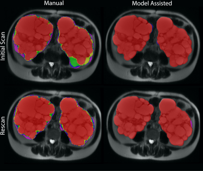 |
2 | Improving Kidney Volume Measurement Reproducibility in ADPKD by Averaging Measurements on Multiple Sequences
Hreedi Dev1, Chenglin Zhu1, Arman Sharbatdaran1, Syed I. Raza1, Sophie Wang1, Dominick J. Romano1, Akshay Goel1, Kurt Teichman1, Mina C. Moghadam1, George Shih1, Jon D. Blumenfeld1,2, Daniil Shimonov2, James Chevalier2, and Martin R. Prince1,3
1Radiology, Weill Cornell Medicine, New York, NY, United States, 2Rogosin Institute, New York, NY, United States, 3Radiology, Columbia University Vagelos College of Physicians and Surgeons, New York, NY, United States Keywords: Kidney, Kidney, MRI, ADPKD Organ volume measurements on MRI are typically performed on a single pulse sequence because of the tedious process of manual contouring. Here we use deep learning to automate kidney segmentations so that kidney volume can be measured on five abdominal MRI sequences. In 17 subjects scanned twice within 3 weeks (when no change in kidney volume was expected), the power of averaging 5 measurements improved reproducibility, achieving 2.5% absolute percent difference compared to 5.9% with manual contouring (p<0.05). Absolute percent error was reduced further to 2.1%, p<0.05 compared to manual segmentations, by excluding outlier measurements. |
|
1646.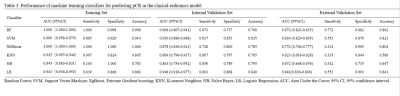 |
3 | MR-based Radiomics Models for Predicting Pathological Complete Response in Locally Advanced Rectal Cancer: A Two-Centre, Multi-Vendor Study
Qiurong Wei1, Weicui Chen1, Liting Mao1, Kan Deng2, Zhaoxian Yan1, Weikang Huang1, and Xian Liu1
1Radiology, The Second Affiliated Hospital of Guangzhou University of Chinese Medicine, Guangzhou, China, 2Philips Healthcare, Guangzhou, China Keywords: Cancer, Radiomics The response to neoadjuvant chemoradiotherapy (nCRT) in patients with locally advanced rectal cancer (LARC) is especially important for prognostic and management decisions. In this study, we used cross-vendor data from two centers to validate the generalization ability of radiomics model based on multiparametric-MRI (MP-MRI) for predicting pCR and to compare the discriminatory performance of different classifiers. Our results demonstrated that radiomics can be used to predict pCR. The clinical-radiomics model had superior performance compared to the radiomics model and clinical model. Furthermore, the RF classifier outperformed the other classifiers in prediction. |
|
1647.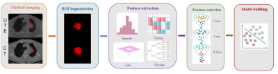 |
4 | Prediction of Histological Subtypes Using Machine-Learning Model Based on UTE-MRI in Non-Small Cell Lung Cancer:A comparative study with CT
Pengyang Feng1, Nan Meng2, Xuan Yu2, Yaping Wu2, Fangfang Fu2, Yu Luo2, Han Jiang3, Ziqiang Li3, Jianmin Yuan4, Yang Yang5, Zhe Wang4, and Meiyun Wang*2
1Department of Medical Imaging, Henan University People’s Hospital & Henan Provincial People’s Hospital, Zhengzhou, China, 2Department of Medical Imaging, Zhengzhou University People’s Hospital & Henan Provincial People’s Hospital, Zhengzhou, China, 3Department of Medical Imaging, Xinxiang Medical University Henan Provincial People’s Hospital, Zhengzhou, China, 4Central Research Institute, UIH Group, Shanghai, China, 5Beijing United Imaging Research Institute of Intelligent Imaging, UIH Group, Beijing, China Keywords: Cancer, Radiomics Three-dimensional ultrashort echo time (3D-UTE) is a novel MRI technique, which yields similar diagnostic results as conventional pulmonary computed tomography (CT). Our results showed that the predication model based on clinical factors and 3D-UTE radiomics features could noninvasively assess the subtype of in non-small cell lung cancer. Compared with the CT model, it has similar diagnostic efficiency but less radiation, which is expected to provide new ideas for related research. |
|
1648.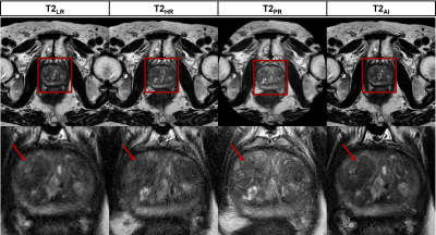 |
5 | AI-based reconstruction of T2-weighted sequences in prostate MRI: clinical evaluation and impact on diagnostic confidence
Leon Bischoff1,2, Alexander Isaak1,2, Christoph Katemann3, Dmitrij Kravchenko1,2, Narine Mesropyan1,2, Christoph Endler1,2, Barbara Wichtmann1,2, Oliver Weber3, Johannes Peeters4, Claus Christian Pieper1, Daniel Kütting1,2, Alois Martin Sprinkart1,2, Ulrike Attenberger1, and Julian Luetkens1,2
1Department of Diagnostic and Interventional Radiology, University Hospital Bonn, Bonn, Germany, 2Quantitative Imaging Lab, University Hospital Bonn, Bonn, Germany, 3Philips GmbH Market DACH, Hamburg, Germany, 4Philips MR Clinical Science, Best, Netherlands Keywords: Prostate, Machine Learning/Artificial Intelligence, Prostate cancer In this prospective study, 56 male patients with suspected prostate cancer were included to evaluate an artificial intelligence (AI) based reconstruction method for T2-weighted sequences in multiparametric MRI (mpMRI). After comparison with conventionally acquired and reconstructed sequences we found the new AI-based method to produce images with higher image sharpness and delineation of lesions, confirmed by both qualitative and quantitative analysis. This is accompanied by a reduction of scan time by 29-37%. Confidence in the assessed PI-RADS scores was significantly higher for the AI-reconstruction. This new technique could therefore potentially increase diagnostic accuracy of mpMRI of the prostate. |
|
1649.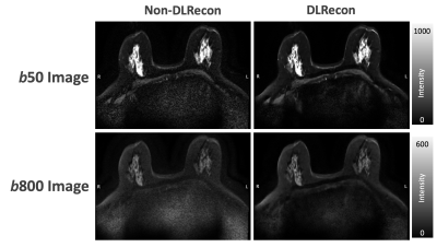 |
6 | Deep Learning Based Reconstruction for Multi-shot DWI of the Breast: A Preliminary Study
Ning Chien1, Cheng-Ya Yeh1, Yi-Chen Chen1, Yeun-Chung Chang2, Chia-Wei Li3, Chien-Yuan Lin3, Patricia Lan4, Xinzeng Wang5, Arnaud Guidon6, and Kao-Lang Liu1
1Department of Medical Imaging, National Taiwan University Cancer Center and National Taiwan University College of Medicine, Taipei, Taiwan, 2Department of Medical Imaging, National Taiwan University Hospital and National Taiwan University College of Medicine, Taipei, Taiwan, 3GE Healthcare, Taipei, Taiwan, 4GE Healthcare, Menlo Park, CA, United States, 5GE Healthcare, Houston, TX, United States, 6GE Healthcare, Boston, MA, United States Keywords: Breast, Machine Learning/Artificial Intelligence, Deep learning reconstruction, Multi-shot DWI Diffusion-weighted imaging (DWI) in the breast is limited by image distortion, which can be improved with multi-shot DWI (MUSE). We conducted a pilot study to investigate the impact of deep-learning reconstruction (DLRecon) on MUSE image quality. Compared with the non-DL MUSE images, the MUSE DLRecon showed higher SNR without altering the mean ADC value. Moreover, the higher shots MUSE DL with reduced NEX could provide less-distortion DWI with comparable SNR and scan time to 2-shot MUSE imaging, which is commonly used in the clinical setting. Preliminary results indicate the feasibility of MUSE-DWI in the breast with higher number of shots. |
|
1650.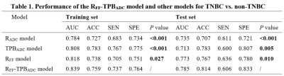 |
7 | Multiparametric MR-based Feature Fusion Radiomics Combined with ADC Maps-based Tumor Proliferative Burden in Distinguishing TNBC vs. non-TNBC
Fangrong Liang1,2, Wanli Zhang1,2, Jiamin Li1,2, Yongzhou Xu3, Aaron Zhang3, Xinqing Jiang1,2, Xin Zhen4, and Ruimeng Yang1,2
1Department of Radiology, The Second Affifiliated Hospital, School of Medicine, South China University of Technology, Guangzhou, China, 2Department of Radiology, Guangzhou First People’s Hospital, Guangzhou, China, 3Philips Healthcare, Guangzhou, China, 4School of Biomedical Engineering, Southern Medical University, Guangzhou, China Keywords: Breast, Diffusion/other diffusion imaging techniques, Tumor proliferative burden; Triple negative breast cancer; Multiparametric magnetic resonance imaging Triple negative breast cancer (TNBC) is highly heterogeneous, with poorer prognosis, higher recurrence rates and severe treatment challenges. Accurate preoperative identification of TNBC is helpful for individualized patient management. Based on multiparametric magnetic resonance imaging (mMRI), we employed whole-tumor ADC maps-based radiomics (RADC) model, tumor proliferative burden (TPBADC) model, mMRI-based feature fusion radiomics (RFF) model and combinational RFF-TPBADC model to investigate their performance in distinguishing TNBC from non-TNBC. Our results showed that the RFF-TPBADC model outperformed the RADC, TPBADC, and RFF models by integrating mMRI radiomics features and TPBs, demonstrating its potential for classification of breast cancer. |
|
1651.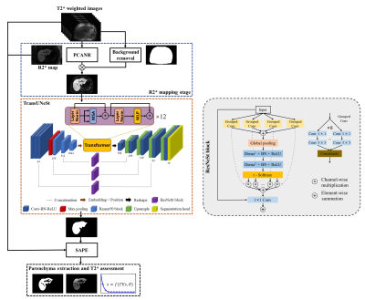 |
8 | A Fully Automatic Data Processing Method for MRI T2* Relaxometry of Iron Loaded Liver
Zifeng Lian1, Qiqi Lu1, and Yanqiu Feng1
1School of Biomedical Engineering, Southern Medical University, Guangzhou, China Keywords: Relaxometry, Liver, T2*, liver iron content MRI T2* relaxometry is a reliable method for assessing liver iron overload. To develop a fully automatic T2* relaxometry data processing method for assessing liver iron overload, we promoted a semi-automatic parenchyma extraction to an automatic approach by introducing a modified TransUNet on R2* map for the segmentation of whole liver. The proposed method showed excellent liver segmentation performance on the internal and external test sets and yielded T2* measurements highly consistent with those by the semi-automatic method. This fully automatic approach will enable an efficient and reliable measurement of liver T2* for assessing hepatic iron content in clinical practice. |
|
1652.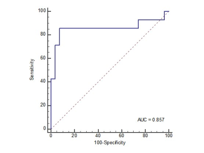 |
9 | Using convolutional neural network predicts microvascular invasion in hepatocellular carcinoma based on Gd-EOB-DTPA-enhanced MRI
Baoer Liu1, Pingjing Wang2, Jianbin Huang1, Wu Zhou2, and Yikai Xu1
1Nanfang Hospital, Southern Medical University, Guangzhou, China, 2Guangzhou University of Chinese Medicine, Guangzhou, China Keywords: Liver, Cancer, Gd-EOB-DTPA-enhanced MRI An accurate preoperative assessment of microvascular invasion (MVI) in patients with hepatocellular carcinoma (HCC) is of great clinical importance in choosing appropriate surgical interventions. We aimed to investigate diagnostic performance of Gd-EOB-DTPA-enhanced MRI for prediction of MVI in HCC using convolutional neural network (CNN). The CNN model based on hepatobiliary phase (HBP) images had great diagnostic efficiency for the prediction of MVI with the AUC of 0.858 (range, 0.854, 0.893). Deep learning with CNN based on Gd-EOB-DTPA-enhanced MRI can be conducive to preoperative prediction of MVI in HCC. |
|
1653. |
10 | Radiomics Nomogram Based on Multi-scale MRI for the Prediction of Microvascular Invasion in Intrahepatic Cholangiocarcinoma
Xianling Qian1, Yunfei Zhang2, Gengyun Miao1, Yongming Dai2, and Mengsu Zeng1
1Zhongshan Hospital, Fudan University, Shanghai, China, 2MR Collaboration, Central Research Institute, United Imaging Healthcare, Shanghai, China Keywords: Liver, Radiomics Microvascular invasion (MVI) is a significant adverse prognostic indicator of intrahepatic cholangiocarcinoma (ICC), and affects the selection of individualized treatment regimen. However, the preoperative imaging-based identification of MVI status is rather difficult. This study sought to establish a multi-sequence and multi-scale MR image-based radiomics nomogram for the presurgical prediction of MVI in ICC. |
|
1654.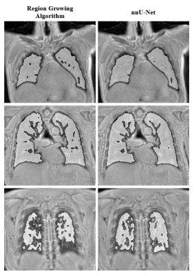 |
11 | Deep Learning Segmentation of Lung Parenchyma For UTE Proton MRI
Christopher Keen1, Peter Šereš1, Justin Grenier1, Robert Stobbe1, Ian Paterson2, Kumar Punithakumar 3, Jacob Jaremko3, and Richard Thompson1
1Department of Biomedical Engineering, University of Alberta, Edmonton, AB, Canada, 2Division of Cardiology, University of Alberta, Edmonton, AB, Canada, 3Department of Radiology and Diagnostic Imaging, University of Alberta, Edmonton, AB, Canada Keywords: Machine Learning/Artificial Intelligence, Lung Accurate segmentation is required to perform quantitative analysis on lung parenchyma in ultrashort echo time (UTE) proton MRI. Deep learning methods offer a solution to this problem, however, previous application to UTE lung MRI is limited. A deep learning model was trained to segment lung parenchyma using fine tuned region growing masks as a reference. To test the generalizability of the model the performance of three different 3D UTE k-space trajectories were compared. Overall, the model produced high quality segmentation for the different acquisition approaches with improvements over training data in areas of high vessel density and high signal intensity. |
|
1655. |
12 | An update of Graded prognostic assessment using molecular markers and Radiomics for estimating survival in NSCLC patients with brain metastases
Lan He1,2 and Zaiyi Liu1,2
1Department of Radiology, Guangdong Provincial People's Hospital , Guangdong Academy of Medical Sciences, Guangzhou, China, 2Guangdong Provincial Key Laboratory of Artificial Intelligence in Medical Image Analysis and Application, Guangdong Provincial People’s Hospital, Guangdong Academy of Medical Sciences, Guangzhou, China Keywords: Lung, Radiomics, Prognosis prediction; Non-small cell lung cancer Rad-molGPA index is prognostic for non-small cell lung cancer patients with brain metastases. The updated Rad-molGPA incorporating radiomics features into the Lung-molGPA is a user-friendly tool to facilitate clinical decision making and appropriate stratification of future clinical trials. |
|
1656.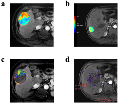 |
13 | Quantitative 3D tumor enhancement on contrast-enhanced MR imaging in patients with HCC after TACE: a consistency evaluation
Xu Hua Gong1, Lei Lv2, and Li Jun Qian1
1Radiology, Renji Hospital, Shanghai Jiao Tong University, School of Medicine, Shanghai, China, 2ShuKun (BeiJing) Technology Co., Ltd., Beijing, China Keywords: Liver, Cancer Quantitative European Association for the Study of the Liver (qEASL) can better evaluate the therapeutic efficacy of hepatocellular carcinoma (HCC) after transcatheter arterial chemoembolization (TACE). Conventional qEASL is often achieved semi-automatically, which is tedious, labor-intensive, and time-consuming. In this study, we provide and assess an automatic qEASL approach based on a VoxelMorph and Faster R-CNN framework named Shukun PortalDoc. We compared the consistency of the proposed method with the widely accepted semi-automatic software (MultiModality Tumor Tracking, Philips IntelliSpace Portal, Philips healthcare) in assessing qEASL of hepatocellular carcinoma after TACE. |
|
1657.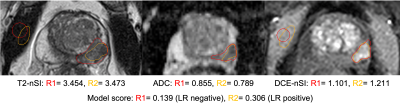 |
14 | The importance of multi-reader assessment for external validation of prostate lesion classification models using quantitative mpMRI.
Tom Syer1, Nikolaos Dikaios2, Thomas Parry3, Giorgio Brembilla3, Mrishta Brizmohun3, Saurabh Singh4, Susan Heavey5, Hayley Pye6, Hayley Whitaker5, Sue Mallett3, David Atkinson3, and Shonit Punwani3
1Centre for Medical Imaging, University College London, London, United Kingdom, 2Mathematics Research Center, Academy of Athens, Athen, Greece, 3Centre for Medical Imaging, Univeristy College London, London, United Kingdom, 4Radiology, Univeristy College London Hospital, London, United Kingdom, 5Department of Targeted Intervention, Univeristy College London, London, United Kingdom, 6National Pathology Imaging Co-operative, Leeds, United Kingdom Keywords: Prostate, Machine Learning/Artificial Intelligence Machine learning for classifying prostate mpMRI lesions may help reduce unnecessary biopsies. However, external validation with multiple scanners and readers is required before the clinical adoption of such models can be considered. Two readers validated a previously published and well-performing logistic regression model on an external cohort. The model performance was not generalisable and offered no advantage to using PSAd cut-offs, and there was marked variation in model score related to contour differences from different readers. This potential variability should be investigated in future models which use quantitative MRI. |
|
1658. |
15 | Development and validation of combined Ki67 status prediction model for intrahepatic cholangiocarcinoma based on MRI radiomics
Xianling Qian1, Changwu Zhou1, Yunfei Zhang2, Yongming Dai2, and Mengsu Zeng1
1Zhongshan Hospital, Fudan University, Shanghai, China, 2MR Collaboration, Central Research Institute, United Imaging Healthcare, Shanghai, China Keywords: Liver, Radiomics In the past decades, the incidence and mortality of intrahepatic cholangiocarcinoma (ICC) have been on the rise. A nuclear antigen named Ki67 is a poor prognostic predictor and an attractive therapeutic target in patients with ICC. Therefore, accurate prediction of Ki67 status in ICC patients is a predictor for treatment efficacy evaluation and outcome prediction. In this study, we established a multiparametric model for predicting Ki67 status in ICC patients preoperatively. |
|
| 1659. | WITHDRAWN | ||
1660.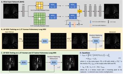 |
16 | Self-supervised Denoising of Pulmonary Perfusion Imaging in Human Subjects and Swine
Changyu Sun1,2, Craig A. Emter3, Darla L. Tharp3, and Talissa A. Altes2
1Biomedical, Biological and Chemical Engineering, University of Missouri Columbia, Columbia, MO, United States, 2Radiology, University of Missouri Columbia, Columbia, MO, United States, 3Biomedical Sciences, University of Missouri Columbia, Columbia, MO, United States Keywords: Lung, Machine Learning/Artificial Intelligence, Self-supervised Denoising Self-supervised learning denoising networks can be applied to noisy only datasets when the clean-noisy pairs are not available, which is suitable for dynamic contrast-enhanced (DCE) pulmonary imaging where SNR is low and no ground truth clean image can be acquired. Blind-spot network with asymmetric pixel-shuffle downsampling (AP-BSN) was trained to utilize the advantages of self-supervised BSN and improve the denoising performance for pixel-wise independent and dependent noise. AP-BSN denoised images showed improved image quality by human reader assessment. AP-BSN showed generalization ability from human subjects to swine and from 1.5T to 3T. |
|
1661.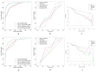 |
17 | Constructing different models to predict the degree of uterine prolapse based on high-resolution MRI radiomics
Qian Wang1, Qiu Bi2, Yijun Zheng1, Yaoxing Wang1, Yuhui Chen2, Chenrong Li1, Xianhong Wang1, Yunzhu Wu3, and Guoli Bi2
1Medical school, Kunming University of Science and Technology, Kunming, China, 2Department of MRI, the First People’s Hospital of Yunnan Province, Kunming, China, 3MR Scientific Marketing, Siemens Healthineers Ltd, Shanghai, China Keywords: Uterus, Radiomics, High-resolution MRI Uterine prolapse has become one of the most common chronic diseases affecting women's health and quality of life. Early detection and intervention would be important factors to delay the progress of uterine prolapse. In this study, high-resolution magnetic resonance imaging(HR-MRI) of pelvic floor combined with radiomics was used to investigate the degree of uterine prolapse. It was found that the HR-MRI radiomics can be used as an effective evaluation method, and the nomogram has the best diagnostic efficacy for predicting the degree of uterine prolapse. |
|
1662.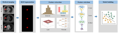 |
18 | An 18F-FDG PET/3D-UTE MRI-based radiomics model facilitates the preoperative assessment of lymph node status in non-small cell lung cancer
Nan Meng1, Pengyang Feng2, Xuan Yu1, Yaping Wu1, Fangfang Fu1, Ziqiang Li3, Yu Luo1, Hongna Tan1, Jianmin Yuan4, Yang Yang5, Zhe Wang4, and Meiyun Wang*6
1Department of Medical Imaging, Zhengzhou University People’s Hospital & Henan Provincial People’s Hospital, Zhengzhou, China, 2Department of Medical Imaging, Henan University People’s Hospital & Henan Provincial People’s Hospital, Zhengzhou, China, 3Department of Medical Imaging, Xinxiang Medical University Henan Provincial People’s Hospital, Zhengzhou, China, 4Central Research Institute, UIH Group, Shanghai, China, 5Beijing United Imaging Research Institute of Intelligent Imaging, UIH Group, Beijing, China, 6Zhengzhou University People’s Hospital & Henan Provincial People’s Hospital, Zhengzhou, China Keywords: Cancer, PET/MR 18F-FDG PET/MRI is one of the most advanced means for the noninvasive diagnosis and evaluation of tumors. Three-dimensional ultrashort echo time (3D-UTE) is a novel MRI technique, which not only does not have ionizing radiation but also yields similar diagnostic results as conventional pulmonary CT. Our results showed that the 18F-FDG PET/MRI model based on clinical factors, 3D-UTE, and PET radiomics features could noninvasively assess the lymph node status in non-small cell lung cancer(NSCLC). Compared with the PET/CT model, it has similar diagnostic efficiency but less radiation, which is expected to provide new ideas for related research. |
|
1663.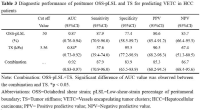 |
19 | 3D MR Elastography-based Prediction of the Histologic VETC Pattern Associated with Aggressive Hepatocellular Carcinoma
Mengsi Li1, Keni Zheng2, Ziying Yin2, Lina Zhang1, Jinhui Zhou1, Dingyue Zhang1, Jun Chen2, Kevin J. Glaser2, Richard L. Ehman2, and Jin Wang1
1The Third Affiliated Hospital of Sun Yat-Sen University, Guangzhou, China, 2Mayo Clinic, Rochester, MN, United States Keywords: Cancer, Elastography, Hepatocellular carcinoma A histologic sign known as “vessels encapsulating tumor clusters” (VETC) has been shown to be a powerful predictor of aggressive hepatocellular carcinoma (HCC) and is associated with unfavorable prognosis. It has been previously demonstrated that MR elastography (MRE)-based stiffness and shear strain mapping are promising in prediction of HCC aggressiveness. We investigated the diagnostic performance of MRE for predicting the VETC finding in HCC. Our results showed that 3D MRE-based peritumor OSS-pLSL and tumor stiffness performed well in predicting VETC status preoperatively, and their combination achieved an AUC of 0.92 in predicting VETC with sensitivity (87.9%) and specificity (83.9%). |
|
 Back to Meeting Home
 Back to the Program-at-a-Glance
The International Society for Magnetic Resonance in Medicine is accredited by the Accreditation Council for Continuing Medical Education to provide continuing medical education for physicians.
 Back to Meeting Home
 Back to the Program-at-a-Glance
 View Presentation Video