Digital Poster
DWI Breast: Clinical Applications & Recent Advances
ISMRM & ISMRT Annual Meeting & Exhibition • 03-08 June 2023 • Toronto, ON, Canada

Digital Poster
DWI Breast: Clinical Applications & Recent Advances
| Computer # | |||
|---|---|---|---|
2549.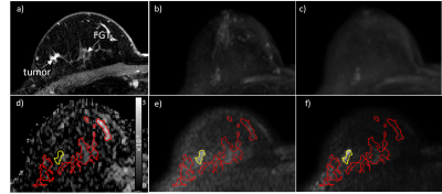 |
21 |
Factors Affecting Cancer Detectability on Standalone Diffusion
Weighted Imaging for Non-contrast Breast Cancer Screening
Debosmita Biswas1,
Jin You Kim1,2,
Isabella Li1,
Michaela R DelPriori3,
Dallas Turley4,
Mary Lynn Bryant1,
Wei Huang5,
Habib Rahbar1,
and Savannah C Partridge1
1Radiology, University of Washington, Seattle, WA, United States, 2Radiology, Pusan National University Hospital, Busan, Korea, Republic of, 3Bio Engineering, University of Washington, Seattle, WA, United States, 4Philips Healthcare, Bothell, WA, United States, 5Advanced Imaging Research Center, Oregon Health & Science University, Portland, OR, United States Keywords: Breast, Diffusion/other diffusion imaging techniques, Diffusion Weighted Imaging Diffusion weighted imaging (DWI) is emerging as a viable tool for non-contrast MRI breast cancer screening, but it is unclear what factors on DWI impact lesion detectability. In this prospective clinical trial, we evaluated lesion and imaging factors that affected cancer detection. Cancers were overall more detectable at higher b=1200 vs b=800 s/mm2, but the background parenchymal signal (BPS) impacted cancer visibility at the higher b value. Cancer histologic type also impacted detectability on DWI. Overall, our findings suggest that interpretation at higher b values and further technical refinements to reduce appearance of BPS may help improve DWI sensitivity. |
|
2550.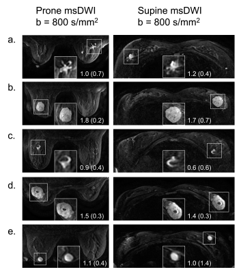 |
22 |
Multi-shot Diffusion-weighted Imaging of the Breasts in the
Supine versus Prone Position
Catherine J. Moran1,
Matthew J. Middione1,
Valentina Mazzoli1,
Jessica A. McKay-Nault1,
Arnaud Guidon2,
Uzma Waheed1,
Eric L. Rosen1,
Steven P. Poplack1,
Jarrett Rosenberg1,
Daniel B. Ennis1,
Brian A. Hargreaves1,
and Bruce L. Dnaiel1
1Radiology, Stanford University, Stanford, CA, United States, 2General Electric Healthcare, Boston, MA, United States Keywords: Breast, Diffusion/other diffusion imaging techniques Diffusion-Weighted Imaging (DWI) allows for the detection of breast cancer without a contrast injection, while supine positioning may improve the comfort and efficiency of a breast MRI screening exam. This work investigates multi-shot DWI of the breasts in the supine versus prone positions, in both asymptomatic volunteers and patients with breast lesions. Supine multi-shot DWI outperformed prone multi-shot DWI based on an image quality observer study, receiving significantly higher ratings for sharpness, aliasing, and overall image quality. Lesion Apparent Diffusion Coefficients (ADCs) were highly correlated between the two positions, while fibroglandular tissue ADCs were significantly higher in the supine position. |
|
2551.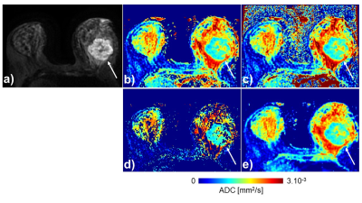 |
23 |
Apparent Diffusion Coefficient in Breast MRI
Stephane Loubrie1,
Maren Andreassen2,
Ana Rodriguez-Soto1,
Michael Carl3,
Summer Batasin1,
Christopher Conlin1,
Tyler Seibert1,4,5,
Michael Hahn1,
Joshua Kuperman1,
Anders Dale1,6,
and Rebecca Rakow-Penner1,5
1Radiology, University of California, San Diego, San Diego, CA, United States, 2Circulation and Medical Imaging, Norwegian University of Science and Technology, Trondheim, Norway, 3Global MR Application and Workflow, GE Healthcare, Boston, MA, United States, 4Radiation medicine, University of California, San Diego, San Diego, CA, United States, 5Bioengineering, University of California, San Diego, San Diego, CA, United States, 6Neurosciences, University of California, San Diego, San Diego, CA, United States Keywords: Breast, Diffusion/other diffusion imaging techniques Diffusion weighted Imaging (DWI) acquisitions are often part of breast cancer MRI protocols. The signal intensity decrease as a function of the b-value in diffusion imaging is non-linear and complex with inconsistent Apparent Diffusion Coefficients. In this abstract, we investigate ADC mapping computation and accuracy. Mapping techniques gave similar performances, with comparable medians and standard deviations. |
|
2552. |
24 |
Diagnostic value of 5b and 2b-value-DWI in breast tumors:
comparison with combined MRI
Aika Okazawa1,
Mami Iima1,2,
Masako Kataoka1,
Ryosuke Okumura3,
Sachiko Takahara4,
Tomotaka Noda5,
Taro Nishi5,
Takayoshi Ishimori3,
Maya Honda1,6,
and Yuji Nakamoto1
1Department of Diagnostic Imaging and Nuclear Medicine, Graduate School of Medicine, Kyoto University, Kyoto, Japan, 2Institute for Advancement of Clinical and Translational Science, Kyoto University Hospital, Kyoto, Japan, 3Department of Diagnostic Radiology, Kitano Hospital, Tazuke Kofukai Medical Research Institute, Osaka, Japan, 4Department of Breast Surgery, Kitano Hospital, The Tazuke Kofukai Medical Research Institute, Osaka, Japan, 5Department of Clinical Radiology Service, Kitano Hospital, The Tazuke Kofukai Medical Research Institute, Osaka, Japan, 6Department of Diagnostic Radiology, Kansai Electric Power Hospital, Osaka, Japan Keywords: Breast, Diffusion/other diffusion imaging techniques We propose a DWI-reading method based on the an adjusted BI-RADS lexicons using five b values of 0, 200, 800, 1000, and 1500 s/mm2 (5b-value-DWI). Diagnostic performance was evaluated in comparison with BI-RADS lexicons from standard BI-RADS DCE MRI and a DWI-reading method based on the adjusted BI-RADS lexicons using two b values of 0, 800 s/mm2 (2b-value-DWI).Our proposed DWI reading methods achieved diagnostic performance comparable to the standard BI-RADS. With higher sensitivity and NPV in 5b-value-DWI compared to 2b-value-DWI, DWI reading with 5b-values has the potential to increase diagnostic confidence in differentiating malignant and benign breast tumors. |
|
2553.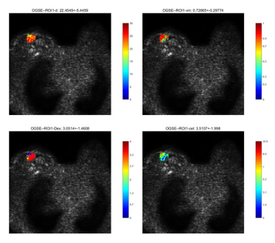 |
25 |
Time-Dependent Diffusion MRI for the Breast to Differentiate
between Benign and Malignant Lesions
Yun Su1,
Xiang Zhang1,
Huijun Hu1,
Lingjie Yang1,
Yu Wang1,
Chen Zhao2,
and Yishi Wang3
1Department of Radiology, Sun Yat-Sen Memorial Hospital, Sun Yat-Sen University, Guangzhou, China, 2Philips Healthcare, Guangzhou, China, 3Philips Healthcare, Beijing, China Keywords: Breast, Breast, Tumor, Diffusion, OGSE, PGSE Defining the nature of benign and malignant breast tumors is essential to reduce unnecessary biopsies of benign tumors. Magnetic resonance imaging (MRI) is the most sensitive imaging modality for evaluating breast carcinoma lesions, but its specificity is relatively low. This study investigated the significance of the parameters for the differential diagnosis of mammary mass using time-dependent diffusion MRI. Results showed significant cell size differences between benign and malignant space-occupying lesions, with high accuracy (85.7%), specificity (83.3%), sensitivity (87.5%) and AUC (0.823). This provides a new direction for noninvasively differentiating benign and malignant lesions of the mammary gland. |
|
2554. |
26 |
The diagnostic performance of time-dependent diffusion technique
in differentiating benign and malignant breast tumors
Jie Ding1,2,3,
Zhen Zhang3,
Yajia Gu1,2,
Yishi Wang4,
Xiuzheng Yue4,
Dazhi Chen3,
Rongrong Zhu3,
and Ruoshui Ha3
1Department of Radiology, Fudan University Shanghai Cancer Center, Shanghai, China, 2Department of Oncology, Shanghai Medical College, Fudan University, Shanghai, China, 3People's Hospital of Ningxia Hui Autonomous Region, Yinchuan, China, 4Philips Healthcare, Beijing, China Keywords: Breast, Cancer Some novel biomarkers have emerged from the fact that the ADC depends strongly on the diffusion time, such as the rate of ADC change using two diffusion times for the differentiation of breast tumor types and cell size modeling. In this study, we propose to use Multiband (MB) SENSE to accelerate the data acquisition of OGSE and PGSE sequences and compare the diagnostic value of ADC at different diffusion times as well as cell size imaging parameters using IMPULSED model for breast tumor. Our data showed vin and cellularity and the rate of ADC change have superior diagnostic performance. |
|
2555.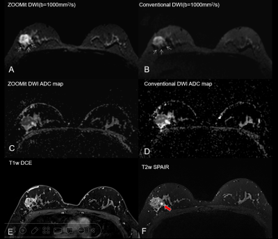 |
27 |
Comparison of ZOOMit DWI and Conventional DWI with the same
geometric parameters at 3T in Patients with Breast Diseases
Zhu Kaiguo1,
Wang Pingping1,
Dang Yanli1,
Wang Lifang1,
Liu Rumei1,
Chen Baoying1,
and shaoyu wang2
1Xi'an International Medical Center Hospital, Xi'an, China, 2MR Scientific Marketing, Siemens Healthineers, Shanghai, China Keywords: Breast, Diffusion/other diffusion imaging techniques DWI performs an important role in the diagnosis of the benign and malignant breast lesions. This study compared the overall image quality, SNR,CNR and the diagnostic performance of ADC values between ZOOMit DWI and conventional DWI in breast cancer with same geometric parameters. The result showed that ZOOMit DWI provided significantly higher image quality and lesion conspicuity than C-DWI with no difference diagnostic performance for ADC values and with almost the same scanning time, which can help to improve the diagnosis efficiency in breast cancer. |
|
2556.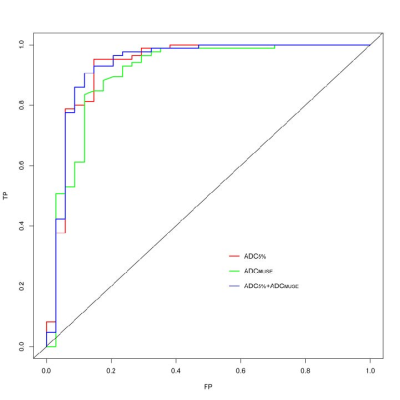 |
28 |
Value of ADC Histogram Analysis in the Differentiation of Benign
and Malignant Breast Lesions Based on Multiplexed Sensitivity
Encoding
jinrui liu1,
Bing Chen2,
mengyin xu2,
zhihao li3,
and zhaohui an2
1Ningxia Medical University, yin chuan, China, 2General Hospital of Ningxia Medical University, yinchuan, China, 3GE Healthcare China, xian, China Keywords: Breast, Cancer The ADC histogram parameters based on the MUSE-DWI are important indicators for breast cancer diagnosis. ADC5% was more accurate for lesion discrimination than ADCMUSE. |
|
2557.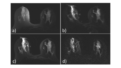 |
29 |
MultiBand Acquisition for Breast Restriction Spectrum Imaging
Stephane Loubrie1,
Ana Rodriguez-Soto1,
Michael Carl2,
Summer Batasin1,
Christopher Conlin1,
Tyler Seibert1,3,4,
Michael Hahn1,
Joshua Kuperman1,
Arnaud Guidon2,
Anders Dale1,5,
Haydee Ojeda-Fournier1,
and Rebecca Rakow-Penner1,4
1Radiology, University of California, San Diego, San Diego, CA, United States, 2Global MR Application and Workflow, GE Healthcare, Boston, MA, United States, 3Radiation medicine, University of California, San Diego, San Diego, CA, United States, 4Bioengineering, University of California, San Diego, San Diego, CA, United States, 5Neurosciences, University of California, San Diego, San Diego, CA, United States Keywords: Breast, Diffusion/other diffusion imaging techniques Diffusion-weighted imaging holds great potential in improving specificity in breast cancer MRI, potentially reducing the number of unnecessary biopsies. Additionally, breast cancer screening protocols would benefit from high-resolution DWI acquisitions, especially in the through-plane direction. In this abstract we explore multi-slice excitation as a promising parallel-imaging tool to improve through-plane image resolution. |
|
2558.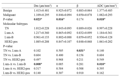 |
30 |
Differentiating Benign from Malignant Breast Lesions and
Molecular Subtypes Using MRI with a Continuous-Time Random-Walk
Diffusion Model
Huan Chang1,
Dawei Wang2,
Yuting Li3,
Shaoxin Xiang4,
Ke Xue4,
Peng Kong5,
and Qingshi Zeng2
1Department of Radiology, Shandong Provincial Qianfoshan Hospital, Shandong University, Shandong, China, Jinan, China, 2Department of Radiology, The First Affiliated Hospital of Shandong First Medical University & Shandong Provincial Qianfoshan Hospital,Shandong, China, Jinan, China, 3Department of Radiology, The First College of Clinical Medicine, Shandong University of Traditional Chinese Medicine, Shandong, China, Jinan, China, 4MR Collaboration, Central Research Institute, United Imaging Healthcare, Shanghai, China, Shanghai, China, 5Department of Breast Surgery, The First Affiliated Hospital of Shandong First Medical University & Shandong Provincial Qianfoshan Hospital,Shandong, China, Jinan, China Keywords: Breast, Diffusion/other diffusion imaging techniques Accurately differentiating breast cancer is crucial for clinical treatment. However, it is challenging due to the high heterogeneity of breast cancer. This study used a continuous-time random-walk (CTRW) diffusion model to characterize breast lesions and their molecular subtypes. The malignant lesions had significantly lower ADC, Dm, and α values than benign lesions. Significant differences were also found between luminal A and triple-negative groups in parameter β. And the combination of CTRW parameters yielded better performance in classifying breast cancer than ADC. Therefore, this study highlights that the CTRW parameters are superior to ADC in distinguishing breast lesions and molecular subtypes. |
|
2559.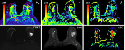 |
31 |
Breast cancer diagnosis and prognosis by using a high b-value
continuous-time random-walk model
Hui Feng1,
Hui Liu1,
Qi Wang1,
Mengyu Song1,
Tianshu Yang2,
Liyun Zheng2,
Dongmei Wu3,
Xian Shao4,
and Gaofeng Shi1
1Department of Radiology, The Fourth Hospital of Hebei Medical University, Shijiazhuang, China, 2Shenzhen United Imaging Research Institute of Innovative Medical Equipment, Shenzhen, China, 3Shanghai Key Laboratory of Magnetic Resonance, School of Physics and Electronics Science, East China Normal University, Shanghai, China, 4Department of Anesthesiology, The Fourth Hospital of Shijiazhuang, Shijiazhuang, China Keywords: Breast, Diffusion/other diffusion imaging techniques Breast cancer is a common cancer that severely threatens the health of women worldwide. The advanced diffusion model using high b-values enables a more comprehensive description of the tumor tissue by cellularity and heterogeneity. In current study, a continuous-time random-walk (CTRW) model was applied to identify malignancy of breast lesions and the association between model-derived parameters and immunohistochemical indices was evaluated. All model-derived parameters could identify tumor malignancy, combined parameter could further discriminate ER+/ER- and PR+/PR- patients, while temporal heterogeneity parameter was significantly correlated with PR expression. The CTRW model has demonstrated potential value in breast cancer diagnosis and prognosis. |
|
| 2560. | 32 |
Prediction of the Prognostic Factors of Breast Cancer Using MRI
with a Continuous-Time Random-Walk Diffusion Model
Huan Chang1,
Dawei Wang2,
Yuting Li3,
Shaoxin Xiang4,
Ke Xue4,
Peng Kong5,
and Qingshi Zeng2
1Department of Radiology, Shandong Provincial Qianfoshan Hospital, Shandong University, Jinan, China, 2Department of Radiology, The First Affiliated Hospital of Shandong First Medical University & Shandong Provincial Qianfoshan Hospital, Jinan, China, 3Department of Radiology, The First College of Clinical Medicine, Shandong University of Traditional Chinese Medicine, Jinan, China, 4MR Collaboration, Central Research Institute, United Imaging Healthcare, Shanghai, China, 5Department of Breast Surgery, The First Affiliated Hospital of Shandong First Medical University & Shandong Provincial Qianfoshan Hospital, Jinan, China Keywords: Breast, Diffusion/other diffusion imaging techniques Breast cancer is a heterogeneous tumor, and its treatment is customized determined by the pathologic and tumor molecular features. This study used a continuous-time random-walk (CTRW) diffusion model to characterize prognostic factors status of breast lesions. The parameter β showed significantly differences in ER and PR status and showed significant correlation with ER and PR expression, and the combination of Dm, α, and β produced the best performance in identifying ER and PR status. Our study demonstrated that the CTRW parameters as potential biomarkers were superior to conventional ADC in predicting prognostic factors of breast cancer. |
|
2561.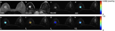 |
33 |
Quantitative Assessment of Restriction Spectrum MR Imaging for
the Diagnosis of Breast Cancer and Association with Prognostic
Factors
Yanjin Qin1,
Yunfei Zhang2,
Yongming Dai2,
and Tao Ai1
1Tongji Hospital, Tongji Medical College, Huazhong University of Science and Technology, Wuhan, China, 2MR Collaboration, Central Research Institute, United Imaging Healthcare, Shanghai, China Keywords: Breast, Cancer This study aimed to assess the diagnostic performance of restriction spectrum imaging (RSI)-derived parameters for distinguishing benign from malignant breast lesions compared to conventional DWI values and further evaluate the associations between RSI-derived parameters and prognostic factors of breast cancer. The results indicated the RSI-derived parameters (f1, f3, and f1f2) may facilitate the differential diagnosis between benign and malignant breast lesions. |
|
2562.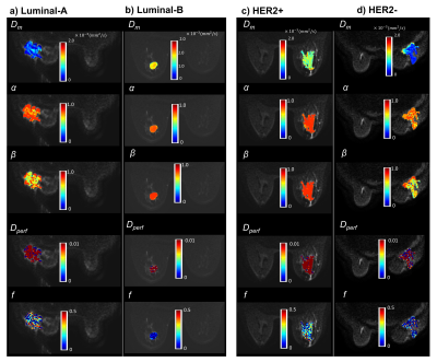 |
34 |
Predicting Molecular Subtypes and Prognostic Factors of Breast
Cancer Using Integrated Diffusion MRI
Muge Karaman1,2,
Yangyang Bu3,4,
Guangyu Dan1,2,
Zheng Zhong1,
Qingfei Luo1,
Shiwei Wang3,4,
Changyu Zhou3,4,
Weihong Hu3,4,
X. Joe Zhou1,2,5,
and Maosheng Xu3,4
1Center for MR Research, University of Illinois at Chicago, Chicago, IL, United States, 2Department of Biomedical Engineering, University of Illinois at Chicago, Chicago, IL, United States, 3The First School of Clinical Medicine of Zhejiang Chinese Medical University, Hangzhou, China, 4The First Affiliated Hospital of Zhejiang Chinese Medical University, Hangzhou, China, 5Departments of Radiology and Neurosurgery, University of Illinois at Chicago, Chicago, IL, United States Keywords: Breast, Breast, molecular subtype prediction, heterogeneity, high-b-value diffusion MRI Breast cancer exhibits a wide spectrum of molecular subtypes and, which has important implications in treatment strategies. In this study, we used an integrated diffusion-weighted imaging approach for simultaneous assessment of tissue cellularity, vascularity, and heterogeneity – DISMANTLE – to predict molecular subtypes and prognostic factors of breast cancer. We investigated the feasibility of using the histogram features of the cellularity-, vascularity-, and heterogeneity-related parameters of DISMANTLE for differentiation between luminal-A and luminal-B and HER2+ and HER2- breast cancer. |
|
2563.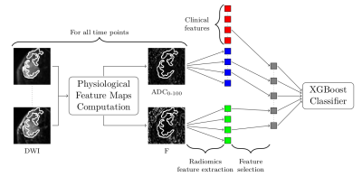 |
35 |
A Physiologically-Decomposed DWI machine-learning model improves
prediction of response to NAC treatment in invasive breast
cancer
Maya Gilad1 and
Moti Freiman2
1Efi Arazi School of Computer Science, Reichman University, Herzliya, Israel, 2Faculty of Biomedical Engineering, Technion-Israel Institute of Technology, Haifa, Israel Keywords: Breast, Diffusion/other diffusion imaging techniques Early prediction of pathological complete response (pCR) following neoadjuvant chemotherapy for breast cancer plays a critical role in surgical planning and optimizing treatment strategies. Recently, machine and deep-learning based methods were suggested for early pCR prediction from multi-parametric MRI data with moderate success. We introduce PD-DWI, a physiologically-decomposed DWI machine-learning model to predict pCR from DWI and clinical data. Our model first decomposes the raw DWI data into the various physiological cues that are influencing the DWI signal and then uses the decomposed data, in addition to clinical variables, as the input features of a radiomics-based XGBoost model. |
|
| 2564. | 36 |
An ADC-based Radiomics model: differential diagnosis of mucinous
breast carcinoma from similar morphological invasive ductal
breast carcinoma
yuanfei li1,
ning ning1,
lina zhang1,
qi wu1,
siqi zhao1,
ailian liu1,
qinhe zhang1,
jianzhou chen2,
jingjing cui2,
and lizhi xie3
1The First Affiliated Hospital of Dalian Medical University, Dalian, China, 2Shanghai United Imaging Intelligence, Co., Ltd., Shanghai, China, 3GE Healthcare, Beijing, China Keywords: Breast, Radiomics Mucinous breast carcinomas (MBCs) always have similar morphological manifestations with invasive ductal breast carcinomas (IDCs) on conventional MRI. MBCs have a better prognosis and early treatment can achieve good results. Radiomics is a novel tool automated data feature extraction algorithms,which has the potential to uncover disease characteristics that are difficult to identify by human vision alone. We develop a radiomics model for explore the preoperative differentiation of combining the intratumoral signal intensity (SI) on T2WI and ADC derived from DWI in MBCs and IDC. The results confirm that the model has potential in the differential diagnosis of MBCs and IDC. |
|
2565.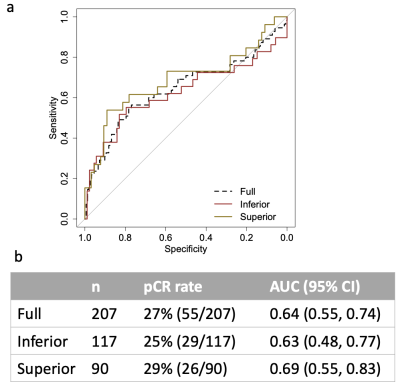 |
37 |
Image quality ranking for breast diffusion-weighted MRI in a
multi-center clinical trial
Wen Li1,
Julia Carmona-Bozo1,
Lisa J Wilmes1,
Natsuko Onishi1,
Jiachao Liang1,
Jessica E Gibbs1,
Nu N Le1,
Judith Zimmermann1,
David C Newitt1,
Bonnie N Joe1,
John Kornak1,
Savannah C Partridge2,
Patrick Bolan3,
I-SPY 2 Investigator Network4,
I-SPY 2 Imaging Working Group1,
and Nola M Hylton1
1University of California, San Francisco, San Francisco, CA, United States, 2University of Washington, Seattle, WA, United States, 3University of Minnesota, Minneapolis, MN, United States, 4Quantum Leap Healthcare, San Francisco, CA, United States Keywords: Breast, Cancer Acquiring breast diffusion-weighted MRI (DWI) with adequate image quality is a challenge in multi-center clinical trials. The most common issues are inadequate fat suppression, image artifacts, and poor signal-to-noise ratio (SNR). We developed and evaluated a DWI quality ranking system for identifying data with quality issues while preserving analyzable data. The identification of artifacts and adequate SNR had the highest and lowest inter-reader agreement, respectively. Removing inferior quality data improved prediction of pathologic outcome following neoadjuvant treatment in a multi-center clinical trial. The quality ranking system standardizes identification of poor quality data and informs recommendations for improvement to clinical sites. |
|
2566.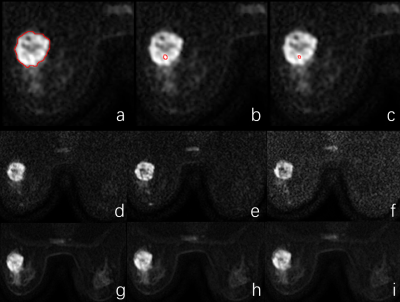 |
38 |
Synthetic high-b-value reduced field-of-view DWIs in breast
cancer elevates lesion clarity and CNR
Qianqian Feng1,
Weiyin Vivian Liu2,
Ling Sang1,
Kejun Wang1,
Xingyao Yu1,
Wen Chen1,
and Lin Xu1
1Department of Radiology,Taihe hospital, Hubei, China, 2GE Healthcare, MR Research, Beijing, China Keywords: Breast, Diffusion/other diffusion imaging techniques Our study showed synthetic DW images (syDWIs) with high b-value provided better lesion conspicuity, image quality and contrast to noise (CNR), reduce scan time of DWI acquisitions especially at very high b-value, and improved lesion visualization particularly in dense breast. Moreover, derived ADC values showed good consistency between scanned and synthetic DWIs. Therefore, syDWIs with b value of 800 s/mm2 have great potential in elevation of diagnosis confidence and efficacy on tumors and high repeatability of ADC values is helpful in replacement of scanned high-b-value DWIs in clinics despite existed intra-modality, but not inter-modality, difference of manual ROI measures. |
|
2567.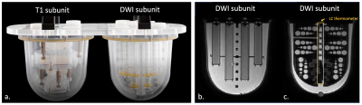 |
39 |
Longitudinal assessment of variability in a multicenter MRI QC
program using the CaliberMRI breast phantom: protocol adherence
and ADC values
Lisa J. Wilmes1,
Jessica E. Gibbs1,
Nu N. Le1,
Todor Karaulanov2,
David C. Newitt1,
Kathryn E. Keenan3,
Alina Tudorica4,
Gwen Bowden5,
Yucheng Liu6,
Lauren Fang7,
Patrick J. Bolan8,
Dariya Malyarenko9,
Thomas L. Chenevert9,
David Van Wie10,
Chelsea Pyle4,
Bethany Niell5,
Richard Ha6,
Rebecca Rakow-Penner7,
Haydee Ojeda-Fournier7,
An Church8,
Bonnie N. Joe1,
and Nola M. Hylton1
1University of California San Francisco, San Francisco, CA, United States, 2CaliberMRI, Boulder, CO, United States, 3National Institute of Standards and Technology, Boulder, CO, United States, 4Oregon Health & Science University, Portland, OR, United States, 5Moffitt Cancer Center, Tampa, FL, United States, 6Columbia University Medical Center, New York, NY, United States, 7University of California San Diego, San Diego, CA, United States, 8University of Minnesota Medical School, Minneapolis, MN, United States, 9University of Michigan Health System, Ann Arbor, MI, United States, 10Boulder Labs, Boulder, CO, United States Keywords: Breast, Diffusion/other diffusion imaging techniques CaliberMRI’s multiparametric quantitative breast MRI phantom was scanned with a standard protocol, including DWI, at seven clinical sites as part of a quality control program. Protocol adherence was assessed. ADC values for fibroglandular and malignant breast tissue DWI mimics were calculated and corrected for temperature using the qCal software, and summary reports were generated. Inter- and intra-site variations in ADC derived from DWI phantom scans acquired longitudinally over the last 16 months provided information that can be used to troubleshoot imaging acquisition issues with sites and vendors as part of a phantom-based QC program. |
|
 Back to Meeting Home
 Back to the Program-at-a-Glance
The International Society for Magnetic Resonance in Medicine is accredited by the Accreditation Council for Continuing Medical Education to provide continuing medical education for physicians.
 Back to Meeting Home
 Back to the Program-at-a-Glance
 View Presentation Video