Digital Poster
Hepatobiliary Imaging: Current Applications & Advances I
ISMRM & ISMRT Annual Meeting & Exhibition • 03-08 June 2023 • Toronto, ON, Canada

Digital Poster
Hepatobiliary Imaging: Current Applications & Advances I
| Computer # | |||
|---|---|---|---|
1838.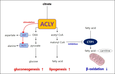 |
21 | HP 13C pyruvate MRS reveals that ATP Citrate Lyase regulates alanine aminotransferase activity in the db/db liver.
Young-Suk Choi1 and Ho-Taek Song2
1Department of Radiology and Research Institute of Radiological Science, Yonsei University College of Medicine, Seoul, Korea, Republic of, 2Yonsei University College of Medicine, Seoul, Korea, Republic of Keywords: Liver, Hyperpolarized MR (Non-Gas) Nonalcoholic fatty liver disease and diabetes are known to be closely related, but the mechanism has not been defined yet. The metabolic feature of the diabetic liver is that gluconeogenesis and fatty acid synthesis increase simultaneously. In this study, we hypothesized that ATP citrate lyase (ACLY) would play an essential role in inducing metabolic contradiction in the diabetic liver and investigate ACLY’s role on carbohydrate metabolism using hyperpolarized (HP) 13C pyruvate magnetic resonance spectroscopy (MRS) and primary mouse hepatocytes. |
|
1839.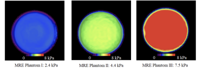 |
22 | Accuracy and test-retest repeatability of stiffness measurement with MR Elastography: a multi-center phantom study
Efe Ozkaya1,2, Paul Kennedy1,2, Jun Chen3, Octavia Bane1,2, Jonathan R. Dillman4, Kartik S. Jhaveri5, Michael Ohliger 6,7, Phillip J. Rossman3, Jean A. Tkach4, Sudhakar K. Venkatesh3, Richard L. Ehman3, and Bachir Taouli1,2
1Department of Diagnostic, Molecular and Interventional Radiology, Icahn School of Medicine at Mount Sinai, New York, NY, United States, 2BioMedical Engineering and Imaging Institute, Icahn School of Medicine at Mount Sinai, New York, NY, United States, 3Department of Radiology, Mayo Clinic, Rochester, MN, United States, 4Department of Radiology, Cincinnati Children’s Hospital Medical Center, University of Cincinnati College of Medicine, Cincinnati, OH, United States, 5Joint Department of Medical Imaging, University Health Network, Mount Sinai Hospital, and Women’s College Hospital,, University of Toronto, Toronto, ON, Canada, 6Department of Radiology and Biomedical Imaging, University of California San Francisco, San Francisco, CA, United States, 7Department of Radiology, Zuckerberg San Francisco General Hospital, San Francisco, CA, United States Keywords: Liver, Elastography, Phantom, Multicenter study, Accuracy, Repeatability The purpose of our multicenter study was to determine the accuracy and test-retest repeatability of stiffness measured with MRE in phantoms. Three phantoms with known stiffness (2.4, 4.4, and 7.5 kPa) were circulated between 5 different centers. 1.5T or 3T systems from the 3 major vendors were used for scanning with 2D GRE and/or SE-EPI MRE sequences. The reference was based on measurements in the reference center made at the start and end of study. For all 3 phantoms the mean accuracy error for the 4 testing centers was 10.2%, while test-retest repeatability error was 2.9% for all centers. |
|
1840.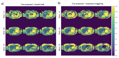 |
23 | Fast multi-slice liver water T1 mapping using single-shot continuous inversion recovery spiral imaging
Elizabeth Huaroc Moquillaza1, Kilian Weiss2, Yoo Jin Lee3, Jonathan Stelter1, Thomas Amthor3, Peter Koken3, Marcus R. Makowski1, Rickmer Braren1, Mariya Doneva3, and Dimitrios C. Karampinos1
1Department of Diagnostic and Interventional Radiology, School of Medicine, Technical University of Munich, Munich, Germany, 2Philips GmbH Market DACH, Hamburg, Germany, 3Philips Research Lab, Hamburg, Germany Keywords: Liver, Relaxometry Liver T1 mapping can characterize liver alterations in diffuse or focal (e.g. metastatic) liver disease, but it is currently limited to acquisitions of a single-slice per breath-hold. We propose a continuous inversion recovery methodology combining a single-shot gradient echo spiral readout, Dixon processing for water-fat separation and dictionary-based analysis for water selective T1 mapping in the liver at 3T. The spiral readout is employed for a high k-space sampling efficiency, enabling acquisitions with only 1.2s per slice. The method allows multi-slice full-liver water T1 mapping either in a single 11s-breath-hold or in a respiratory-triggered acquisition. |
|
1841.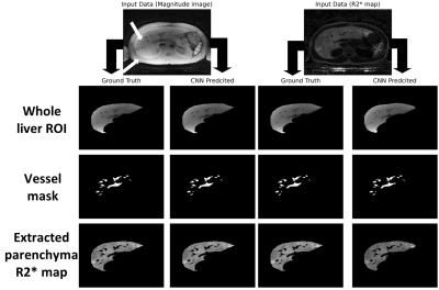 |
24 | Automatic Extraction of Liver Parenchyma and R2* Estimation using Deep Learning for UTE Imaging for Assessment of Hepatic Iron Overload
Utsav Shrestha1, Cara Morin2,3, Ralf Loeffler4, Zachary R. Abramson2, Jane Hankins2, Claudia Hillenbrand4, and Aaryani Tipirneni-Sajja1,2
1The University of Memphis, Memphis, TN, United States, 2St. Jude Children's Research Hospital, Memphis, TN, United States, 3Cincinnati Children’s Hospital Medical Center, CINCINNATI, OH, United States, 4University of New South Wales, Sydney, Australia Keywords: Liver, Liver, Deep Learning, Vessel Segmentation, R2*, HIC Ultra-short echo time (UTE) imaging increases the accuracy of R2*-based hepatic iron content (HIC) quantification in cases of high iron overload when conventional GRE sequences can fail due to rapid signal decay. Segmenting whole liver to estimate liver R2* requires human expert and is time consuming. In this study, we trained a convolutional neural network (CNN) to automatically segment the liver parenchyma on radial UTE acquisitions using magnitude images and R2* maps. Our results show an excellent agreement between manual and CNN-based liver segmentation and mean R2* values, hence demonstrating the potential of our proposed method for automated HIC assessment. |
|
1842.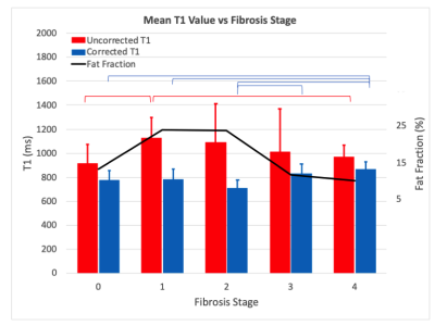 |
25 | Towards Non-Invasive Diagnosis of NASH: Retrospective Fat Correction of MP2RAGE T1 Improves Correlation with Liver Fibrosis Stage
Andrew Duffy1, Parth Maheta2, Andreu F. Costa3, Sharon Clarke3,4, Ashley Stueck5, Magnus McLeod6, and James Rioux2,3,4
1Medicine, Dalhousie University, Halifax, NS, Canada, 2Physics and Atmospheric Science, Dalhousie University, Halifax, NS, Canada, 3Diagnostic Radiology, Dalhousie University, Halifax, NS, Canada, 4Biomedical Translational Imaging Centre, Nova Scotia Health Authority, Halifax, NS, Canada, 5Pathology, Dalhousie University, Halifax, NS, Canada, 6General Internal Medicine, Dalhousie University, Halifax, NS, Canada Keywords: Liver, Fat, Fibrosis There is significant interest in non-invasive biomarkers for staging of liver fibrosis in non-alcoholic fatty liver disease (NAFLD) to reduce the need for biopsy. T1 correlates with fibrosis but can also be influenced by fat, and while Dixon fat-water separation can remove this effect, such methods are uncommon in clinical T1 mapping sequences. In this work, we demonstrate a retrospective correction to estimate the T1 of liver tissue based on MP2RAGE without fat-water separation. This enables fat compensation in a wider range of acquisitions and improves the potential clinical utility of T1 as a biomarker for liver fibrosis. |
|
1843.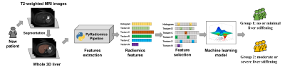 |
26 | Machine Learning Stratification of Liver Stiffness using T2-weighted MRI Radiomic Data: A Multi-Site Study
Hailong Li1, Ziang Chen1, Jinzhao Qian1, Wen Pan1, Scott B. Reeder2, David T. Harris2, William R. Masch3, Anum Alsam3, Krishna P. Shanbhogue4, Anas Bernieh1, Sarangarajan Ranganathan1, Nehal A. Parikh1, Jonathan R. Dillman1, and Lili He1
1Cincinnati Children's Hospital Medical Center, Cincinnati, OH, United States, 2University of Wisconsin-Madison, Madison, WI, United States, 3Michigan Medicine, University of Michigan, Ann Arbor, MI, United States, 4NYU Langone Health, New York, NY, United States Keywords: Liver, Liver, Liver stiffness MR elastography (MRE) offers a non-invasive approach to quantify liver stiffening, a surrogate for hepatic fibrosis. However, it has drawbacks, including long exam time, patient discomfort, and the need for additional hardware. The objective of this multi-site study is to develop a machine learning model to categorically stratify the severity of liver stiffness using clinical, routinely collected T2-weighted MRI data from pediatric and adult patients from four study sites. With radiomic features extracted from MRI data, our model achieved an AUROC of 0.72 for stratifying liver stiffness, demonstrating the potential of such a machine learning strategy for clinical utilization. |
|
1844.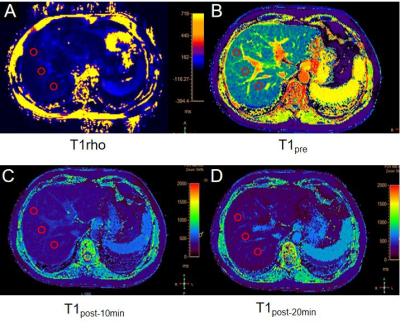 |
27 | Evaluation the value of T1rho and Gd-EOB-DTPA-Enhanced T1 Mapping for liver function reserve in patients with chronic liver disease
Jie Zou1,2,3, Yanli Jiang1,2,3, Fengxian Fan1,3, Pin Yang1,3, Yuan Ding1,3, Jing Zhang1,3, Kai Ai4, and Zhigang Wu5
1Department of Magnetic Resonance, Lanzhou University Second Hospital, Lanzhou, China, 2Second Clinical School, Lanzhou University, Lanzhou, China, 3Gansu Province Clinical Research Center for Functional and Molecular Imaging, Lanzhou, China, 4Philips Healthcare, Xi'an, China, 5Philips Healthcare, Shenzhen, China Keywords: Liver, Liver, T1rho, chronic liver disease, T1 mapping The purpose of this study is to evaluate the diagnostic performance of T1 rho and Gd-EOB-DTPA-Enhanced T1 Mapping in patients with chronic hepatitis B in liver function reserve. For the two groups of Child-Pugh A vs. B/C and Child-Pugh A/ B vs. C, the AUC of T1 rho and T1post-10min is higher than other metrics. The AUC value combined with these two indicators is the highest (0.861; 0.912). Δ T1, Δ R1 and ECV and T1 rho quantitative techniques may be helpful for staging liver function reserve, and the combination of T1 rho and T1post-10min can provide higher diagnostic efficiency. |
|
1845.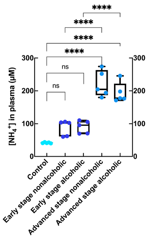 |
28 | Ammonium quantification in human plasma by 1H-NMR for staging of liver fibrosis in alcohol and non-alcoholic-related liver disease
Marc Azagra1, Elisa Pose2, Francesco De Chiara3, Martina Pérez2, Emma Avitabile2, Joan-Marc Servitja4, Laura Brugnara4, Javier Ramón-Azcón3, and Irene Marco-Rius1
1MIPMED, IBEC, Barcelona, Spain, 2Liver Unit, Hospital Clínic, Barcelona, Spain, 3Biosensors for Bioengineering, IBEC, Barcelona, Spain, 4IDIBAPS, Barcelona, Spain Keywords: Liver, Blood, Ammonium quantification, chronic liver disease, hepatic dysfunction, disease biomarker, NMR Invasive and painful liver biopsy is the gold standard for liver disease diagnosis. Non-invasive methods to assess liver fibrosis in patients with alcohol-related liver disease (ArLD) and non-alcoholic fatty liver disease (NAFLD) are an unmet clinical need. We have developed a robust and reliable 1H NMR protocol to quantify the endogenous ammonium concentration in biological fluids. The measurement of ammonium in blood plasma samples of ArLD and NAFLD patients discerned between some stages of the disease retrospectively and showed that ammonium readout is a robust diagnostic marker of fibrosis stage, more so than current clinically assessed blood hepatic biomarkers. |
|
| 1846. | 29 | Test-Retest Repeatability and Cross-Platform Reproducibility of Optimized 2D MRE and 3D MRE Liver Protocols with Automated Analysis
Bogdan Dzyubak1, Kay M Pepin1, Jun Chen1, Yuan Le1, Kyle Kalutkiewicz2, Roger Grimm1, Jeremy A Heilman1, Scott Kruse1, Jennifer Kugel1, Meng Yin1, Kevin J Glaser1, and Ehman L. Richard1
1Radiology, Mayo Clinic, Rochester, MN, United States, 2Engineering, Resoundant, Rochester, MN, United States Keywords: Liver, Elastography Magnetic Resonance Elastography is a technique used to effectively stage liver fibrosis by measuring tissue stiffness. This work optimized several acquisition and processing parameters, and performed a comprehensive repeatability/reproducibility validation across the three types of MRE (GRE, EPI, 3DEPI), 1.5T and 3T field strengths, and the three major manufacturers (GE, Siemens, Philips). When using the optimized methods and automated analysis, the repeatability coefficients were 11.8% for GRE, 10.8% for EPI, and 7.7% for 3DEPI, all superior to QIBA’s 19% benchmark. Agreement between manufacturers was within 2%. |
|
1847.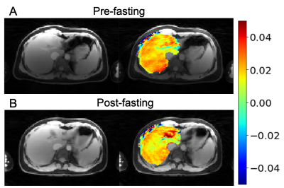 |
30 | Glycogen Imaging of The Human Liver Using GraspNOE-Dixon
Xiang Xu1, Rodolphe Leforestier1, Ding Xia1, and Li Feng1
1BioMedical Engineering and Imaging Institute, Icahn School of Medicine at Mount Sinai, New York, NY, United States Keywords: Liver, Magnetization transfer We aim to translate the glycogen imaging approach to clinical scanners for human liver imaging. By combining saturation transfer preparation and Golden-angle Radial Sparse Parallel (GRASP) imaging, we can achieve steady-state saturation suitable for generating glycogen nuclear Overhauser enhancement (NOE) contrast and free-breathing imaging readout. Furthermore, we incorporated multi-echo acquisition, enabling NOE quantification in water-only images without fat influence. In this project, we demonstrated the effect of fat signal in z spectra from the composite images and the removal of such effect in z spectra from water only images. We also validated the glycoNOE signal using a fasting protocol. |
|
1848.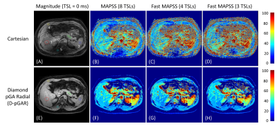 |
31 | Fast Free-Breathing 3D T1ρ Abdominal Imaging Using an Efficient Diamond Radial Sampling Strategy at 3T
Can Wu1, Qi Peng2, Ramin Jafari3, Yansong Zhao3, Victoria Yu1, and Ricardo Otazo1,4
1Department of Medical Physics, Memorial Sloan Kettering Cancer Center, New York, NY, United States, 2Department of Radiology, Albert Einstein College of Medicine and Montefiore Medical Center, Bronx, NY, United States, 3MR Clinical Science, Philips Healthcare, Cambridge, MA, United States, 4Department of Radiology, Memorial Sloan Kettering Cancer Center, New York, NY, United States Keywords: Liver, Relaxometry An efficient diamond radial sampling strategy was proposed for free-breathing 3D T1ρ abdominal imaging at 3T. The phantom experiment shows that diamond radial sampling provides T1ρ measurement values comparable to that of 3D Cartesian and radial stack-of-stars sampling. In-vivo volunteer studies illustrate that diamond radial sampling is superior to Cartesian sampling, where image quality is significantly compromised by breathing motion artifacts. In addition, scan time can be drastically reduced using the fast MAPSS method. This work demonstrates the feasibility of quantitative free-breathing 3D T1ρ imaging with diamond radial sampling in the abdomen. |
|
1849.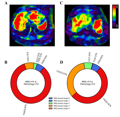 |
32 | Heterogeneity of liver stiffness in chronic HBV infection: Influence on concordance between MRE-based fibrosis staging and biopsy staging
Chao Li1, Mingkai Li2, Xin Jin1, Mengshi Dong1, Yu Han1, Meng Yin3, Jun Chen1, Kevin J Glaser3, Richard L Ehman3, and Jin Wang1
1Department of Radiology, The Third Affiliated Hospital, Sun Yat-sen University (SYSU), Guangzhou, China, 2Department of Gastroenterology, The Third Affiliated Hospital, Sun Yat-sen University (SYSU), Guangzhou, China, 3Department of Radiology, Mayo Clinic, Rochester, MN, United States Keywords: Liver, Elastography, MR elastography; Liver fibrosis; Heterogeneity Chronic hepatitis B (CHB) viral infection can lead to liver fibrosis. Liver biopsy, the gold standard for liver fibrosis assessment, is invasive and carries risks of complications and sampling errors. MR elastography (MRE) is regarded as the most accurate noninvasive tool for evaluating liver fibrosis. In this study we characterized the spatial heterogeneity of liver stiffness depicted by MRE in patients with CHB and found heterogeneity at all pathological fibrosis stages. The severity of spatial heterogeneity affected the concordance between MRE-based and pathology-based fibrosis staging. |
|
1850.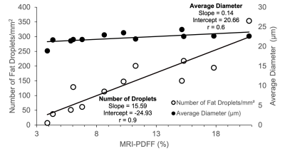 |
33 | Quantitative Digital Histological Analysis and Comparison to Proton Density Fat Fraction in Patients Undergoing Weight Loss Surgery
Devashish Joshi1, David Harris2, Nikolaos Panagiotopoulos2, Danielle Batakis3, Eduardo Grunvald4, Xiaofei Zhang5, Rashmi Agni5, Santiago Horgan6, Luke Funk1, Claude Sirlin3, and Scott Reeder2,7,8,9,10
1Surgery, University of Wisconsin - Madison, Madison, WI, United States, 2Radiology, University of Wisconsin - Madison, Madison, WI, United States, 3Radiology, Liver Imaging Group, University of California San Diego, La Jolla, CA, United States, 4Medicine, University of California San Diego, La Jolla, CA, United States, 5Pathology, University of Wisconsin - Madison, Madison, WI, United States, 6Surgery, University of California San Diego, La Jolla, CA, United States, 7Emergency Medicine, University of Wisconsin - Madison, Madison, WI, United States, 8Medicine, University of Wisconsin - Madison, Madison, WI, United States, 9Biomedical Engineering, University of Wisconsin - Madison, Madison, WI, United States, 10Medical Physics, University of Wisconsin - Madison, Madison, WI, United States Keywords: Liver, Fat The goal of our study was to correlate quantitative digital microscopic histological markers of hepatic steatosis with MRI-PDFF and demonstrate that microenvironment characterization can be used to evaluate hepatic microstructure in NAFLD for future MRI-based studies. Patients were recruited to undergo weight loss surgery during which a liver biopsy was performed. The biopsies were analyzed for steatosis proportionate area (SPA) and characterization of fat droplets. The SPA strongly correlated to the pathologist grading and MRI-PDFF and the liver fat content strongly correlated with the number of fat droplets. Our work demonstrated the feasibility of using MRI to characterize liver microstructure. |
|
1851. |
34 | Detecting Hepatic Flexibility in patients with Non-Alcoholic Fatty Liver Disease (NAFLD) using 1H Magnetic Resonance Spectroscopy
Dragana Savic1, Ferenc E. Mózes1, Leanne Hodson1, Stefan Neubauer1, Michael Pavlides1, and Ladislav Valkovič1
1University of Oxford, Oxford, United Kingdom Keywords: Liver, Liver, L-carnitine, acetylcarnitine This study investigated serum carnitine species and in-vivo hepatic acetylcarnitine levels as measured with MRS in healthy volunteers, and patients with low-risk NAFLD and high-risk NAFLD. L-carnitine facilitates transport of fatty acids into the mitochondria. We show that a single injection of L-carnitine modulated acetylcarnitine levels in the liver in healthy volunteers and low-risk NAFLD, but that this mechanism was blunted in the high-risk NAFLD group. This was accompanied by changed serum medium-chain and long-chain carnitine species, but only in the high-risk NAFLD group. Further studies are needed to understand the plasma and hepatic metabolic changes of carnitine species. |
|
1852.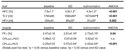 |
35 | Effect of isocaloric ketogenic diet on fat content in the liver and muscle and volume of adipose tissue
Petr Sedivy1, Barbora Setinova1, Martin Burian1, Dita Pajuelo1, Milan Hajek1, Viktor Sebo2, Michal Koc2, Michaela Siklova2, Marina Henikova2, Eva Krauzova3, Jan Gojda2, Lenka Rossmeislova2, Jan Kovar4, and Monika Dezortova1
1MR-Unit, Institute for Clinical and Experimental Medicine, Prague, Czech Republic, 2Department of Pathophysiology, Third Faculty of Medicine, Charles University, University Hospital Královské Vinohrady, Prague, Czech Republic, 3Department of Internal Medicine, Third Faculty of Medicine, Charles University, University Hospital Královské Vinohrady, Prague, Czech Republic, 4Centre for Experimental Medicine, Institute for Clinical and Experimental Medicine, Prague, Czech Republic Keywords: Liver, Metabolism, fat fraction, liver steatosis, intra- and extramyocellular fat, calf muscle The effect of a 28-day isocaloric ketogenic diet followed by a 2-day carbohydrate realimentation on adipose tissue and fat content in the liver and calf muscle in 22 obese women was studied using MRS and MRI methods. During the ketogenic diet, there was a significant decrease in liver fat volume (69±20 ml, i.e. decrease 43±4% of basal value) and liver fat content (2.7±0.6 %, i.e. 35±5% of basal value) and reduction cross-section in both subcutaneous (6 %) and visceral (7 %) fat. Contrary, the intramyocellular fat increased during the diet. |
|
1853. |
36 | Evaluation of Confounding Influence of Fat on Hepatic Quantitative Parametric Mapping
Anandh Kilpattu Ramaniharan1, Mary Kate Manhard1,2, Jean A Tkach1,2, Andrew T Trout1,2, Jonathan R Dillman1,2, and Amol Pednekar1,2
1Radiology, Cincinnati Children's Hospital Medical Center, Cincinnati, OH, United States, 2Radiology, University of Cincinnati College of Medicine, Cincinnati, OH, United States Keywords: Liver, Quantitative Imaging, Relaxometry Quantitative parametric mapping is an increasingly important tool for non-invasive assessment of liver disease. The extent of change in both water and fat content in the liver is dependent on the type and stage of the disease. Thus, proton density fat fraction (PDFF%) can have a confounding influence on the longitudinal change in estimated hepatic T1, T2, and T2* measurements. In 14 research participants, T1 was proportionately underestimated (4.70 (2.85, 6.54) ms/PDFF%) and T2 was proportionately overestimated (-0.60 (-0.76, -0.43) ms/PDFF%) with non-fat-suppressed parametric map compared to analogous estimates with fat-suppressed parametric maps, while T2* estimates were insensitive to PDFF. |
|
1854. |
37 | In Vivo Evaluation of a Novel Deep Learning-based MR Image Reconstruction for Liver Fat Quantification
Nikolaos Panagiotopoulos1,2, Nathan T Roberts3, David Harris1, Daiki Tamada1, Ty Cashen3, Thekla H Oechtering1,2, Diego Hernando1,4,5,6, Claude B Sirlin7, and Scott B Reeder1,4,5,8,9
1Department of Radiology, University of Wisconsin-Madison, Madison, WI, United States, 2Department of Radiology and Nuclear Medicine, Universität zu Lübeck, Lübeck, Germany, 3GE Healthcare, Waukesha, WI, United States, 4Department of Medical Physics, University of Wisconsin-Madison, Madison, WI, United States, 5Department of Biomedical Engineering, University of Wisconsin-Madison, Madison, WI, United States, 6Department of Departments of Electrical & Computer Engineering, University of Wisconsin-Madison, Madison, WI, United States, 7Department of Radiology, University of California San Diego, San Diego, CA, United States, 8Department of Medicine, University of Wisconsin-Madison, Madison, WI, United States, 9Department of Emergency Medicine, University of Wisconsin-Madison, Madison, WI, United States Keywords: Liver, Quantitative Imaging Deep learning (DL)-based MR reconstruction methods show promise to reduce image noise compared to conventional image reconstruction while maintaining quantitative accuracy. The purpose of this work is to apply and evaluate the performance of DL reconstruction to chemical shift-encoded MRI for quantification of proton density fat-fraction (PDFF) in the liver. We compared quantitative PDFF results, test-retest repeatability, and standard deviation within regions of interest in nine patients with a wide range of PDFF (1-31%), for different levels of DL denoising. PDFF between reconstructions showed excellent agreement and constant test-retest repeatability. Standard deviation decreased with increasing DL denoising levels. |
|
1855.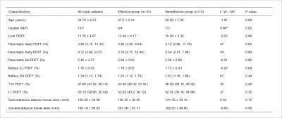 |
38 | The relationship between the prognosis of NAFLD and abdominal organ fat fraction
Xingchen Pan1, Jiahui Fu1, Lei Zhang1, Yueluan Jiang2, Ye Sun1, Xin Shi1, and Fan Yang1
1Department of Radiology, First Hospital of Jilin University, changchun, China, 2MR Scientific Marketing, Siemens Healthineers, Beijing, China Keywords: Liver, Liver, NAFLD; prognosis; fat fraction; abdominal organ The relationship between the prognosis of non-alcoholic fatty liver disease (NAFLD) treatment and the fat fraction or fat area of other abdominal organs has not been explored. In this study, we measured the proton density fat fraction of four subsegments of the liver, pancreas, renal cortex, thoracic 12 and lumbar 1 vertebral body, and the area of subcutaneous adipose tissue and visceral adipose tissue at the level of lumbar 3, and explored their relationship with the prognosis of NAFLD. |
|
1856.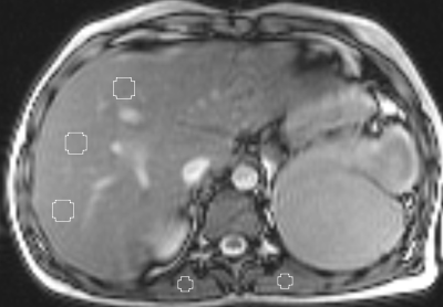 |
39 | MRI-based liver iron quantification using refocused gradient-echo (bSSFP): initial results
Arthur Peter Wunderlich1,2, Holger Cario3, Michael Götz2, Meinrad Beer1, and Stefan Andreas Schmidt1
1Diagnostic and Intervnetional Radiology, Ulm University, Medical Center, Ulm, Germany, 2Section for Experimental Radiology, Ulm University, Medical Center, Ulm, Germany, 3Department of Pediatrics and Adolescent Medicine, Ulm University, Medical Center, Ulm, Germany Keywords: Liver, Hematologic, Iron overload Purpose: To evaluate the feasibility of using a balanced steady-state free precession sequence (bSSFP) to determine liver iron content (LIC). Methods: Thirty-five consecutive patients with liver iron overload were examined with bSSFP. Signal intensity ratios between liver parenchyma and paraspinal muscles were correlated with LIC reference values obtained using Ferriscan. Results: LIC values ranged from 24 to 756 µmol/g. The best SIR-to-LIC correlation was obtained with 3.5 ms repetition time and 17° excitation flip angle. Conclusion: bSSFP is suitable to determine LIC. Its advantages are high SNR efficiency and the ability to acquire the entire liver in a breath hold. |
|
1857.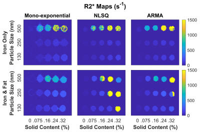 |
40 | Impact of Iron Particle Sizes on R2* Estimation in Phantoms Mimicking Hepatic Iron Overload and Steatosis Using MRI
Sarah Brasher1, Annie Chan1, Ayaz Khan2, Zachary Abramson2, Cara Morin3, and Aaryani Tipirneni-Sajja1,2
1Biomedical Engineering, University of Memphis, Memphis, TN, United States, 2Diagnostic Imaging, St. Jude Children's Research Hospital, Memphis, TN, United States, 3Radiology, Cincinnati Children's Hospital Medical Center, Cincinnati, OH, United States Keywords: Liver, Phantoms Size and concentration of iron will affect the dephasing of an MRI signal. However, iron particle size is often unaccounted for in phantom studies investigating iron overload. In this study, phantoms utilizing iron nanoparticles of different diameters were used to mimic in vivo iron deposits, and R2* quantification was analyzed using different signal models. Our results show that R2* was higher and more unstable in phantoms with iron particles of 500nm diameter in comparison to those with 250nm and 130nm particles potentially due to clustering. High iron concentrations and large iron particle sizes were also shown to confound fat quantification. |
|
 Back to Meeting Home
 Back to the Program-at-a-Glance
The International Society for Magnetic Resonance in Medicine is accredited by the Accreditation Council for Continuing Medical Education to provide continuing medical education for physicians.
 Back to Meeting Home
 Back to the Program-at-a-Glance
 View Presentation Video