Digital Poster
Advances in Prostate Imaging
ISMRM & ISMRT Annual Meeting & Exhibition • 03-08 June 2023 • Toronto, ON, Canada

| Computer # | |||
|---|---|---|---|
1858. |
41 | ADC as a quantitative value: classifying lymph node dignity in prostate cancer and inter-scanner variability
Jochen Bauer1, Maria Eveslage2, and Benjamin Noto1
1Department of Clinical Radiology, University of Muenster, Münster, Germany, 2Institute of Biostatistics and Clinical Research, University of Muenster, Muenster, Germany Keywords: Prostate, Diffusion/other diffusion imaging techniques, lymph node, reproducibility Apparent diffusion coefficient (ADC) has potential as a quantitative imaging biomarker for differentiation of malignant from benign tissue. Here, we present a phantom and in vivo measurement of ADC reproducibility and a restrospective study in over 100 patients with prostate cancer to evaluate ADC as a classifier. |
|
1859.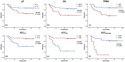 |
42 | Prognostic Value of ADC Histogram Analysis in Men with High Risk Prostate Cancer Receiving Adjuvant Hormonal Therapy after Radical Prostatectomy
Kangwen He1, Xiaoyan Meng1, and Zhen Li1
1Radiology, Tongji Hospital, Tongji Medical College, Huazhong University of Science and Technology, Wuhan, China Keywords: Prostate, Cancer, high-risk prostate cancer, biochemical recurrence 直方图分析是医学图像的定量后处理方法,被认为在癌症管理中很有价值。我们使用表观扩散系数 (ADC) 图的直方图参数分析了在根治性前列腺切除术 (RP) 后接受辅助激素治疗 (AHT) 的高危前列腺癌 (PCa) 患者的结果。我们观察到,较低的 ADC 50 和较高的峰度可以独立预测较差的生化无复发生存期 (BCR-fs),并且当添加直方图参数时,预后模型的性能会有所提高。我们的研究结果显示了 ADC 直方图分析在这一特定的 PCa 患者亚组中的增量预后价值。 |
|
1860.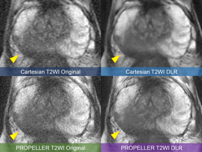 |
43 | High-resolution PROPELLER T2-weighted imaging of the prostate with deep learning reconstruction: a phantom and clinical preliminary study
Atsushi Nakamoto1, Hiromitsu Onishi1, Takahiro Tsuboyama1, Takashi Ota1, Hideyuki Fukui1, Keigo Yano1, Kengo Kiso1, Toru Honda1, Shohei Matsumoto1, Mitsuaki Tatsumi1, Hiroyuki Tarewaki2, Yoshihiro Koyama2, Tetsuya Wakayama3, Xinzeng Wang4, and Noriyuki Tomiyama1
1Osaka University Graduate School of Medicine, Suita, Japan, 2Osaka University Hospital, Suita, Japan, 3GE Healthcare, Hino, Japan, 4GE Healthcare, Houston, TX, United States Keywords: Prostate, Image Reconstruction, Deep learning reconstruction The image quality of high-resolution PROPELLER T2WI combined with deep learning reconstruction (DLR) in prostate MRI was evaluated by the phantom and clinical studies. In the phantom study, noise reduction was achieved by DLR, and spatial resolution was remarkably improved in PROPELLER T2WI compared to Cartesian T2WI. In the clinical study, DLR showed a significant improvement in signal-to-noise ratio, and the qualitative analyses showed reduced image noise and improved spatial resolution, with PROPELLER T2WI DLR images showing the highest overall image quality. PROPELLER T2WI with DLR would be promising technique to improve the image quality of T2WI in prostate MRI. |
|
1861.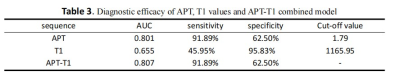 |
44 | To evaluate the value of APT combined with T1 mapping in the differential diagnosis of prostate cancer and benign prostatic hyperplasia
Xiwei Li1, Lihua Chen1, Nan Wang1, Liangjie Lin2, Jiazheng Wang2, and Ailian Liu1
1The First Affiliated Hospital of Dalian Medical University, Dalian, China, 2Philips Healthcare,Beijing, Beijing, China Keywords: Cancer, Prostate At present, the prostate specific antigen (PSA) detection is a common method for screening prostate cancer. However, the specificity of PSA is very low, and it is easy to appear false positive. In recent years, multiparameter imaging has great value in the diagnosis of prostate diseases. Amide proton weighted(APTw) imaging and T1 mapping imaging can more accurately reflect the information of tumor tissue cell metabolism and pathological cell microstructure.The purpose of this study was to evaluate the value of APT and T1 measurements in the diagnosis of prostate cancer and benign prostatic hyperplasia. |
|
1862. |
45 | Sub-differentiation of PI-RADS 3 lesions in TZ by advanced diffusion-weighted imaging to aid the biopsy decision process
Kun-Peng Zhou1, Jie Bian2, Hua-Bin Huang1, Chao Bu1, and Li-Zhi Xie3
1Radiology, The Seventh Affiliated Hospital, Sun Yat-sen University, Shenzhen, China, 2Radiology, the second hospital of dalian medical university, dalian, China, 3GE Healthcare, Beijing, China, Beijing, China Keywords: Prostate, Prostate The aim of this study was to investigate the use of advanced diffusion-weighted imaging (IVIM, stretched exponential model and DKI) to sub-differentiation of TZ PI-RADS 3 lesions and aid the biopsy decision process. Finally, the logistic model could correctly classify 89.39% of the subjects. The results of ROC analysis showed that AUC was 0.9197, CI 95%: 0.8736-0.9659. Sensitivity, specificity, positive predictive value and negative predictive value were 92.1%, 80.4%, 93.9% and 75.5%, respectively. Therefore, advanced diffusion-weighted imaging can predict PCa in TZ PI-RADS 3 lesions and inform the decision-making process of whether or not to perform a biopsy. |
|
1863.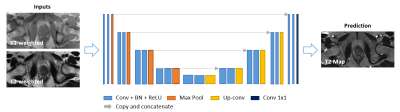 |
46 | Retrospective T2 quantification from conventional weighted MRI of the prostate based on deep learning
Haoran Sun1,2, Lixia Wang1, Timothy Daskivich3, Shihan Qiu1,2, Fei Han1, Alessandro D'Agnolo4, Rola Saouaf5, Hyung Kim3, Debiao Li1,2, and Yibin Xie1
1Biomedical Imaging Research Institute, Cedars-Sinai Medical Center, Los Angeles, CA, United States, 2Bioengineering, University of California, Los Angeles, Los Angeles, CA, United States, 3Minimally Invasive Urology, Cedars-Sinai Medical Center, Los Angeles, CA, United States, 4Imaging/Nuclear Medicine, Cedars-Sinai Medical Center, Los Angeles, CA, United States, 5Imaging, Cedars-Sinai Medical Center, Los Angeles, CA, United States Keywords: Prostate, Quantitative Imaging Prostate cancer (PCa) is one of the most common types of cancer with a considerable morbidity and mortality. Multiparametric MRI as a noninvasive imaging tool in PCa diagnosis has limitations. Recent studies suggest that quantitative T2 information is helpful in PCa diagnosis and lesion characterization but is not generally available due to the need for additional scans. Here, we developed a DL-based method to estimate T2 maps retrospectively from clinically acquired T1- and T2-weighted images. The developed technique has the potential to improve PCa diagnosis and lesion characterization using quantitative T2 information estimated from conventional clinical scans. |
|
1864.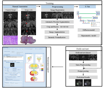 |
47 | Study of deep-learning models for detection and localization of early-stage clinically significant prostate cancer on mpMRI
Zhaonan Sun1, Xiaoying Wang2, and Kexin Wang3
1Department of Radiology, Peking University First Hospital, Beijing, China, 2Department of Radiology, Peking University First Hospital, Beijing, China, 3School of Basic Medical Sciences, Capital Medical University, Beijing, China Keywords: Prostate, Cancer The purpose of this study was to explore the best sequences for fully automated detection and localization of early-stage csPCa with a PSA range of 4-10 ng/mL.1628 prostate mpMRI examined with seven scanners were retrospectively enrolled. A training dataset (n=1428) was used to train models, i.e., a diffusion model was trained with diffusion-weighted imaging, and a biparametric model was trained with diffusion-weighted imaging and T2-weighted imaging. A hold-out test dataset (n=200) was reserved for validation. Our research shows that the diffusion model trained using ADC and DWI achieved best performance in detection and localization of early-stage csPCa. |
|
1865. |
48 | Positive Predictive Value of Multiparametric MRI in Detection of Prostate cancer: A Tertiary Center Retrospective Study
Zachary Franks1, Jarrett Rosenberg1, Richard E. Fan1, Geoffrey Sonn1, and Pejman Ghanouni1
1Stanford Health Care, Stanford, CA, United States Keywords: Prostate, Cancer Prostate Imaging Reporting and Data System (PIRADS) guidelines are used as a standard to help radiologists diminish variation in the acquisition, interpretation, and reporting of multiparametric magnetic resonance imaging (mpMRI) for prostate cancer diagnosis. However, the performance of mpMRI has been shown to vary across radiologists. A retrospective study was performed at a tertiary care center to assess the range of positive predictive values (PPV) across individual radiologists interpreting mpMRI in detecting any prostate cancer compared to targeted MR-US guided fusion biopsy pathology results. |
|
1866.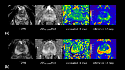 |
49 | Estimated quantitative relaxation mapping calculated from Multiple-Repetition Multiple-Echo (MRME) based DWI acquisition in prostate
Yu Ueda1, Tsutomu Tamada2, Ayumu Kido2, Kazunori Moriya2, Shigeru Shibata2, Mitsuru Takeuchi3, Akira Yamamoto2, Makoto Obara1, and Marc Van Cauteren4
1Philips Japan, Ltd., Tokyo, Japan, 2Department of Radiology, Kawasaki Medical School, Kurashiki, Japan, 3Department of Diagnostic Radiology, Radiolonet Tokai, Nagoya, Japan, 4Philips Healthcare, Business Unit MR, Tokyo, Japan Keywords: Prostate, Quantitative Imaging It has been reported that synthetised DW images with short TR and zero TE, generated from quantitative relaxation parameters, improved lesion contrast in prostate. In this study, we investigate the added value of the estimated T1 and T2 values in differentiating benign from malignant lesion in suspicious cancers with PI-RADS category 3, potentially reducing unnecessary biopsies. In addition to the conventional DW images at b-values of 1000 and 2000, ADC map, and estimated T1 and T2 maps, synthetised DW images can be acquired in approximately 4 minutes, an acceptable scan time in clinical practice. |
|
1867.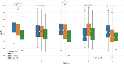 |
50 | Studying variation in DWI MRI derived ADC for Prostate Cancer (PCa) with respect to Gleason Grade and location of PCa lesions
Sudhanya Chatterjee1, Dattesh Dayanand Shanbhag1, Aanchal Mongia1, Uday Patil1, Adele Courot2, Nicolas Gogin2, and Rakesh Mullick1
1GE Healthcare, Bangalore, India, 2GE Healthcare, Buc, France Keywords: Prostate, Cancer, DWI, ADC, Gleason grade Diffusion weighted MRI (DWI) derived apparent diffusion coefficient (ADC) is a marker of the extent of restrictive diffusion in tissues. Higher Gleason Grade (GG) prostate cancer (PCa) lesions are known to exhibit higher restrictive diffusion. In this study we investigated whether ADC as a tissue biomarker follows the known histopathological behavior of PCa lesions by studying variations in ADC with respect to GG of PCa lesions and its location. We observe that ADC values of lesion voxels, although belonging to same GG, vary based on its location. |
|
1868.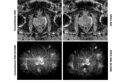 |
51 | Accelerated diffusion-weighted imaging of the prostate employing Echo Planar Imaging with Compressed SENSE based reconstruction (EPICS)
Yannik Christian Layer1, Petra Muertz1, Leon Bischoff1, Christoph Katemann2, Kilian Weiss2, Julian Luetkens1, Ulrike Attenberger1, and Claus Christian Pieper1
1Department of Diagnostic and Interventional Radiology, University Hospital Bonn, Bonn, Germany, 2Philips GmbH Market DACH, Hamburg, Germany Keywords: Prostate, Tumor The aim of this single-center, prospective study was to evaluate an accelerated diffusion-weighted imaging sequence of the prostate using Echo Planar Imaging with Compressed SENSE based reconstruction and assess its performance in comparison to conventional DWI. The evaluated sequences considerably reduced the total DWI acquisition time by 43% and achieved overall good image quality without significant differences compared to the conventional DWI sequence. The reduced scan time allows for an increase of the overall number of scanned patients as well as increased patient convenience with less potential of motion artifacts and therefore benefits care of patients with suspected prostate cancer. |
|
1869.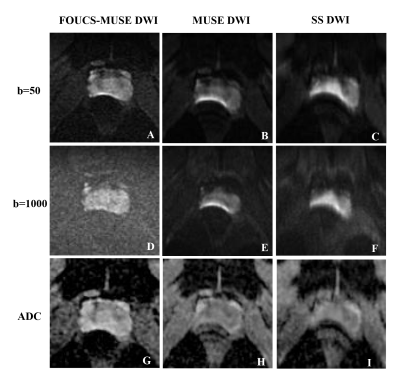 |
52 | Improving Image Quality While Reducing Geometric Distortion of Diffusion-weighted MRI in prostate: Comparison of FOCUS-MUSE, MUSE, and SS DWI
Yanling Chen1, Huanjun Wang 1, and Yan Guo1
1Department of Radiology, The First Affiliated Hospital, Sun Yat-Sen University, Guangzhou, China Keywords: Prostate, Diffusion/other diffusion imaging techniques The current study investigated the qualitative and quantitative image quality of three DWI sequences of the prostate: SS DWI, MUSE DWI and a new DWI sequence named FOCUS-MUSE DWI. Twenty-three healthy volunteers were enrolled. The diagnostic image quality score was assessed by two radiologists on the three DWI sequences. And the ADC values in the peripheral and transitional zone were measured together with the Dice Coefficient calculated for quantitative comparison. Our results indicated that FOCUS-MUSE DWI had a superior image quality and more stable ADC measurement compared with SS DWI and MUSE DWI. The geometric distortion level was also reduced. |
|
1870. |
53 | Sub-differentiation of intermediate risk prostate cancer by advanced diffusion-weighted imaging
Kun-Peng Zhou1, Hua-Bin Huang1, Long Liu1, Jie Bian2, and Li-Zhi Xie3
1Radiology, The Seventh Affiliated Hospital, Sun Yat-sen University, Shenzhen, China, 2Radiology, The second hospital of Dalian medical university, Dlian, China, 3GE Healthcare, Beijing, Beijing, China Keywords: Prostate, Prostate The aim of this study was to investigate the use of advanced diffusion-weighted imaging to sub-differentiation of intermediate risk group PCa and aid treatment decision process. Finally, the logistic model was statistically significant, χ2=90.969, p<0.001. The model could correctly classify 86.40% of the subjects. The results of ROC analysis showed that area AUC was 0.9429, CI 95%: 0.9054-0.9805. Sensitivity, specificity, positive predictive value and negative predictive value were 88.06%, 84.31%, 88.06% and 84.31%, respectively. The results of this research may indicate that advanced diffusion-weighted imaging can sub-differentiate intermediate risk group PCa and aid the decision process of treatment. |
|
1871.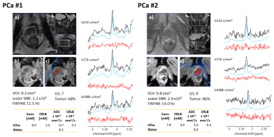 |
54 | Initial exploration of the potential of diffusion-weighted MRS for the evaluation of prostate pathology
Rudy Rizzo1,2, Angeliki Stamatelatou3, Kadir Simsek4, Jack J.A. Van Asten3, Roland Kreis1,2, Arend Heerschap3, and Tom Scheenen3
1Magnetic Resonance Methodology, Institute of Diagnostic and Interventional Neuroradiology, University of Bern, Bern, Switzerland, 2Translational Imaging Center (TIC), Swiss Institute for Translational and Entrepreneurial Medicine, Bern, Switzerland, 3Department of Medical Imaging, Radboud University Medical Center, Nijmegen, Netherlands, 4Cardiff University Brain Research Imaging Centre (CUBRIC), Cardiff University, Cardiff, United Kingdom Keywords: Prostate, Cancer Prostate pathologies like cancer and prostatitis cause alterations in tissue microstructure that are reflected in signal variations on diffusion-weighted MRI or MR relaxometry. Diffusion-weighted MR Spectroscopy (DW-MRS) allows specific characterization of tissue microstructure by quantifying both concentration and diffusion properties of MR-observable metabolites. In this work, an optimized DW-MRS protocol tailored to overcome the inherent challenges of prostate measurements is deployed. Initial results on potential alterations of metabolite concentrations and diffusivities (ADCs) are reported for prostate cancer and prostatitis. The preliminary findings are tentatively explained by microstructure alterations of the prostate tissue. |
|
1872. |
55 | Reverse Encoding Distortion Correction for DWI: Improving Image Quality and Differention Capability of Malignant from Benign Prostatic Areas
Takahiro Ueda1, Yoshiharu Ohno1,2, Maiko Shinohara3, Kaori Yamamoto3, Masato Ikedo3, Masao Yui3, Akiyoshi Iwase4, Minami Furuta1, Yuki Obama1, Hiroyuki Nagata2, Hirotaka Ikeda1, Yoshiyuki Ozawa1, and Hiroshi Toyama1
1Radiology, Fujita Health University School of Medicine, Toyoake, Japan, 2Joint Research Laboratory of Advanced Medical Imaging, Fujita Health University School of Medicine, Toyoake, Japan, 3Canon Medical Systems Corporation, Otawara, Japan, 4Fujita Health University Hospital, Toyoake, Japan Keywords: Prostate, Cancer We hypothesize that RDC is useful for image quality and diagnostic performance improvements on DWI with b value at 1500 s/mm2 in suspected prostatic cancer patients, although there was little influence of RDC on DWI at in vitro study. The purpose of this study was to determine the influence of RDC for ADC measurement at in vitro study and its’ utility for improving image quality and diagnostic performance of malignant from benign prostatic areas on prostatic DWI as in vivo study. |
|
1873.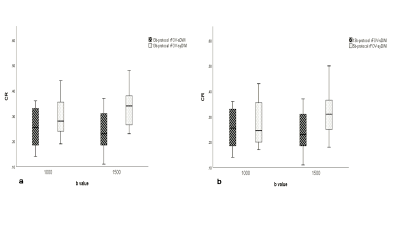 |
56 | A clinical validation study of synthetic high b-value reduced filed-of-view diffusion weighted imaging on prostate
Qiqi Zhou1, Weiyin Vivian Liu2, Qian Tang3, Ling Sang1, Wen Chen1, and Lin Xu1
1Department of Radiology,Taihe Hospital,Hubei University of Medicine, Hubei, China, 2GE Healthcare, MR Research, Beijing, China, 3Department of Biomedical Engineering,Hubei University of Medicine, Hubei, China Keywords: Prostate, Cancer High b-value diffusion weighted imaging is clinically challenging due to its inherently low SNR and prone to presence of severe eddy current distortion due to utility of large diffusion sensitization gradient, not to mentioned high-resolution prostate DWI .In our study, 5b-protocol rFOV-syDWIswith pros of short scan time provided better lesion clarity and higher image quality.Moreover, synthetic ADCs offered reliable and satisfactory diagnostic value as scanned 13b-protocol DWIs despite significant difference of mean and median were found between syADCb=1500 and sADCb=1500 in PCa but not in hyperplasia, indicating diagnosis withrFOV-syADCs should be careful when referring to rFOV-sADCs. |
|
1874.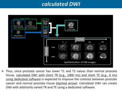 |
57 | Comparison of calculated DWI and single-shot EPI DWI for prostate cancer detection at 3T
TSUTOMU TAMADA1, YUICHI KOJIMA1, AYUMU KIDO1, YU UEDA2, MITSURU TAKEUCHI3, KENTARO ONO1, MIDORI YAMAMOTO1, AKIRA YAMAMOTO1, and YOSHIHIKO FUKUKURA1
1Radiology, Kawasaki Medical School, Kurashiki-city, Japan, 2Philips Japan, Tokyo, Japan, 3Radiology, Radiolonet Tokai, Nagoya, Japan Keywords: Prostate, Prostate We compared diagnostic performance of clinically significant PC (csPC) between calculated DWI (calDWI) which enable to synthesize DW images with any TR and TE and native acquired single-shot EPI DWI (nDWI). The calDWI was generated with TR of 1000 and TE of 0 ms. Three radiologists independently assessed eight regions of each prostate by PI-RADS v 2.1 DWI score. For diagnostic performance of csPC, AUC was comparable between calDWI and nDWI in two readers. The diagnostic specificity was significantly higher in calDWI than in nDWI in two readers. Compared with nDWI, calDWI had similar or higher diagnostic performance in csPC. |
|
1875.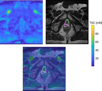 |
58 | Primary prostate cancer characterisation using internal references for 23Na MRI quantification
Anne Adlung1,2, Fabian Tollens3, Anika Strittmatter1,2, Niklas Westhoff4, Daniel Hausmann5, Stefan O Schoenberg3, Lothar R Schad1,2, Dominik Nörenberg3, and Frank G Zöllner1,2
1Computer Assisted Clinical Medicine, Medical Faculty Mannheim, Heidelberg University, Mannheim, Germany, 2Mannheim Institute for Intelligent Systems in Medicine, Medical Faculty Mannheim, Heidelberg University, Mannheim, Germany, 3Department of Radiology and Nuclear Medicine, Mannheim University Medical Centre, Mannheim, Germany, 4Department of Urology and Urosurgery, Mannheim University Medical Centre, Mannheim, Germany, 5Department of Radiology, Kantonsspital Baden, Baden, Switzerland Keywords: Prostate, Tumor Aim of this prospective study was to evaluate 23Na MRI of 36 patients with suspected prostate cancer and quantify tissue sodium concentration (TSC) based on internal references. TSC reference regions were defined within both femoral blood vessels (FBV), where TSC showed no significant differences between both sides (3.3±2.2 mM, p=0.076). TSC in the prostate was 39.0±5.3 mM; significantly higher in the peripheral than in the transitional zone (p=0.0004, mean difference 4.7±3.5 mM). Nine lesions were segmented and showed mean TSC of 32.2±5.5, significantly lower than contralateral (p=0.0181). TSC might represent a quantitative biomarker that could improve prostate lesion characterization. |
|
1876.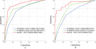 |
59 | Magnetic Resonance Imaging-based Radiomics for Clinically Significant Prostate Cancer in PSA Gray Zone to Reduce Unnecessary Biopsies
Xiaomeng Qiao1, Chenhan Hu1, Jie Bao1, Ximing Wang1, Yang Song2, and Yunzhu Wu2
1Department of radiology, First Affiliated Hospital of Soochow University, Suzhou, China, 2MR Scientific Marketing, Siemens Healthineers Ltd, Shanghai, China Keywords: Prostate, Radiomics, Clinically significant prostate cancer; Prostate-specific antigen Radiomics was a promising method for csPCa with PSA levels in the gray zone and outperformed PI-RADS assessments. |
|
1877.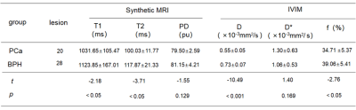 |
60 | The diagnostic value of synthetic MRI combined with rFOV IVIM in transition zone prostate cancer
Qiqi Zhou1, Weiyin Vivian Liu2, Qian Tang3, Ling Sang1, Wen Chen1, and Lin Xu1
1Department of Radiology,Taihe Hospital,Hubei University of Medicine, Hubei, China, 2GE Healthcare, MR Research, Beijing, China, 3Department of Biomedical Engineering,Hubei University of Medicine, Hubei, China Keywords: Prostate, Cancer This study investigated the value of relaxation time in synthetic MRI imaging combined with intravoxel incoherent motion (IVIM) imaging in differentiating transitional zone prostate cancer from benign prostatic hyperplasia (BPH). Finally, it was found that T2 value in synthetic MRI imaging and D value in IVIM imaging had higher diagnostic sensitivity and specificity, and their combined diagnostic performance was higher. It is helpful for the differential diagnosis of BPH and PCa in transitional zone. |
|
 Back to Meeting Home
 Back to the Program-at-a-Glance
The International Society for Magnetic Resonance in Medicine is accredited by the Accreditation Council for Continuing Medical Education to provide continuing medical education for physicians.
 Back to Meeting Home
 Back to the Program-at-a-Glance
 View Presentation Video