Digital Poster
Updates in Oncologic Liver Imaging I
ISMRM & ISMRT Annual Meeting & Exhibition • 03-08 June 2023 • Toronto, ON, Canada

Digital Poster
Updates in Oncologic Liver Imaging I
| Computer # | |||
|---|---|---|---|
2234.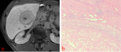 |
61 | Imaging findings and differential diagnosis of hyperintense hepatocellular carcinoma on hepatobiliary phase of Gd-EOB-DTPA-enhanced MRI
Ji yong Gu1, Fei Xing2, Yi shi Wang3, and Xian ce Zhao4
1Nantong Haimen People’s Hospital, Nantong, China, 2the Third Affiliated Nantong Hospital of Nantong University, Nantong, China, 3Philips Healthcare, Beijing, China, 4Philips Healthcare, Shanghai, China Keywords: Liver, Cancer, HCC This study evaluates imaging features that can help to differentiate hyperintense HCC from non-HCC benign and malignant lesions. On HBP images, hyperintense nodules can be differentiated with several imaging characteristics, especially HBP hypointense rim, and a focal defect in contrast uptake, that indicate the diagnosis of hyperintense HCCs. |
|
2235.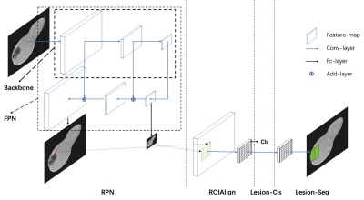 |
62 | A clinically applicable deep-learning model for automatic detection of focal liver lesions on Gd-DTPA-enhanced MRI
Jiahui Jiang1, Lixue Xu1, Xiaolan Zhang2, Niange Yu2, Dawei Yang1, Hui Xu1, and Zhenghan Yang1
1Department of Radiology, Beijing Friendship Hospital, Capital Medical University, Beijing, China, 2Shukun (Beijing) Technology Co., Ltd, Beijing, China Keywords: Liver, Liver Accurate detection of focal liver lesions in Gd-DTPA-enhanced magnetic resonance imaging (MRI) necessitates a high level of skill and experience. This task is typically performed by radiologists through visual inspection, which is time-consuming, labor-intensive, and subject to intra- and inter-observer variation. Convolutional neural networks (CNNs) have demonstrated significant potential in detecting lesions on medical imaging. Our study presents a unified multi-sequence lesion detector model for automatically detecting focal liver lesions on Gd-DTPA-enhanced MR images to aid in treatment decision-making. |
|
2236.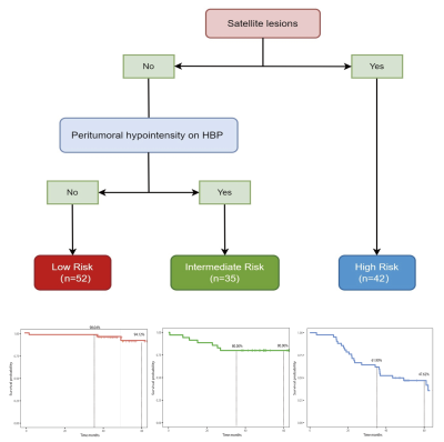 |
63 | Preoperative prognostic Stratification for Patients with HCC Using Gadoxetic acid-enhanced MR Imaging
Feifei Gao1, Yi Wei2, Qian Li2, Xiaocheng Wei3, Lisha Nie3, Miaoqi Zhang3, and Bin Song2
1Radiology, West China Hospital, Chengdu, China, 2West China Hospital, Chengdu, China, 3MR Research China,GE Healthcare, Beijing, China, Beijing, China Keywords: Liver, Cancer Accurate preoperative risk stratification is of critical importance for determine patients who will benefit from liver resection. Our study developed three subgroups of patients with HCC associated with different prognosis (RFS and OS) after resection based on preoperative MR imaging features using the Survival Classification and Regression Tree (CART) method. The generated subgroups using CART method demonstrated good discriminatory for preoperatively stratification of the prognosis. Therefore, the CART model of this present study may help clinicians to consider non-surgical treatment more often in worse prognostic patients and as well as inform more intense surveillance following resection. |
|
2237.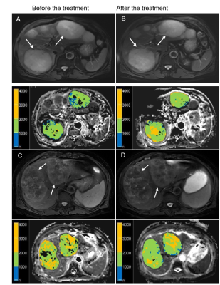 |
64 | Magnetic Resonance Diffusion Imaging for Colorectal Liver Metastases: A Prospective Study for Model Comparison and Early Response Biomarker
Yue Li1, Huan Zhang1, Lei Yue1, Caixia Fu2, Robert Grimm3, Wenhua Li4, Weijian Guo4, and Tong Tong1
1Department of Radiology, Fudan University Shanghai Cancer Center, Shanghai, China, 2MR Application Development, Siemens Shenzhen Magnetic Resonance Ltd, Shenzhen, China, Shenzhen, China, 3MR Applications Predevelopment, Siemens Healthineers Ltd., Erlangen, Germany, Erlangen, Germany, 4Fudan University Shanghai Cancer Center, Shanghai, China Keywords: Liver, Diffusion/other diffusion imaging techniques This study investigated and compared the feasibility of whole tumor based texture analysis of various magnetic resonance diffusion imaging as early predictors of the clinical response to chemotherapy in patients with colorectal liver metastases (CRLM). The results showed that baseline DWI parameters and the follow-up changes of IVIM and DKI parameters can be conductive to predict the chemotherapeutic response of patients with CRLM. And Changes in D-parameters (Δ% DMean, Δ% D5th percentile, and Δ% DDiff-entropy) are superior to other diffusion related parameters. This suggests that DKI-related parameters could effectively predict the response in short period after treatment (2-3 weeks). |
|
2238.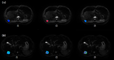 |
65 | Application of whole-tumor histogram analysis of restriction spectrum imaging in the diagnosis of benign and malignant liver lesions
Yu Luo1, Nan Meng1, Lei Shen1, Pengyang Feng2, Ziqiang Li3, Han Jiang3, Han Jiang3, Han Jiang3, Yunfei Zhang4, Yongming Dai4, and Meiyun Wang1
1Zhengzhou University People’s Hospital & Henan Provincial People’s Hospital, Henan, China, 2Henan University People’s Hospital & Henan Provincial People’s Hospital, Henan, China, 3Xinxiang Medical University & Henan Provincial People's Hospital, Henan, China, 4MR Collaboration, Central Research Institute, United Imaging Healthcare, Shanghai, China Keywords: Liver, Cancer Restricted spectrum imaging (RSI) is an advanced technique, which can detect the signal fractions of restricted, hindered and free diffusion of water molecules in the water compartments inside and outside the cell. However, the application of RSI in the assessment of liver disease is rare. Therefore, this study was to evaluate the diagnostic performance of RSI and DWI models in distinguishing benign and malignant liver lesions. Our results showed that the RSI model has better discriminative performance, and the combination of RSI parameters can further improve diagnostic performance. RSI provides a new perspective for exploring the information of tissue microenvironment, and its clinical application has broad prospects. |
|
2239.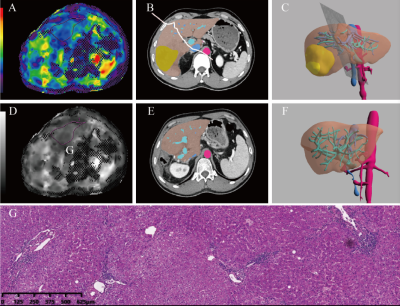 |
66 | Preoperative evaluation of liver regeneration following hepatectomy in hepatocellular carcinoma using magnetic resonance elastography
Tong Zhang1, Qian Li1, Yi Wei1, Hehan Tang1, Lisha Nie2, Xiaocheng Wei2, and Bin Song1,3
1West China Hospital, Sichuan University, Cheng Du, China, 2MR Research China,GE Healthcare, Bei Jing, China, 3Sanya People’s Hospital, San Ya, China Keywords: Liver, Elastography, liver regeneration, elasticity imaging techniques, hepatocellular carcinoma, hepatectomy The liver stiffness (LS) value based on MRE may be a useful preoperative predictor of liver regeneration in patients with HCC. The LS derived from MRE shows a significant negative correlation with fibrosis, an important predictor of liver regeneration. In the low and high parenchymal hepatic resection rate (PHRR) groups, there was no significant relationship between regeneration index (RI) and LS values of MRE. However, a negative relationship was shown between LS values and RI in the intermediate PHRR group. |
|
2240.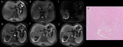 |
67 | Multiparametric MRI manifestations of spontaneous intratumoral coagulative necrosis in hepatocellular carcinoma
Dexin Yu1, Liping Zuo1, Mingyuan Hou1, Jinlei Fan1, Yueming An1, and Bowen Wang1
1Qilu Hospital of Shandong University, Jinan, China Keywords: Liver, Cancer Problem: To illustrate the MRI manifestations of spontaneous intratumoral coagulative necrosis (iCN) in hepatocellular carcinoma (HCC) and its value in predicting postoperative early recurrence. Methods: 263 patients with HCC confirmed by surgical pathology were enrolled. The qualitative assessments of MRI features were performed. Results: The iCN in HCC was characterized by a similar appearance to the tumor on pre-contrast MRI but with non-enhancement on post-contrast MRI. Presence of iCN on MRI was associated with early tumor recurrence. Conclusions: Presence of iCN on MRI is associated with early tumor recurrence and might assist to guide clinical treatment. |
|
2241.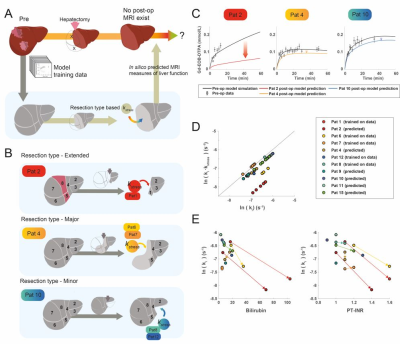 |
68 | Pre-Operative In Silico Risk Assessment for Hepatectomy Patients, Using MRI and Pharmacokinetic Modeling
Christian Simonsson1,2,3, Wolf Claus Bartholomä1,2, Anna Lindhoff Larsson4, Markus Karlsson1, Jens Tellman1, Gunnar Cedersund2,3, Bengt Norén1, Nils Dahlström1,2, Per Sandström4, and Peter Lundberg1,2
1Department of Radiation Physics, Radiology, Department of Medical and Health Sciences, Linköping University, Linköping, Sweden, 2Center for Medical Image Science and Visualization (CMIV), Linköping University, Linköping, Sweden, Linköping University, Linköping, Sweden, 3Department of Biomedical Engineering, Linköping University, Linköping, Sweden, 4Department of Surgery, Department of biomedical and clinical sciences, Linköping University, Linköping, Sweden Keywords: Liver, Liver, Hepatectomy, Gd-DPTA-EOB, Pharmacokinetic Modeling , Risk assessment For a range of late-stage liver disease the only curative treatment option may be hepatectomy surgery, which can have fatal complications. Therefore, a pre-operative risk assessment is vital. However, usually the assessment only investigates global liver function. For a more precise assessment, we investigate the possibility of using DCE-MRI in combination with pharmacokinetic modeling to quantify both global- and regional liver function. Also, we show a novel eight-compartment hepatic model, capable of performing an in-silico resection. We show the tentative predictive capabilities of this approach. This approach could lead to a more precise pre-operative assessment. |
|
2242.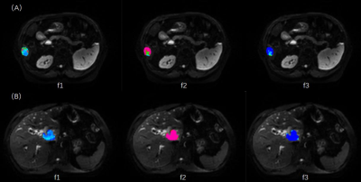 |
69 | Application value of restriction spectrum imaging in differentiation of hepatocellular carcinoma and intrahepatic cholangiocarcinoma
Nan Meng1, Yu Luo1, Lei Shen1, Pengyang Feng2, Ziqiang Li3, Han Jiang3, Yunfei Zhang4, Yongming Dai4, Yan Bai1,5, and Meiyun Wang1
1Zhengzhou University People’s Hospital & Henan Provincial People’s Hospital, Henan, China, 2Henan University People’s Hospital & Henan Provincial People’s Hospital, Henan, China, 3Xinxiang Medical University & Henan Provincial People's Hospital, Henan, China, 4MR Collaboration, Central Research Institute, United Imaging Healthcare, Shanghai, China, 5Department of Medical Imaging, Zhengzhou University People’s Hospital & Henan Provincial People’s Hospital, Zhengzhou, China Keywords: Liver, Cancer Diffusion-weighted imaging (DWI) can be used to evaluate the degree of tumor cell proliferation and tumor necrosis. The diffusion motion of water molecules is assumed to be Gaussian distribution, which has some limitations. In recent years, some advanced models have emerged that can provide more parameters to explore complex biological behaviors. Restricted spectral imaging (RSI) is one of them. However, the application of this model in liver disease is rare. The purpose of this study was to explore the potential value of the RSI model in the preoperative non-invasive differential diagnosis of hepatocellular carcinoma (HCC) and intrahepatic cholangiocarcinoma (ICC). |
|
2243. |
70 | A Radiomics Nomogram for the Prediction of Microvascular Invasion of HCC and Patients’ Benefit from Postoperative Adjuvant TACE
Kun Zhang1, Zhi Wei Shen2, and Wen Shen1
1Department of Radiology, Tianjin First Central Hospital, Tianjin Institute of imaging medicine, Tianjin, China, 2Philips healthcare,Beijing,China, Beijing, China Keywords: Liver, Cancer, Hepatocellular carcinoma · Neoplasm invasion • The novel radiomics nomogram developed based on Gd-EOB-DTPA MRI achieved preoperative non-invasive MVI risk prediction.• An m-score based on the radiomics nomogram could stratify HCC patients and further identify individuals who may benefit from the PA-TACE.• The radiomics nomogram could help clinicians to implement more appropriate interventions and perform individualized precision therapies. |
|
2244. |
71 | Comparison of the diagnostic performance in the HCC with cirrhosis between the 2017 and 2018 versions of LI‑RADS based on Gd‑EOB‑DTPA enhanced MRI
Fei Xing1, Yi shi Wang2, and Xian ce Zhao3
1the Third Affiliated Nantong Hospital of Nantong University, Nantong, China, 2Philips Healthcare, Beijing, China, 3Philips Healthcare, Shanghai, China Keywords: Liver, Cancer, HCC The Liver Imaging Reporting and Data System (LI-RADS) is widely adopted for noninvasive diagnosis of hepatocellular carcinoma (HCC). It’s updated to version 2018 recently, with some major changes compared with v2017.Therefore, the aim of this study was to compare the diagnostic performances of LI-RADS for diagnosing HCC between v2017 and v2018 based on Gd-EOB-DTPA enhanced MRI. |
|
2245. |
72 | Value of texture analysis based on R2* map for predicting early recurrence of HCC after hepatectomy
Qihao Xu1, Ailian Liu1,2, Ying Zhao1, Yue Wang1, Tao Lin1, Xue Ren1, Qingwei Song1, Yan Guo3, Xin Li3, and Tingfan Wu3
1Department of Radiology, The First Affiliated Hospital of Dalian Medical University, Dalian, China, 2Engineering Research Center for Artificial Intelligence in Medical Imaging, Dalian, China, 3GE Healthcare, Shanghai, China Keywords: Liver, Cancer Hepatectomy is an important therapeutic method for hepatocellular carcinoma (HCC). The overall survival rate of patients with early recurrence is often lower than that of patients without early recurrence. Therefore, this worked aimed at investigating the value of texture analysis based on R2* map for predicting early recurrence of HCC after hepatectomy. Eleven optimal texture features were obtained to predict early recurrence of HCC. This research suggested that R2* map texture analysis had certain predictive value for early recurrence of HCC after hepatectomy, which was valuable for noninvasively, preoperatively and accurately predicting the prognostic factors during clinical practice. |
|
2246.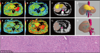 |
73 | The value of IVIM for preoperative evaluation of liver regeneration after hepatectomy in hepatocellular carcinoma
Qian Li1, Tong Zhang1, Lisha Nie2, Xiaocheng Wei2, Yi Wei1, and Bin Song1,3
1Department of radiology, West China hospital, Sichuan Univeristy, Chengdu, China, 2MR Research China, GE Healthcare, Beijing,China, Beijing, China, 3Department of radiology, Sanya People’s Hospital, Sanya, China Keywords: Liver, Diffusion/other diffusion imaging techniques, Liver regeneration; Intravoxel incoherent motion; Carcinoma, Hepatocellular; Hepatectomy The D value derived from IVIM diffusion-weighted imaging may be a useful marker for the preoperative prediction of liver regeneration in patients with HCC. and the D value derived from IVIM diffusion-weighted imaging shows a significant negative correlation with fibrosis, an important predictor of liver regeneration. No IVIM parameters were associated with liver regeneration in patients who underwent major hepatectomy, but the D value was a significant predictor of liver regeneration in patients who underwent minor hepatectomy. |
|
2247.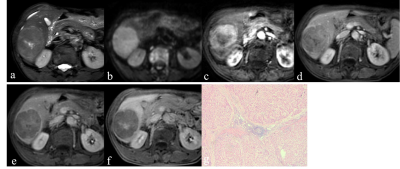 |
74 | Hepatocellular carcinoma with central scar: imaging findings of gadoxetic acid-enhanced MRI and pathological features
Fei Xing1, Yi shi Wang2, and Xian ce Zhao3
1the Third Affiliated Nantong Hospital of Nantong University, Nantong, China, 2Philips Healthcare, Beijing, China, 3Philips Healthcare, Shanghai, China Keywords: Liver, Cancer, HCC central scar is not a rare feature of conventional non-fifibrolamellar HCC. The dynamic enhancement mode of Gd-EOB-DTPA was helpful to distinguish inflammatory, collagenous, and vascular scars. The EOB “target sign” had a certain characteristic but non-specific, which still needed to be combined with MRI features of the mass. |
|
2248.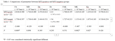 |
75 | Predicion of Microvascular Invasion in Hepatocellular Carcinoma using Diffusion Kurtosis Imaging
Yanxi Xiong1, Zhao Ying1, Ailian Liu1, Ren Xue1, Qihao Xu1, and Qingwei Song1
1The First Affiliated Hospital, Dalian Medical University, Dalian, China Keywords: Liver, Blood vessels Microvascular invasion (MVI) is an independent factor leading to poorer prognosis after liver resection and transplantation. In this retrospective study, we evaluated the discrimination power of Diffusion Kurtosis Imaging for predicting microvascular invasion Microvascular Invasion in hepatocellular carcinoma Hepatocellular Carcinoma (HCC). Our resuts showed that DKI-based quantitative parameters might have a certain value in identifying MVI status in HCC. |
|
2249.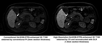 |
76 | Gd-EOB-DTPA Enhanced 3D T1WI: Utility of Compressed Sensing with Deep Learning Reconstruction for Liver Tumor Detection Improvement
Hiroyuki Nagata1, Yoshiharu Ohno1,2, Takeshi Yoshikawa2,3, Kaori Yamamoto4, Masato Ikedo4, Masao Yui4, Maiko Shinohara4, Akiyoshi Iwase5, Takahiro Matsuyama2, Takahiro Ueda2, Hirotaka Ikeda2, Yoshiyuki Ozawa2, and Hiroshi Toyama2
1Joint Research Laboratory of Advanced Medical Imaging, Fujita Health University School of Medicine, Toyoake, Japan, 2Radiology, Fujita Health University School of Medicine, Toyoake, Japan, 3Diagnostic Radiology, Hyogo Cancer Center, Akashi, Japan, 4Canon Medical Systems Corporation, Otawara, Japan, 5Fujita Health University Hospital, Toyoake, Japan Keywords: Liver, Cancer We hypothesize that CS with DLR can improve spatial resolution, image quality and tumor detection on Gd-EOB-enhanced 3D T1WI in suspected liver tumor patients. The purpose of this study was to determine the utility of CS and DLR for image quality and liver tumor detection improvements on high-resolution 3D CE-T1WI (HR-CE-T1WI) as compared with conventional 3D CE-T1WI with PI (conventional CE-T1WI) in patients with suspected liver tumors. |
|
2250.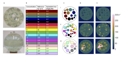 |
77 | Development of Hepatocellular Carcinoma phantom and its use for assessing R2* mapping sequences for detecting iron sparing
Elisabeth Pickles1,2, Eleanor Cox3,4, Alison Telford2, Ferenc Mozes5, Gabriela Belsley5, Elizabeth Tunnicliffe5, Michael Brady2, Michael Pavlides5,6,7, Daniel Bulte1, and Susan Francis3,4
1Institute of Biomedical Engineering, University of Oxford, Oxford, United Kingdom, 2Perspectum Ltd, Oxford, United Kingdom, 3Sir Peter Mansfield Imaging Centre, University of Nottingham, Nottingham, United Kingdom, 4NIHR Nottingham Biomedical Research Centre, Nottingham University Hospitals NHS Trust and the University of Nottingham, Nottingham, United Kingdom, 5Oxford Centre for Magnetic Resonance, University of Oxford, Oxford, United Kingdom, 6NIHR Nottingham Biomedical Research Centre, University of Oxford, Oxford, United Kingdom, 7Translational Gastroenterology Unit, University of Oxford, Oxford, United Kingdom Keywords: Liver, Cancer Survival rates for hepatocellular carcinoma (HCC) are poor, and improved methods for early detection are required. One such method may be the detection of iron sparing in HCC compared to the liver tissue using R2* mapping. We optimised sequences for the assessment of whole liver 3D R2* mapping, and developed a HCC phantom comprising small spheres with a range of R2* values to assess them. Agreement of the phantom R2* with reference values and between vendors, as well as the ability to detect small differences in R2* indicated the sequences have strong potential to detect iron sparing. |
|
2251.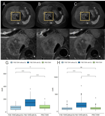 |
78 | Deep learning reconstruction for liver T2-weighted and diffusion-weighted imaging:Improvement of image quality and lesion delineation
Qian Chen1, Huimin Lin1, Shu Fang1, Jiankun Dai2, Guifeng Fu2, Ruokun Li1, and Fuhua Yan1
1Department of Radiology, Ruijin Hospital Affiliated to Shanghai Jiao Tong University, Shanghai, China, 2MR Research, GE Healthcare, Beijing, China Keywords: Liver, Tumor, Magnetic resonance imaging, T2-weighted imaging, DIffusion weighted imaging In this study, deep learning reconstruction (DLR) of liver fast spin echo T2-weighted (FSE-T2WI) and diffusion-weighted (DWI) was performed. The results showed the liver SNR, and lesion CNR were dramatically increased for both the FSE-T2WI and DWI with DLR compared with conventional reconstruction. The image quality of DLR FSE-T2WI even surpassed that of the routinely used PROPELLER T2WI. DLR didn’t impact the quantitative apparent coefficient derived from DWI. T2WI and DWI with DLR also improved the delineation of lesion structure due to improvement of image quality. Our study indicated DLR FSE-T2WI and DWI would be beneficial for liver disease diagnosing. |
|
2252.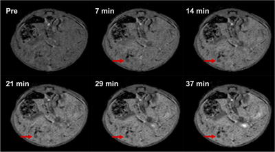 |
79 | Tumor burden assessment in a SB CCA.Mdr2-/- cholangiocarcinoma mouse model with gadoxetate disodium-enhanced MRI at 9.4 T
Qianhui Dou1, Pinzhu Huang2, Jesse Kirkpatrick3,4, Disha Skelton-Badlani2, Gopal Varma1, Aaron K. Grant1, Yury Popov2, and Leo Lee Tsai1
1Radiology, Beth Israel Deaconess Medical Center, Boston, MA, United States, 2Division of Gastroenterology, Beth Israel Deaconess Medical Center, Boston, MA, United States, 3Koch Institute for Integrative Cancer Research, Massachusetts Institute of Technology, Cambridge, MA, United States, 4Harvard-MIT Division of Health Sciences and Technology, Massachusetts Institute of Technology, Cambridge, MA, United States Keywords: Liver, Tumor, preclinical models, cholangiocarcinoma Cholangiocarcinoma (CCA) is a primary biliary malignancy with very poor prognosis. Early diagnostic detection and effective targeted therapies remain challenging. A novel hepatic fibrosis/CCA mouse model provides an investigative platform, but in vivo tumor detection/tracking is needed. Here we trial the use of in vivo MRI with gadoxetate disodium to detect CCA tumors and to differentiate infiltrative CCA from fibrosis in these models. |
|
2253.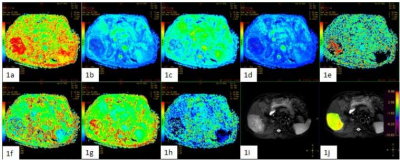 |
80 | Differentiation between HCC and ICC using 3D amide proton transfer weighted imaging and diffusion kurtosis imaging
Qihao Xu1, Ailian Liu1,2, Tao Lin1, Dandan Zheng3, Xiaoxiao Zhang3, Lihua Chen1, Qingwei Song1, Renwang Pu1, Ying Zhao1, and Xue Ren1
1Department of Radiology, The First Affiliated Hospital of Dalian Medical University, Dalian, China, 2Engineering Research Center for Artificial Intelligence in Medical Imaging, Dalian, China, 3Clinical & Technical Support, Philips Healthcare, Beijing, China Keywords: Liver, CEST & MT Hepatocellular carcinoma (HCC) and intrahepatic cholangiocarcinoma (ICC) are the two most common primary intrahepatic malignant tumors, and the prognosis and treatment methods are significantly different between them. The purpose of this study was to explore the diagnostic ability of APTw and DKI in distinguishing ICC from HCC. When the APTw values were combined with DKI parameters, the AUC was improved from 0.760 to 0.784 and the sensitivity reached 100%. Our preliminary study showed that APTw combined with the mean diffusivity and kurtosis anisotropy values drived from DKI demonstrated a better performance in the differential diagnosis of HCC and ICC. |
|
 Back to Meeting Home
 Back to the Program-at-a-Glance
The International Society for Magnetic Resonance in Medicine is accredited by the Accreditation Council for Continuing Medical Education to provide continuing medical education for physicians.
 Back to Meeting Home
 Back to the Program-at-a-Glance
 View Presentation Video