Digital Poster
Hepatobiliary Imaging: Current Applications & Advances II
ISMRM & ISMRT Annual Meeting & Exhibition • 03-08 June 2023 • Toronto, ON, Canada

Digital Poster
Hepatobiliary Imaging: Current Applications & Advances II
| Computer # | |||
|---|---|---|---|
2036.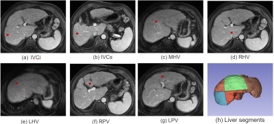 |
41 | Automatic Couinaud segmentation in Intravenous-Phase Enhanced MRI images by key point detection with deep neural network
Dong Miao1,2, Xue Ren3,4, Ying Zhao3,4, AiLian Liu3,4, Yu Yao1,2, Qihao Xu3,4, and Qingwei Song3,4
1Chengdu Institute of Computer Application, Chinese Academy of Sciences, Chengdu, China, 2School of Computer Science and Technology, University of Chinese Academy of Sciences, Beijing, China, 3Department of Radiology, First Affiliated Hospital of Dalian Medical University, Dalian, China, 4Dalian Engineering Research Center for Artificial Intelligence in Medical Imaging, Dalian, China Keywords: Liver, Liver, Couinaud segmentation This study presents an effective framework for automatic Couinaud liver segmentation in Intravenous-Phase Enhanced MRI images, without the time-consuming delineation of each segment or identifying different branches of the vessel system. We define seven key points located at the bifurcations of the vascular system to divide liver into Couinaud segments II-VIII. We train a key point detection model to locate the coordinate of key point. The overall dice score is 81.17% and average surface distance is 2.35mm in test set. |
|
2037.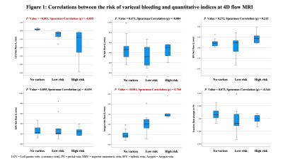 |
42 | Noninvasive direct assessment of the left gastric vein using 4D flow MRI for risk stratification of esophageal variceal bleeding
Atsushi Higaki1, Yoshihiko Fukukura1, Akihiko Kanki1, Akira Yamamoto1, kazunori Moriya1, and Tsutomu Tamada1
1Radiology, Kawasaki Medical School, Okayama, Japan Keywords: Liver, Liver This study focused on the feasibility of 4D flow MRI with low-velocity encoding sensitivity as a non-invasive tool for direct estimation of the left gastric vein and stratification of bleeding risk of esophageal varices. Our study showed that the development of esophageal varices was associated with decreased and increased net flow volumes in the left gastric and azygos veins, respectively. These results suggest the promise of 4D flow MRI with low-velocity encoding sensitivity as a non-invasive and accurate method for stratifying the bleeding risk of esophageal varices. |
|
2038.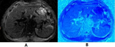 |
43 | Assessing Liver Fibrosis with Diffusion-weighted MRI-based Virtual Elastography
Li Yang1, Caixia Fu2, and Mengsu Zeng3
1Department of Radiology, Zhongshan Hospital,Fudan University, Shanghai, China, 2MR Application Development, Siemens Shenzhen Magnetic Resonance Ltd, Shenzhen, China, 3Zhongshan Hospitai,Fudan University, shanghai, China Keywords: Liver, Diffusion/other diffusion imaging techniques Recently, diffusion-weighted (DW) imaging–based elastography was proposed for noninvasive liver staging by converting a shifted apparent diffusion coefficient (sADC) to tissue elasticity on the basis of two b values (200 and 1500sec/mm2). We retrospectively calculated sADC from DW MRI and converted it to DW MRI–based virtual shear modulus (µDiff) for evaluating liver fibrosis in 90 patients. Our results showed the µDiff was significantly correlated with liver fibrosis stage, and the areas under the curves were 0.864 and 0.821 for significant and advanced fibrosis, respectively, indicating DW imaging–based elastography is a promising method for predicting liver fibrosis. |
|
2039.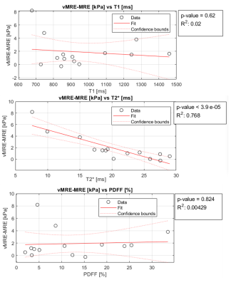 |
44 | Magnetic Resonance Elastography: A Comparison with Virtual Diffusion Derived Elastography
Jonatan Eriksson1,2, Wolf Bartholomä2,3, Nils Dahlström2,3, Jens Tellman1,2, Stergios Kechagias4, Patrik Nasr4, Mattias Ekstedt2,4, Peter Lundberg1,2, and Johan Kihlberg2,3
1Department of Medical Radiation Physics, and Department of Health, Medicine and Caring Sciences, Linköping University, Linköping, Sweden, 2Center for Medical Imaging Science and Visualization, Linköping University, Linköping, Sweden, 3Department of Radiology in Linköping and Department of Health Medicine and Caring Sciences, Linköping University, Linköping, Sweden, 4Division of Diagnostics and Specialist Medicine, Department of Health, Medicine and Caring Sciences, Linköping University, Linköping, Sweden, Linköping, Sweden Keywords: Liver, Elastography, MRE, Diffusion A virtual manner of determining apparent stiffness values from diffusion images, so called ‘vMRE’, was evaluated in this study which included both healthy subjects and patients with Non-Alcoholic Fatty Liver Disease (NAFLD). No association was observed between true MR-Elastography (MRE) and vMRE. However, the difference between MRE and vMRE was more strongly correlated with T2*. Iron deposition is not uncommon in patients with NAFLD, and for that reason, a spin echo-MRE sequence was used, instead of the gradient-based sequence that has been used previously. Iron deposition in liver shortens T2*, which can also affect diffusion images and the vMRE. |
|
2040. |
45 | Whole Upper Abdominal T1 Mapping on Free-Breathing 3D Look-Looker Sequence using Radial Sampling Acquisition: A Phantom and Volunteer Study
Tomohiro Noda1, Keitaro Sofue2, Ryuji Shimada1, Yu Ueda3, Yoshiko Ueno2, Yuichiro Somiya1, Shintaro Horii1, Akiko Kusaka1, and Takamichi Murakami2
1Center of Radiology and Radiation Oncology, Kobe University Hospital, Kobe, Japan, 2Department of Radiology, Kobe University Graduate School of Medicine, Kobe, Japan, 3Philips Japan MR Clinical Science, Tokyo, Japan Keywords: Liver, Quantitative Imaging, T1mapping We developed a free-breathing 3D Look-Locker sequence with radial sampling acquisition (3D-Radial LL) to acquire T1 map of the whole upper abdomen. To evaluate the quantitative accuracy, T1 values obtained from 3D-Radial LL and modified Look-Locker inversion recovery (MOLLI) in a phantom and volunteers were compared. The phantom study showed excellent correlation regarding T1 values between on the MOLLI and 3D-Radial LL. In the volunteer study, the difference in T1 values were within 6% for the liver, pancreas, spleen, and paraspinal muscle. The 3D-Radial LL enables accurate measurement of T1 values in the upper abdomen. |
|
2041.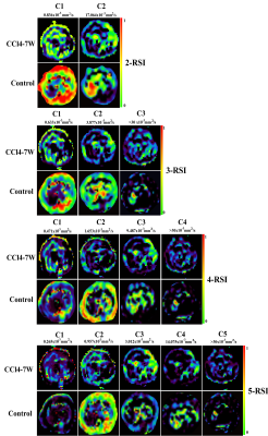 |
46 | Multicompartmental restriction spectrum imaging model for characterizing liver fibrosis in a mouse model
Yeyu Cai1, Jiayi Liu2, Ziyi Zhou3, Tian Chai1, Liyun Zheng4, and Yongming Dai5
1Department of Radiology, Second Xiangya Hospital, Central South University, Changsha, China, 2Department of Oncology, Second Xiangya Hospital, Central South University, Changsha, China, 3Department of Radiology, Maternal and Child Health Hospital of Hubei Province, Wuhan, China, 4Shenzhen United Imaging Research Institute of Innovative Medical Equipment, Shenzhen, China, 5MR Collaboration, Central Research Institute, United Imaging Healthcare, Shanghai, China Keywords: Liver, Animals Liver fibrosis is a serious medical issue and noninvasive technique, such as magnetic resonance imaging (MRI), is required to identify and quantify liver fibrosis. Recently, several advanced diffusion MRI signal models that employ additional parameters to describe diffusion signals from complex microstructural compartments have been developed. The purpose of this study was to apply the multicompartmental restriction spectrum imaging (RSI) model to evaluate carbon tetrachloride (CCl4)-induced liver fibrosis in mice. The optimal model to characterize liver fibrosis was the 4-compartment RSI model. Compared to control groups, mice with liver fibrosis exhibited significantly lower ADC and C3, but higher C1. |
|
2042.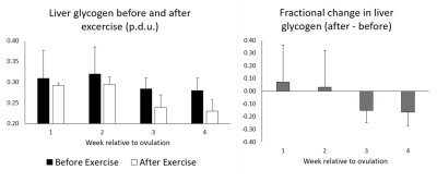 |
47 | The Effects of Menstrual Cycle on Hepatic Glycogen Stores Before and After Exercise: Preliminary Data From a 13C MRS Study
Stephen Bawden1,2, Louise Dexter2, Tomoka Matsuda3, Mehri Kaviani2, Penny Gowland2, and Guruprasad P Aithal1
1Nottingham Digestive Diseases Centre, NIHR Nottingham Biomedical Research Centre, University of Nottingham, Nottingham, United Kingdom, 2Sir Peter Mansfield Imaging Centre, School of Physics and Astronomy, University of Nottingham, Nottingham, United Kingdom, 3Graduate School of Health and Sport Science, Nippon Sport Science University, Tokyo, Japan Keywords: Liver, Metabolism The aim of this study was to investigate the effects of hormonal changes throughout the menstrual cycle on moderate exercise induced liver glycogen changes in healthy women. Preliminary results show variation in hepatic glycogen stores and changes following exercise throughout the menstrual cycle. |
|
2043.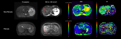 |
48 | Evaluating liver fibrosis in chronic hepatitis B patients using multi-compartment restriction spectrum imaging
Jing Zhang1, Kaifan Yang1, Yiyuan Zhu1, Liyun Zheng2, Yongming Dai3, and Yikai Xu1
1Department of Medical Imaging Center, Nanfang Hospital, Southern Medical University, Guangzhou, China, 2Shenzhen United Imaging Research Institute of Innovative Medical Equipment, Shenzhen, China, 3MR Collaboration, Central Research Institute, United Imaging Healthcare, Shanghai, China Keywords: Liver, Diffusion/other diffusion imaging techniques This study evaluated the feasibility of the restriction spectrum imaging (RSI) model in the characterization and staging of liver fibrosis. The results demonstrated that the four-compartment model emerged as the best option and the C4 derived from the four-compartment model outperformed conventional apparent diffusion coefficient (ADC) values. Therefore, the four-compartment RSI model could be a noninvasive and in vivo diagnostic technique in the evaluation of chronic hepatitis, which is conducive to treatment decisions and patient management. |
|
2044. |
49 | Repeatability and reliability of liver quantitative maps in differentiation of hepatic fibrosis stage
Tianzhu Liu1, Lesheng Huang1, Weiyin Vivian Liu2, and Jun Chen1
1Guangdong Hospital of Traditional Chinese Medicine, Zhuhai, China, 2MR Research China, GE Healthcare, Beijing, China Keywords: Liver, Data Analysis Grading liver fibrosis using intravoxel incoherent motion diffusion-weighted imaging (IVIM-DWI) have been shown inconsistent results possibly for differences between liver segments and recruited types of patients. We systematically evaluated all measurements retrieved based on liver segments and classified by stage of liver fibrosis, and inconsistent repeatability and reliability of IVIM-derived values were found, attributing to inter- and intra- manual measurement bias and inhomogeneous distribution of tissue components such as fibrosis. ROC curve analysis showed moderate diagnostic efficiency between early hepatic fibrosis (EHF) and advanced HF (AHF), but not HCs from EHF based on IVIM-derived parameters with acceptable repeatability and reliability. |
|
2045.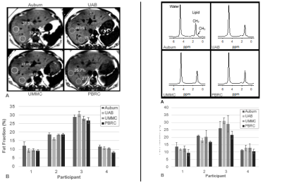 |
50 | Harmonized multi-site MRI-based quantification of human liver fat and stiffness: a pilot study
Maninder Singh1, Owen T Carmichael1, Adil Bashir2, Anne M Russel3, Mark Bolding4, David T Redden3, Judd Storr5, William R Willoughby6, Candace Howard Claudio5, Daniel S Hsia7, Robert P Kimberly8, Meagan E Gray6, Eric Ravussin7,9, and Thomas S Denny2
1Biomedical Imaging Center, Pennington Biomedical Research Center, Baton Rouge, LA, United States, 2Auburn University, Auburn, AL, United States, 3University of Alabama, Birmingham, AL, United States, 4University of Alabama Medical Center, Birmingham, AL, United States, 5The University of Mississippi Medical Center, Jackson, MS, United States, 6The University of Alabama Medical Center, Birmingham, AL, United States, 7Pennington Biomedical Research Center, Baton Rouge, LA, United States, 8The University of Alabama School of Medicine, Birmingham, AL, United States, 9The University of Alabama, Birmingham, AL, United States Keywords: Liver, Liver, Nonalcoholic Fatty Liver Disease; Magnetic Resonance Spectroscopy; Elasticity Imaging Techniques; Magnetic Resonance Imaging Non-alcoholic fatty liver disease (NAFLD) is a leading cause of end-stage liver disease. NAFLD diagnosis and follow-up relies on a combination of clinical data, liver imaging, and/or liver biopsy. However, inter-site imaging differences impede diagnostic consistency and reduce the repeatability of the multi-site clinical trials necessary to develop effective treatments. The goal of this pilot study was to harmonize commercially available 3T magnetic resonance imaging (MRI) measurements of liver fat and stiffness in human participants across academic sites and MRI vendors. |
|
2046.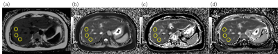 |
51 | Influence of fat deposition in T1 measurement of the liver: The value of T1 mapping with water component only using the 2D DIXON Look-Locker method
Mayumi Higashi1, Masahiro Tanabe1, Masatoshi Yamane2, Mahesh B. Keerthivasan3, Hiroshi Imai 4, Teppei Yonezawa2, and Katsuyoshi Ito1
1Department of Radiology, Yamaguchi University Graduate School of Medicine, Ube, Yamaguchi, Japan, 2Department of Radiological Technology, Yamaguchi University Hospital, Ube, Yamaguchi, Japan, 3MR R&D Collaborations, Siemens Medical Solutions USA Inc, New York, NY, United States, 4MR Research and Collaboration, Siemens Healthcare K.K., Tokyo, Japan Keywords: Liver, Liver The purpose of this study was to elucidate the clinical importance of T1 mapping of the liver with the water component only using the 2D DIXON Look-Locker sequence. T1 values of the liver were compared among T1 maps obtained by IP, OP and DIXON Look-Locker sequences. T1 values of the liver on the T1 maps obtained from IP- and OP-based sequences were significantly influenced by an increased fat component. In contrast, the T1 value of the liver on the T1 maps with the water component only calculated from DIXON water images was unlikely to be influenced by hepatic steatosis. |
|
2047.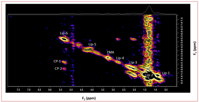 |
52 | In vivo investigation of a Healthy Liver Lipid composition by 2D L-COSY Using 3T
Rajakumar Nagarajan1, Andres Saucedo 2, and M Albert Thomas2
1Human Magnetic Resonance Center, University of Massachusetts, Amherst MA, Amherst, MA, United States, 2Radiological Sciences, University of California Los Angeles, Los Angeles, CA, United States Keywords: Liver, Spectroscopy In vivo magnetic resonance spectroscopy has the potential to provide information about both the quantity and composition of fat within the liver. We have demonstrated the feasibility of detecting cross peaks using the 2D L-COSY technique at 3T in a healthy liver. This can be used to study fatty liver to derive quantitative indexes. |
|
2048.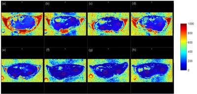 |
53 | Feasibility of fat quantification of in vivo mouse liver at 9.4T
Zhongmiao Wang1, Tingting Bo2,3,4, Chen Chen5, Yuqin Min6, Xinxin Cai3, Huimin Lin3, Hongxia Lei1, Renkuan Zhai1, Jiqiu Wang2, Fuhua Yan3, and Guang Ning2
1Wuhan United Imaging Life Science Instrument Co., Ltd., Wuhan, China, 2Department of Endocrine and Metabolic Diseases, Shanghai Institute of Endocrine and Metabolic Diseases, Ruijin Hospital, Shanghai Jiao Tong University School of Medicine, Shanghai, China, 3Department of Radiology, Ruijin Hospital, Shanghai Jiao Tong University School of Medicine, Shanghai, China, 4Clinical Neuroscience Center, Ruijin Hospital Luwan Branch, Shanghai Jiao Tong University School of Medicine, Shanghai, China, 5United imaging healthcare, Shanghai, China, 6Institute for medical imaging technology, Department of Radiology, Ruijin Hospital, Shanghai Jiao Tong University School of Medicine, Shanghai, China Keywords: Liver, Liver The feasibility of determination of fat in living mouse liver is demonstrated at 9.4T using gradient-echo sequence and the Fat Analysis and Calculation Technique (FACT)—Transition Region Extraction (TREE) algorithm. The proton density fat fraction map is obtained, which is positively correlated with steatosis. The proposed technique is a promising non-invasive tool for diagnosing fatty liver diseases. |
|
2049.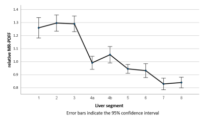 |
54 | MR-based segmental quantification of hepatic lipid content in patients with suspected iron overload
Arthur Peter Wunderlich1,2, Holger Cario3, Stephan Kannengießer4, Veronika Grunau1, Michael Götz2, Meinrad Beer1, and Stefan Andreas Schmidt1
1Diagnostic and Intervnetional Radiology, Ulm University, Medical Center, Ulm, Germany, 2Section for Experimental Radiology, Ulm University, Medical Center, Ulm, Germany, 3Department of Pediatrics and Adolescent Medicine, Ulm University, Medical Center, Ulm, Germany, 4MRI Development, Siemens Healthcare GmbH, Erlangen, Germany Keywords: Liver, Fat, segmental hepatic fat distribution Purpose To investigate the segmental distribution of hepatic fat fraction in hematologic patients. Methods The liver of 44 patients examined with 3D MR multi-echo gradient-echo was segmented semiautomatically and subdivided into nine segments. Segmental fat content was determined on PDFF maps and tested for statistically significant differences. Results Highly significant differences were detected. Segments 1 to 3 had the highest fat content, segments 7 and 8 the lowest. Conclusions Our results suggest that liver segments may represent functional aspects in fat metabolism and/or storage in addition to their anatomical significance. Fat distribution in hematologic patients was similar to living donors. |
|
2050.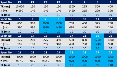 |
55 | Simulation of Short Breath-hold Steady State MultiParameter MRS in Liver.
Gavin Hamilton1, Walter C Henderson1, Danielle N Batakis1, Lael Ceriani1, Ashley L Louie1, Yesenia Covarrubias1, Celene Gonzalez1, Michael S Middleton1, Scott B Reeder2, Kathryn J Fowler1, and Claude B Sirlin1
1Liver Imaging Group, Department of Radiology, University of California San Diego, San Diego, CA, United States, 2Department of Radiology, University of Wisconsin, Madison, Madison, WI, United States Keywords: Liver, Spectroscopy To permit simultaneous T1w, T1f, T2w, T2f and proton density fat fraction (PDFF) estimation, we developed a single-average, 21 s, single-breath-hold, Steady-state, Multi-Parameter MRS (SMP MRS) sequence. However, a 21 s breath-hold may be challenging especially children, such as pediatric subjects, so we simulated a shorter 12 s breath-hold sequence by removing the longer TR and TE values. We found good agreement for T1w, T2w and PDFF between the full 21 s SMP and simulated 21 s sequences, suggesting the shorter 12 s SMP MRS can be run instead of the full 21 s sequence. |
|
2051.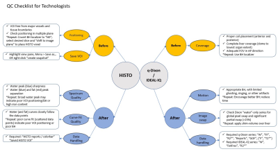 |
56 | Performing Quality Control of your MRI Liver Fat/Iron Quantification Studies: A Critical Requirement
Farid Hajibonabi1, Sadhna Nandwana1, Puneet Sharma1, Patricia Balthazar1, Amir Hossein Davarpanah1, Courtney Coursey Moreno1, and Melina Pectasides1
1Radiology and Imaging Sciences, Emory University, Atlanta, GA, United States Keywords: Liver, Quantitative Imaging, Quality Control Liver fat/iron quantification with MRI is essential for detection of iron/fat overload that can lead to NAFLD and cirrhosis. The two widely used tools for this purpose are chemical shift-encoded MR sequences and HISTO. Both methods can fail due to various preventable technical factors. We analyzed various quality factors of technical acceptability for these studies and determined that >25% of all quantification studies performed over six months at our institution were technically unacceptable or had data handling errors. We then developed a checklist for MR technologists to confirm acceptability of the study and decrease potential errors before sending for interpretation. |
|
2052.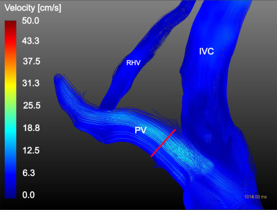 |
57 | Association Between NAFLD and Blood Flow: Portal 4D-flow, 2D-flow, Hepatic Elasticity and Spleen Volume
Jonatan Eriksson1,2, Wolf Bartholomä2,3, Nils Dahlström2,3, Jens Tellman1,2, Stergios Kechagias4, Patrik Nasr4, Mattias Ekstedt2,4, Johan Kihlberg2,3, and Peter Lundberg1,2
1Department of Medical Radiation Physics, and Department of Health, Medicine and Caring Sciences, Linköping University, Linköping, Sweden, 2Center for Medical Imaging Science and Visualization, Linköping University, Linköping, Sweden, 3Department of Radiology in Linköping and Department of Health Medicine and Caring Sciences, Linköping University, Linköping, Sweden, 4Division of Diagnostics and Specialist Medicine, Department of Health, Medicine and Caring Sciences, Linköping University, Linköping, Sweden, Linköping, Sweden Keywords: Liver, Blood vessels, 4D Flow, 2D Flow, MRE, Spleen A group of 6 healthy and 9 NAFLD-patients underwent a multi-parametric MRI examination, including 4D-, 2D-flow, MRE and relaxometry. 2D-flow results were successfully extracted from 4D-flow data, showing good match with 2D-flow, indicating that 4D-flow provides the opportunity to both provide regular flow values as well as additional information e.g. flow visualizations. Correlation between 4D-flow and spleen volume indicate that 4D-flow could provide markers for early portal hypertension. |
|
2053.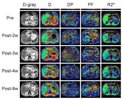 |
58 | Liver regeneration of residual right lobe after left lateral sectionectomy in living liver donors: a pilot study based on IVIM and T2* mapping MRI
shuangshuang xie1, caixin qiu1, and wen shen1
1Tianjin First Central Hospital, Tianjin, China Keywords: Liver, Liver, liver regeneration This study investigated the dynamic changes of residual right liver lobe after left lateral sectionectomy (LLS) and the potential value of IVIM and T2* mapping in evaluation of liver regeneration in living liver donors. All IVIM indexes of right liver lobe showed no obvious changes after surgery, but liver T2* values increased significantly at 2 weeks after surgery, and kept increasing gradually from week 2 to 8. Liver T2* values correlated moderately with liver volume, it may be a good substitute to liver volume in evaluating liver regeneration |
|
2054.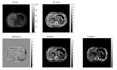 |
59 | 3D liver R2, R2* and quantitative susceptibility maps in a single breath-hold
Chao Li1, Jiahao Li2, Jinwei Zhang2, Pascal Spincemaille3, Thanh Nguyen3, and Yi Wang3
1Applied and engineering physics, Cornell university, New York, NY, United States, 2Biomedical engineering, Cornell university, New York, NY, United States, 3Weill Cornell Medicine, New York, NY, United States Keywords: Liver, Multi-Contrast In this work, a 3D stack-of-spiral spoiled gradient echo sequence was implemented using T2 prepared single and multi-echo gradient echo acquisition for simultaneous R2, R2* and QSM mapping in a single breath hold within 24 secs. |
|
2055.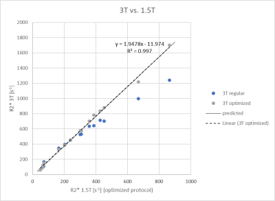 |
60 | Liver R2* with magnitude fitting in iron-overload patients – initial results on agreement between protocol settings and between 1.5T and 3T
Stephan A.R. Kannengiesser1, Arthur P. Wunderlich2,3, Xiaodong Zhong4, Dominik Nickel1, Holger Cario5, Michael Götz2,3, Meinrad Beer3, and Stefan A. Schmidt2
1MR Application Pevelopment, Siemens Healthcare GmbH, Erlangen, Germany, 2Department of Diagnostic and Interventional Radiology, Ulm University Medical Center, Ulm, Germany, 3Section for Experimental Radiology, Ulm University Medical Center, Ulm, Germany, 4MR R&D Collaborations, Siemens Medical Solutions, Inc., Los Angeles, CA, United States, 5Department of Pediatrics and Adolescent Medicine, Ulm University Medical Center, Ulm, Germany Keywords: Liver, Quantitative Imaging, R2*, iron content Purpose: Preliminary liver R2* comparison in patients across field strengths and protocols. Methods: 3T R2* from 3D multi-gradient echo data with magnitude fitting was mapped to 1.5T R2*. Limits of agreement were calculated. Results: Converted 3T R2* agreed well with 1.5T R2*. Best 95% limits of agreement were [-4.9% 7.6%] for 1.5T R2* [166s-1 865s-1] and [-34.5% 41.7%] for [49s-1. 865s-1]. Conclusion: 3T R2* agrees well with 1.5T R2* and can be used interchangeably for 3T R2* between 328s-1 and 1699s-1 with an optimized protocol. For low R2* values, protocols with longer echo times should be used. |
|
 Back to Meeting Home
 Back to the Program-at-a-Glance
The International Society for Magnetic Resonance in Medicine is accredited by the Accreditation Council for Continuing Medical Education to provide continuing medical education for physicians.
 Back to Meeting Home
 Back to the Program-at-a-Glance
 View Presentation Video