Digital Poster
Women's Cancer Imaging
ISMRM & ISMRT Annual Meeting & Exhibition • 03-08 June 2023 • Toronto, ON, Canada

| Computer # | |||
|---|---|---|---|
3448.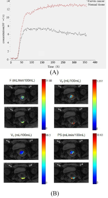 |
41 |
A Multicenter Study of Cervical Cancer using Dynamic
Contrast-Enhanced MRI: Are Measurements of Quantitative Imaging
Reproducible in Clinic?
Xue Wang1,
Shujian Li2,
Zhijun Ye3,
Tang-San Koh4,
Zujun Hou5,
Zhihan Yan1,
and Lu Han6
1The Second Affiliated Hospital and Yuying Children′s Hospital of Wenzhou Medical University, Wenzhou, China, 2The First Affiliated Hospital of Zhengzhou University, Zhengzhou, China, 3The Second Affiliated Hospital of Sichuan University, Chengdu, China, 4National Cancer Center, Singapore, Singapore, Singapore, 5Suzhou Institute of Biomedical Engineering and Technology, Chinese Academy of Sciences, Suzhou, China, 6Philips Healthcare, Shanghai, Shanghai, China Keywords: Uterus, Reproductive, Dynamic Contrast-Enhanced Imaging By comparing the distributions of measured DCE kinetic parameters in cervical cancer tissue by three MRI scanners in different clinical centers. It was demonstrated that (i) Each of the three scanners consistently showed similar findings on the characterization of cervical cancer microenvironment. (ii) The difference in MRI3 stemmed from keyhole imaging in the DCE protocol which could lead to underestimation of concentration of contrast media in viable tumor region. (iii) The close distribution between MRI1 and MRI2 indicated that consistent DCE quantitative metrics could be attained in scanners from different vendors if imaging protocol and data postprocessing could be standardized. |
|
3449.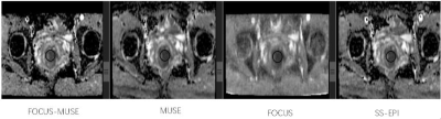 |
42 |
Less-distorted cervical ADC maps improved subjective diagnosis
efficacy on cervical cancer and reliability of measurements
liu hui1,
liu wei yin2,
and ou xiao rong1
1xiangya hospital, changsha, China, 2GE, taiwan, China Keywords: Pelvis, Cancer, DWI Differentiation of patients with IB-IIA from those with IIB-IIC assist track treatment selection for cervical cancer. A diffusion-weighted imaging (DWI) sequence based on field-of-view optimized and constrained undistorted single shot (FOCUS) combined with multiplexed sensitivity-encoding (MUSE) technique (named FOCUS-MUSE DWI) was used in our study to explore the differentiation performance on stages of cervical cancer. We found the reliability of apparent diffusion coefficient (ADC) for FOCUS MUSE-DWI is the highest among single-shot DWI (ss-DWI), FOCUS-DWI and MUSE-DWI, and better image quality, SNR, and CNR than the former two. There was no significant difference of ADC values for four DWI sequence between IB-IIA group and IIB-IIIC, but ADC values were consistent with previous studies. |
|
3450.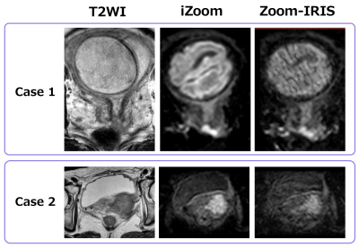 |
43 |
iZoom applying tilted 2D Echo-Planar RF excitation improved the
image quality of reduced FOV DWI for uterine cervical cancer: a
preliminary study
Akiyo Takada1,
Hajime Yokota2,
Ryuna Kurosawa1,
Takayuki Sada1,
Koji Matsumoto1,
Namiki Takashi3,
Masami Yoneyama3,
Adam Wu4,
and Takashi Uno2
1Department of Radiology, Chiba University, Chiba-shi, Japan, 2Department of Diagnostic Radiology and Radiation Oncology, Graduate School of Medicine, Chiba University, Chiba-shi, Japan, 3Philips Japan, Tokyo, Japan, 4Philips Healthcare (Shanghai) Ltd., Shanghai, China Keywords: Uterus, Diffusion/other diffusion imaging techniques, uterine cervical cancer Reduced field-of-view (rFOV) DWI is the essential sequence for the diagnosis and staging of uterine cervical cancer (CC). We compared the image quality of rFOV DWI taken with iZoom, which applies tilted 2D Echo-Planar RF excitation, and that of conventional Zoom combined with Reconstruction with Image-space Sampling (Zoom-IRIS) for CC patients. Reduced FOV DWI with iZoom showed higher image quality than Zoom-IRIS both in quantitative and qualitative evaluation. Anatomical details display and lesion conspicuity were mainly improved in quantitative evaluation. iZoom can be helpful for the diagnosis and staging of CC. |
|
3451.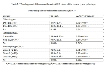 |
44 |
Feasibility of Accelerated T2 Mapping for the Quantitative and
Qualitative Assessment of Endometrial Carcinoma
Zanxia Zhang1 and
Shujian Li1
1The First Affiliated Hospital of Zhengzhou University, Zhengzhou, China Keywords: Uterus, Uterus The objectives of this study were to investigate the feasibility of accelerated T2 mapping in the clinical diagnosis of the pathologic subtypes and histologic grades of endometrial carcinoma (EMC), and to compare synthetic T2-weighted and diffusion-weighted (sT2W/DW) images to conventional T2W and diffusion-weighted (cT2W/DW) images for the myometrial invasion depth. 56 EMC patients underwent T2mapping and DW. Distinct T2 values were obtained for the various pathologic and histologic grades of EMC. Furthermore, pathologic EA grading was more accurate with the T2 values than ADC values. Additionally, in terms of detecting deep myometrial invasion, sT2W/DW showed similar accuracy to cT2W/DW. |
|
3452.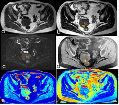 |
45 |
Preliminary Application performance of synthetic MRI
quantitative imaging in stage I endometrial cancer
Yin YunJuan1,
Chen Sujing1,
Chang Jun1,
Guan Rongping1,
Ge Yuxi1,
Pylypenko Dmytro2,
and hu Shudong1
1Medical Imaging, Affiliated Hospital of Jiangnan University, Wuxi, China, 2GE Healthcare, Beijing, China Keywords: Pelvis, Cancer The purpose of this study was to investigate the feasibility of using synthetic MRI to identify and differentiate stage I endometrial cancer. A total of 51 patients with endometrial carcinoma were recruited. Quantitative MRI, including T1, T2 and PD mapping were used. The endometrial carcinoma had significantly different T1, T2 and PD values compared to normal uterus tissue, therefore quantitative MRI is an effective method for detection of endometrial cancer. There was no statistical difference of synthetic MRI in differentiation grades or risk stratification, which needs to be further verified. |
|
3453.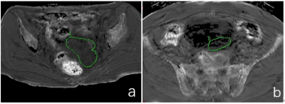 |
46 |
The application of R2 star map of enhanced T2 star weighted
angiography (ESWAN) in diagnosis of ovarian cancer
Qingling Song1,
Ye Li1,
and Ailian Liu1
1Radiology, The First Affiliated Hospital of Dalian Medical University, Dalian, China Keywords: Pelvis, Cancer Ovarian cancer is the leading cause of death of gynecological malignant tumors, however, most of BEOTs can be completely cured. The treatment and prognosis of ovarian cancer and BEOTs are very different, so it is very important to accurately distinguish ovarian cancer from BEOTs |
|
3454.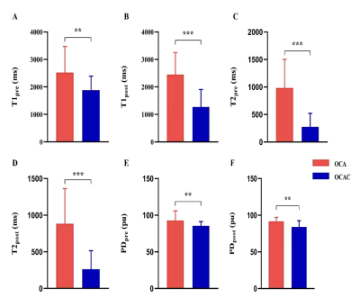 |
47 |
The diagnostic value of synthetic MRI in ovarian epithelial cell
neoplasms and its correlation with Ki-67 expression: a
preliminary study
Lingjie Zhang1,
Meiying Cheng1,
Teng Pan2,
Kaiyu Wang3,
Shifang Tan1,
Lin Lu1,
Junjie Liao1,
and Xin Zhao1
1Department of Radiology, the third affiliated Hospital of Zhengzhou University, Zhengzhou, China, 2Office of Scientific Research, the third affiliated Hospital of Zhengzhou University, Zhengzhou, China, 3MR Research China, GE Healthcare, Beijing, China Keywords: Pelvis, Quantitative Imaging This study aimed to evaluate the diagnostic value of synthetic MRI in ovarian epithelial cell neoplasms and to explore the correlation between the quantitative parameters like T1, T2, and proton density derived from synthetic MRI and Ki-67 expression. Our preliminary study shows that the values of these quantitative parameters were significantly lower in the malignant group than in the benign group, and the parameters were negatively correlated with Ki-67 expression, which can indirectly reflect the degree of tumor invasion. |
|
3455.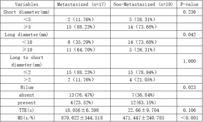 |
48 |
Diagnostic Value of Ultrafast Dynamic Contrast-Enhanced MRI in
Assessing the Status of Axillary Lymph Node Metastasis in Breast
cancer
Lincheng He1,
Ping Yan1,
Wenhong Liu1,
Weijia Song1,
Huiting Zhang2,
Robert Grimm3,
Hong Zhou1,
and Guanghua Luo1
1The First Affiliated Hospital, Hengyang Medical School, University of South China, HengYang, China, 2MR Scientific Marketing, Siemens Healthineers Ltd, Wuhan, China, 3MR Application Predevelopment, Siemens Healthcare GmbH, Erlangen, Germany Keywords: Breast, Cancer This study investigated the feasibility of ultra-fast DCE-MRI in accurately assessing axillary lymph node (ALN) metastasis in breast cancer. Our results showed that maximum slope (MS) accurately estimated the axillary lymph node status using the early-stage data of ultra-fast DCE-MRI. Furthermore, larger size (≥10mm diameter) of ALN and absence of hilum in the ultrafast DCE-MRI was associated with ALN metastasis in breast cancer patients. Our study demonstrated that MS from ultrafast breast MRI is a potential imaging biomarker of ALN metastasis in breast cancer. |
|
3456.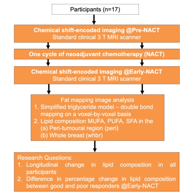 |
49 |
Peri-tumoural lipid composition mapping of the breast for early
response to neoadjuvant chemotherapy using chemical
shift-encoded imaging
Sai Man Cheung1,
Kwok-Shing Chan2,
Nicholas Senn1,
Ravi Sharma3,
Trevor McGoldrick3,
Tanja Gagliardi1,4,
Ehab Husain5,
Yazan Masannat6,
and Jiabao He1,7
1Institute of Medical Sciences, University of Aberdeen, Aberdeen, United Kingdom, 2Donders Institute for Brain, Cognition and Behaviour, Radboud University, Nijmegen, Netherlands, 3Department of Oncology, Aberdeen Royal Infirmary, Aberdeen, United Kingdom, 4Department of Radiology, Royal Marsden Hospital, London, United Kingdom, 5Department of Pathology, Aberdeen Royal Infirmary, Aberdeen, United Kingdom, 6Breast Unit, Aberdeen Royal Infirmary, Aberdeen, United Kingdom, 7Translational and Clinical Research Institute, Newcastle University, Aberdeen, United Kingdom Keywords: Breast, Fat Peri-tumoural lipid composition plays a major role in the disease progression of breast cancer, and novel chemical shift-encoded imaging (CSEI) allows rapid lipid mapping of the whole breast. We have previously observed an imbalance of peri-tumoural lipid composition in postmenopausal women with breast cancer using CSEI. Monitoring of lipid composition for early sign of tumour regression might be crucial for an accurate differentiation of patients responding to neoadjuvant chemotherapy (NACT). We hence set out to elucidate the longitudinal change in lipid composition in the peri-tumoural region and the whole breast in patients with breast cancer using CSEI. |
|
3457.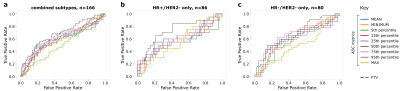 |
50 |
Comparison of diffusion-weighted imaging ADC metrics for early
prediction of neoadjuvant treatment response in HER2-negative
breast cancer
Judith Zimmermann1,
Julia Carmona-Bozo1,
Nu N Le1,
Natsuko Onishi1,
Lisa J Wilmes1,
Jessica Gibbs1,
Jiachao Liang1,
David C Newitt1,
Savannah C Partridge2,
Patrick J Bolan3,
John Kornak4,
Elissa Price1,
Bonnie N Joe1,
Barbara LeStage5,
I-SPY 2 Investigator Network6,
I-SPY 2 Imaging Working Group1,
Wen Li1,
and Nola Hylton1
1Radiology and Biomedical Imaging, University of California, San Francisco, San Francisco, CA, United States, 2Radiology, University of Washington, Seattle, WA, United States, 3Radiology, University of Minnesota, Minneapolis, MN, United States, 4Epidemiology and Biostatistics, University of California, San Francisco, San Francisco, CA, United States, 5I-SPY 2 Advocacy Group, San Francisco, CA, United States, 6Quantum Leap Healthcare, San Francisco, CA, United States Keywords: Breast, Diffusion/other diffusion imaging techniques, treatment response prediction, biomarkers, neoadjuvant chemotherapy In breast imaging, diffusion-based (DW) MRI delivers complementary quantitative value to conventional dynamic contrast enhanced MRI. ADC maps of tumor tissue calculated from DW-MRI, specifically mean ADC of the tumor region of interest, have shown promise for measuring early response to neoadjuvant chemotherapy (NAC). However, depending on intra-tumoral heterogeneity, other histogram-based ADC metrics may provide even better predictive performance than mean ADC, but are yet to be studied thoroughly. Here, we investigated nine ADC-based metrics regarding their predictive performance for NAC treatment response. |
|
3458.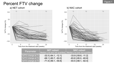 |
51 |
Breast MRI during neoadjuvant endocrine therapy for patients
with estrogen-receptor positive/HER2 negative breast cancer
Natsuko Onishi1,
Jessica E Gibbs1,
Ella F Jones1,
Teffany Joy Bareng1,
Jiachao Liang1,
Julia Carmona-Bozo1,
Wen Li1,
David C Newitt1,
Kimberly M Ray1,
Courtney Lawhn Heath1,
Rita Mukhtar2,
Bonnie N Joe1,
the I-SPY 2 Investigator Network3,
the I-SPY 2 Imaging Working Group1,
Laura J Esserman 2,
A. Jo Chien4,
and Nola M Hylton1
1Department of Radiology & Biomedical Imaging, University of California, San Francisco, San Francisco, CA, United States, 2Department of Surgery, University of California, San Francisco, San Francisco, CA, United States, 3Quantum Leap Healthcare Collaborative, San Francisco, CA, United States, 4Department of Medicine, University of California, San Francisco, San Francisco, CA, United States Keywords: Breast, Cancer This study aimed to investigate the longitudinal changes in functional tumor volume (FTV) and background parenchymal enhancement (BPE) derived from dynamic contrast-enhanced breast MRI during neoadjuvant endocrine therapy (NET). For comparison, we also evaluated a similar cohort of patients who received standard neoadjuvant chemotherapy (NAC). Similar to the NAC cohort, the NET cohort showed early changes in FTV and BPE as early as three weeks of treatment. In the NET cohort, early BPE decrease was prominent especially for patients with higher baseline BPE and premenopausal patients. These finding illustrate the potential of MRI metrics as early imaging biomarkers for NET. |
|
3459.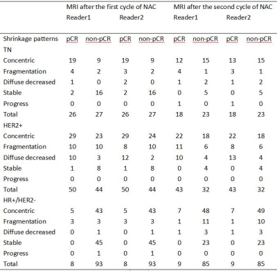 |
52 |
MRI-based tumor shrinkage patterns after early neoadjuvant
therapy in breast cancer: Correlation with subtypes and
pathological response
Mengfan Wang1,
Siyao Du1,
Lizhi Xie2,
Min Zhao2,
and Lina Zhang1
1The First Hospital of China Medical University, Shenyang, China, 2GE Healthcare, Beijing, China Keywords: Breast, Cancer, breast cancer, neoadjuvant chemotherapy We analyzed the correlation between MRI-based tumor shrinkage patterns after the early stage (first and second cycles) of NAC and pathological response in different subtypes. Fragmentation and diffuse decrease are collectively referred to as non-CS. We found that non-CS could be observed in some patients after the early stage of NAC, and non-CS and pCR were significantly correlated in HR+/HER2 subtypes after the first cycle of NAC. |
|
3460.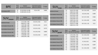 |
53 |
Early parenchymal changes on MRI and fluoroestradiol db-PET
during neoadjuvant endocrine therapy in patients with ER +/HER2-
breast cancer
Julia Carlota Carmona1,
Natsuko Onishi2,
Jiachao Liang2,
Jessica Gibbs2,
Ella Jones3,
Teffany Joy Bareng2,
Wen Li2,
David Newitt2,
Kim Ray2,
Courtney Lawhn1,
Bonnie Joe2,
Rita Mukhtar2,
I-SPY 2 Network2,
ISPY 2 Imaging Working group2,
Laura Esserman2,
Jo Chien2,
and Nola Hylton2
1Radiology, UCSF, San Francisco, CA, United States, 2UCSF, San Francisco, CA, United States, 3NIH, San Francisco, CA, United States Keywords: Breast, Cancer, MRI, fluoroestradiol, PET, endocrine, therapy The aim of our study was to assess early parenchymal changes during neoadjuvant endocrine therapy (NET) using both DCE-MRI and dedicated breast PET with [18F]-fluoroestradiol (FES-dbPET). We investigated background parenchymal enhancement (BPE) on MRI and background parenchymal uptake on FES-dbPET for seventeen patients with ER+/HER2- breast cancers. Statistically significant decrease in BPE was shown after 3 weeks of NET. Estimated SUVs (mean, max and peak) were lower after 3 weeks of NET, although the difference did not reach statistical significance. Our results illustrate the potential of DCE-MRI and FES-dbPET to detect early change in breast parenchyma during NET. |
|
3461.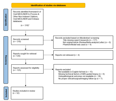 |
54 |
Performance benchmark metrics and clinicopathologic outcomes of
MRI-guided breast biopsies: a systematic review and
meta-analysis
Berat Bersu Ozcan1,
Justin Yan1,2,
Yin Xi1,
Serine Baydoun3,
Marion E. Scoggins4,
and Basak E. Dogan1
1Radiology, University of Texas Southwestern, Dallas, TX, United States, 2Radiology, HCA Houston Healthcare Kingwood, Houston, TX, United States, 3Radiology, Cleveland Clinic Foundation, Cleveland, OH, United States, 4Radiology, University of Texas MD Anderson Cancer Center, Houston, TX, United States Keywords: Breast, Breast, MRI-guided breast biopsy MRI-guided breast biopsy (MRBB) methods and clinicopathological outcomes may vary between institutions. To identify benchmark metrics to help define a successful MRBB program, we identified and systematically reviewed articles on MRBB outcomes through 04.01.2021. Random intercept logistic regression model was used to pool the data. We found that MRBB is an efficient, highly accurate technique with high technical success (99.05%, 95%CI:97.75%-99.60%), low false-negative (0.65%, 95%CI:0.29%-1.44%), and complication (2.45%, 95%CI:2.13%-2.81%) rates. Our findings can assist the development of evidence-based clinical guidelines on follow-up recommendations in benign-concordant lesions and a transparent discussion with patients on the consequences of having an MRBB. |
|
3462.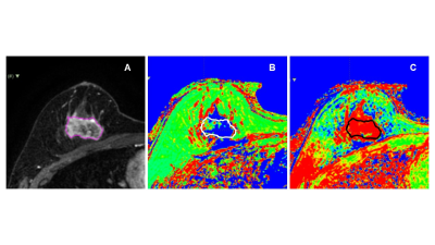 |
55 |
Feasibility of 3D Quantitative Synthetic MRI for Discriminating
Immunohistochemical Status in Invasive Ductal Carcinoma of the
Breast
Maki Amano1,2,
Shohei Fujita1,3,
Naoyuki Takei4,
Katsuhiro Sano1,
Akihiko Wada1,
Kanako Sato1,
Junko Kikuta1,
Yoshiki Kuwatsuru1,
Rina Tachibana1,5,
Towa Sekine1,5,
Yoshiya Horimoto6,
and Shigeki Aoki1
1Radiology, Juntendo University, Tokyo, Japan, 2Radiology, Nihon University Hospital, Tokyo, Japan, 3Radiology, The University of Tokyo Graduate School of Medicine, Tokyo, Japan, 4MR Applications and Workflow, GE Healthcare Japan, Tokyo, Japan, 5Radiological Sciences, Graduate School of Human Health Sciences, Tokyo Metropolitan University, Tokyo, Japan, 6Breast Oncology, Juntendo University, Tokyo, Japan Keywords: Breast, Cancer The present study aimed to evaluate the feasibility of 3D quantitative synthetic MRI for discriminating the immunohistochemical (IHC) status of invasive ductal carcinoma (IDC) of the breast. We found that volumetric T1 and T2 values of IDC varied according to IHC status, including those for hormone receptors and Ki-67, indicating the feasibility of this method for discriminating the IHC status of IDC of the breast. |
|
3463. |
56 |
Prediction of breast cancer response to neoadjuvant chemotherapy
via early changes in quantitative DCE-MRI
Mengyi Wang1,
Xiaoling Zhang1,
Shaofu ling He1,
Yangling Hu1,
and LiJuan Mao1
1Department of Radiology, Sun Yat-sen University First Affiliated Hospital, Guangzhou, China Keywords: Breast, Cancer, Neoadjuvant chemotherapy Our study suggests that the percentage change for second visits relative to baseline of enhanced tumor volume-based imaging metrics of DCE-MRI can quantify heterogeneous changes within the tumor as an indicator of therapy response and improve prediction of PCR as early as the first treatment time point in NAC. |
|
3464.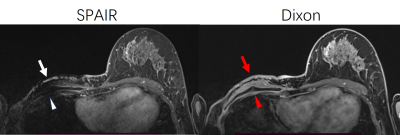 |
57 |
Use of Dixon technique in breast contrast-enhanced T1WI for
post-mastectomy patients at 3T
Qing FU1,
Jie Liu1,
Ding-xi Liu1,
Ting Yin2,
Peng Sun3,
Zi-qiao Lei1,
and Fan Yang1
1Union Hospital, Tongji Medical College, Huazhong University of Science and Technology, Wuhan, China, 2MR Collaborations, Siemens Healthineers Ltd., Chengdu, China, 3Philips Healthcare, Beijing, China Keywords: Breast, Breast, MRI; Dixon; fat suppression; 3T For patients with breast lesion resection, conventional contrast enhanced T1WI images with SPAIR provides poor fat suppression in the post-mastectomy breasts side due to the uneven skin surface, inhomogeneous tissues environment, and the frequency-selective feature of SPAIR scheme. Our study aimed to investigate the performance of fat suppression with the Dixon method in single side post-mastectomy patients. Subjective and objective evaluation suggested the Dixon method is superior to conventional SPAIR fat suppression in depicting the bilateral breast. Dixon method provided better image uniformity and higher fat suppression efficiency, and showed significant advantages in delineating the anatomical structures, with better axillary and lesion visibilities, especially in the completely removed breast side. |
|
3465.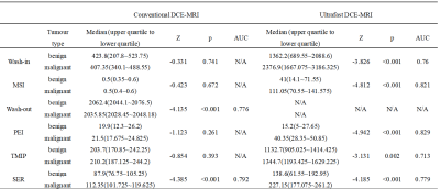 |
58 |
Feasibility of ultrafast DCE-MRI in differentiating benign and
malignant breast tumors
Junhui Huang1,2,
Lihua Qiu3,
Yongsheng Ao3,
Lan Mu4,
Zhanli Hu5,
Dong Liang5,
Xin Liu5,
Hairong Zheng5,
and Na Zhang5
1Paul C. Lauterbur Research Center for Biomedical Imaging, Shenzhen Institute of Advanced Technology, Chinese Academy of Sciences, ShenZhen, China, 2Faculty of Information Engineering and Automation, Kunming University of Science and Technology, Kunming, China, 3Yibin Second People's Hospital, Yibin, China, 4Chengdu Pidu District People's Hospital, Chengdu, China, 5Paul C. Lauterbur Research Center for Biomedical Imaging, Shenzhen Institute of Advanced Technology, Chinese Academy of Sciences, Shenzhen, China Keywords: Breast, Data Analysis, Conventional DCE-MRI, ultrafast DCE-MRI, quantitative parameters, semiquantitative parameters The conventional dynamic contrast-enhanced magnetic resonance imaging (DCE-MRI) technique has been shown to be very sensitive in detecting benign and malignant breast cancer lesions. However, it requires a relatively long acquisition time and hence a semiquantitative imaging technique. To reduce the acquisition time, ultrafast DCE-MRI techniques have been proposed for breast tumor imaging. Our study aims to investigate the feasibility of the ultrafast DCE-MRI technique in differentiating benign and malignant breast tumors. The results show that the semiquantitative and quantitative parameters obtained with ultrafast DCE-MRI are superior to those obtained with conventional DCE-MRI in differentiating benign and malignant breast tumors. |
|
3466.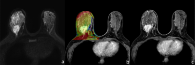 |
59 |
Assessment of breast lesions using amide proton
transfer-weighted imaging and dynamic contrast-enhanced MR
imaging: a pilot study
Lulu Zhuang1,
Chun Lian1,
Zehao Wang1,
Ximin Zhang1,
Zhigang Wu2,
Qingping Gu3,
and Rong Huang1
1Department of Radiology, Peking University Shenzhen Hospital, ShenZhen, China, 2Clinical & Technical Support, Philips Healthcare Ltd, ShenZhen, China, 3Clinical Application, Philips Healthcare Ltd, ShenZhen, China Keywords: Breast, Breast MRI is a non-invasive method for breast lesions diagnosis.However, some imaging features of benign and malignant lesions in breast are still overlap. APT-weighted imaging (APTw) is a endogenous biomarker that allows non-invasive visualization of tissue composition and microscopic information for mobile proteins and peptides. The present study explores the feasibility of the combination of APT and dynamic contrast-enhanced MR imaging to assess of breast lesions. Results of this study indicate the APT value has significant difference between benign and malignant breast lesions. |
|
3467.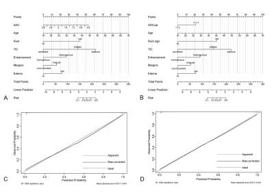 |
60 |
Improved differentiation of benign and malignant breast lesions
based on Kaiser score descriptors and apparent diffusion
coefficient
Lingsong Meng1,
Xin Zhao1,
Jinxia Guo2,
Lin Lu1,
Meiying Cheng1,
Qingna Xing1,
Honglei Shang1,
Yafei Guo1,
Shifang Tan1,
Lingjie Zhang1,
and Xiao-an Zhang1
1The Third Affiliated Hospital of Zhengzhou University, Zhengzhou, China, 2General Electric (GE) Healthcare, MR Research China, Beijing, Beijing, China Keywords: Breast, Breast Breast cancer is the most frequently diagnosed malignant tumor in women and badly threatens the female's health. Kaiser score (KS) shows excellent and robust performance in evaluating breast lesions. This scoring system doesn’t include quantitative image features and clinical information, however, which may result in false-negative diagnoses, especially for cancers showing atypical morphological characteristics. In this study, we evaluated the importance of five features in KS, apparent diffusion coefficient (ADC), and patient age respectively and combined them to build new models to explore if there will be an improvement in the differential diagnosis of breast lesions compared with KS. |
|
 Back to Meeting Home
 Back to the Program-at-a-Glance
The International Society for Magnetic Resonance in Medicine is accredited by the Accreditation Council for Continuing Medical Education to provide continuing medical education for physicians.
 Back to Meeting Home
 Back to the Program-at-a-Glance
 View Presentation Video