Digital Poster
Gynecological Cancer: Diffusion & APT
ISMRM & ISMRT Annual Meeting & Exhibition • 03-08 June 2023 • Toronto, ON, Canada

Digital Poster
Gynecological Cancer: Diffusion & APT
| Computer # | |||
|---|---|---|---|
3253.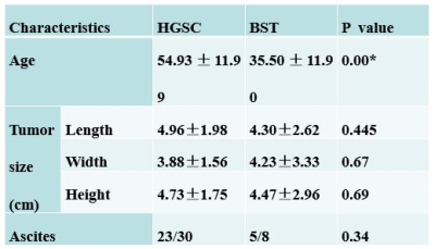 |
21 |
Diagnostic ability of APT in differentiating ovarian high-grade
serous carcinomas from borderline serous tumors
Jiacheng Song1,
Aining Zhang1,
Xiance Zhao2,
Yishi Wang3,
Zhiwei Shen3,
and Ting Chen1
1the first affiliated hospital of Nanjing Medical University, Nanjing, China, 2Philips Healthcare, Shanghai, China, 3Philips Healthcare, Beijing, China Keywords: Urogenital, CEST & MT The precise assessment of ovarian tumor, which is complex in presentation and classification, is important for clinical decision making. The value of amide proton transfer weighted (APTw) imaging in the assessment of ovarian lesion has not been comprehensively researched. In this study, we investigated the value of APTw imaging in differentiating ovarian high-grade serous carcinomas from borderline serous tumors. It was found that APT value of high-grade tumor was significantly lower than that of borderline tumor. Our finding indicates the potential of using APTw imaging as a novel method for the differentiation diagnosis of ovarian cancers. |
|
3254.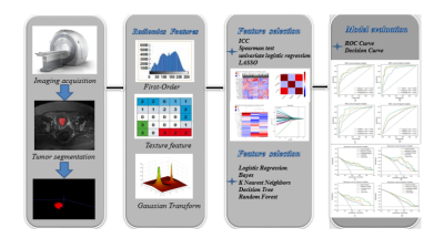 |
22 |
Multiparametric MRI-based Radiomics for Preoperative Prediction
of Multiple Biological Characteristics in Endometrial Cancer
Ma Changjun1,
Song Qingling1,
Tia Shifeng1,
Chen Lihua2,
Wang Nan2,
Lin Liangjie3,
Wang Jiazheng3,
and Liu Ailian2
1Department of Radiology,, The First Affiliated Hospital of Dalian Medical University, Dalian, China, China, 2Department of Radiology, The First Affiliated Hospital of Dalian Medical University, Dalian, China, China, 3Clinical & Technical Support, Philips Healthcare, Beijing, China, China Keywords: Pelvis, Uterus Different biological characteristics of endometrial cancer (EC) may lead to different treatment efficacies and prognoses, so stratification of biological characteristics in EC is important for treatment planning. |
|
3255.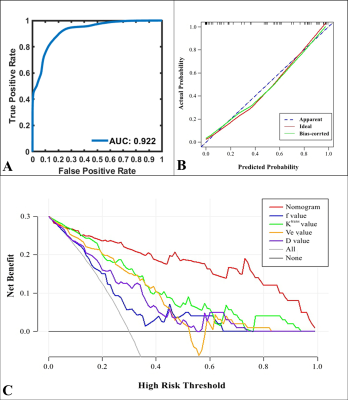 |
23 |
Quantitative MRI of DCE andIVIM for differentiating low-risk
from non-low-risk early-stage endometrial carcinoma
Hongxia Wang1,
Ruifang Yan1,
Beiran Wang1,
Zhong Li1,
Xingxing Jin1,
Zhenfang Guo2,
Wangyi Liu1,
Kaiyu Wang3,
Jinxia Guo3,
and Dongming Han1
1Department of MR, the First Affiliated Hospital, Xinxiang Medical University, Weihui, China, 2Department of Neurology, the First Affiliated Hospital, Xinxiang Medical University, Weihui, China, 3MR Research China, GE Healthcare, Beijing, China Keywords: Pelvis, Cancer Finding a noninvasive and effective means for the risk stratification of early-stage EC is of great benefit to patients. This study aim to investigate the value of dynamic contrast-enhanced MRI (DCE-MRI) and intravoxel incoherent motion (IVIM) in differentiating low-risk from non-low-risk early-stage endometrial carcinoma (EC). Results showed that both DCE-MRI and IVIM facilitate the preoperative identification of low-risk and non-low-risk early-stage EC. Compared with each single parameter, the combination of Ktrans, Ve, and f provided better predictive power and may serve as a superior imaging marker. |
|
3256.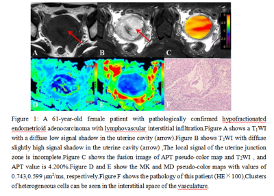 |
24 |
Amide proton transfer weighted and diffusion kurtosis imaging in
evaluating lymphovascular space invasion of endometrial
carcinoma
Xie Zongyuan1,
Tian Shifeng1,
and Liu Ailian1
1Department of Radiology, the First Affiliated Hospital of Dalian Medical University, Dalian, China Keywords: Uterus, Cancer Lymphovascular space invasion(LVSI)is one of the important factors for poor prognosis of endometrial carcinoma(EC).Quantitative parameters of amide proton transfer weighted(APTw)and diffusion kurtosis imaging(DKI) is a novel MRI tool to evaluating LVSI in EC. The APT and MK values of EC with LVSI were higher than those without LVSI.The AUC after combination was higher than that of MK alone.Both APTw and DKI can effectively evaluate EC LVSI.APTw combined DKI application can improve the evaluation efficiency . |
|
3257.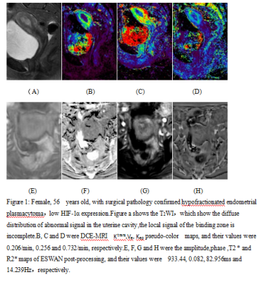 |
25 |
Quantitative assessment of HIF-1a expression in endometrial
cancer by DCE-MRI combined ESWAN multiparameter imaging
Xie Zongyuan1,
Tian Shifeng1,
and Liu Ailian1
1Department of Radiology, the First Affiliated Hospital of Dalian Medical University, Dalian, China Keywords: Uterus, Cancer Hypoxia -inducible factor (HIF-1α) is a major transcriptional factor regulating gene expression under hypoxic conditions. High expression of HIF-1α contributes to high aggressiveness or poor prognosis of cervical carcinoma. Multimodal MRI techniques DCE-MRI and ESWAN are new tools to determine the expression of HIF-1α.Its quantitative parameters Ktrans , Kep R2 * and T2* values were correlated with HIF-1a expression . Therefore,both DCE-MRI and ESWAN techniques can effectively evaluate HIF-1a expression in endometrial cance. |
|
3258.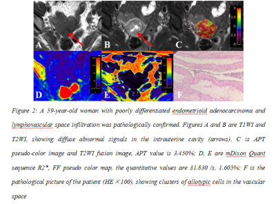 |
26 |
Evaluation of lymphovascular space invasion in endometrial
carcinoma by APTw and mDIXON-Quant
Xie Zongyuan1,
Tian Shifeng1,
and Liu Ailian1
1Department of Radiology, the First Affiliated Hospital of Dalian Medical University, Dalian, China Keywords: Uterus, Cancer Lymphovascular space invasion(LVSI)is one of the important factors for poor prognosis of endometrial carcinoma(EC).Quantitative parameters of amide proton transfer weighted(APTw)and Modified Dixon quantification of fat(mDIXON-Quant) is a novel MRI tool to evaluating LVSI in EC. The APT and R2* values of LVSI(+)EC were higher than those of LVSI(-)EC.APT value was moderately positively correlated with R2* value, and APT value was weakly positively correlated with FF value . Both APTw and Mdixon-Quant techniques can effectively evaluate EC LVSI, and their combined application can improve the evaluation efficiency. |
|
3259.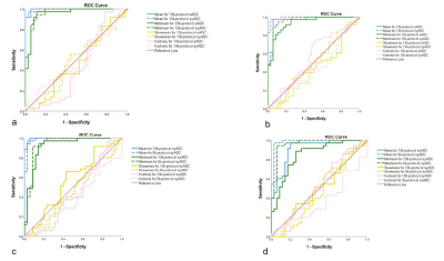 |
27 |
Feasibility of synthetic diffusion weighted images using
computed with 5b protocol in uterine tumors
Qian Tang1,
Weiyin Vivian Liu2,
Qiqi Zhou3,
Wen Chen3,
Ling Sang3,
and Lin Xu3
1Department of Biomedical Engineering, Hubei University of Medicine, Hubei, China, 2GE Healthcare, Beijing, China, 3Department of Radiology,Taihe Hospital, Hubei University of Medicine, Hubei, China Keywords: Pelvis, Cancer High-b-value DWI has been widely applied in clinical practices, but it is challenging to acquire several high b-value images for long acquisition time and more eddy distortion as b-value increases, leading to patient discomfort, increased motion artifacts and decreased SNR. High-b-value SyDWIs showed better SNR and less image distortion than scanned high-b-value DWIs. Our study demonstrate the feasibility of 5b-value synthetic high-b-value reduced full-of-view diffusion weighted imaging with the pros of short scan time, better lesion clarity and higher image quality in comparison of 13b-protocol rFOV-syDWIs and 5b-protocol synthetic ADCmean and ADCminimum offered reliably diagnostic value compared to 13b-protocol ones. |
|
3260. |
28 |
Diffusion kurtosis imaging with multiple quantitative parameters
for predicting microsatellite instability status of endometrial
carcinoma
Qingling Song1,
Wan Dong2,
Shifeng Tian3,
Lihua Chen3,
and Ailian Liu3
1Radiology, the First Affiliated Hospital of Dalian Medical University, Dalian, China, 2Radiology, Wuhan children's Hospital, Tongji Medical College of Huazhong University of Science & Technology, Wuhan, China, 3Radiology, The First Affiliated Hospital of Dalian Medical University, Dalian, China Keywords: Pelvis, Cancer Microsatellite instability (MSI), which results from the failure of mismatch repair (MMR) proteins to fix a DNA replication error, causes insertions, mismatches, or deletions during the DNA replication process, thus having an essential role in maintaining the stability of the genome and regulating gene expression |
|
3261.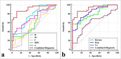 |
29 |
Application of diffusion imaging and dynamic contrast-enhanced
MRI in evaluating the proliferation status of endometrial
carcinoma
Xuejia Wang1,
Wangyi Liu 1,
Gaiyun Zhang1,
Ruifang Yan1,
Jie Shang1,
Kaiyu Wang2,
Jinxia Guo2,
and Dongming Han1
1Department of MR, the First Affiliated Hospital, Xinxiang Medical University, Weihui, China, 2MR Research China, GE Healthcare, Beijing, China Keywords: Pelvis, Cancer Intravoxel incoherent motion (IVIM) is able to reflect the true diffusion, microcirculatory perfusion, average diffusion, and structural complexity in tissues. Dynamic contrast-enhanced MRI (DCE-MRI) can be used to analyze the dynamic distribution of contrast agents through the pharmacokinetic model to quantitatively detect the blood supply of biological tissues. Our results showed that IVIM and DCE-MRI-derived parameters such as D, α, Ktrans, and Kep were associated with Ki-67 status in EC, and the combination of D and Kep may serve as a superior imaging marker for the identification of low-proliferation and high-proliferation EC. |
|
3262.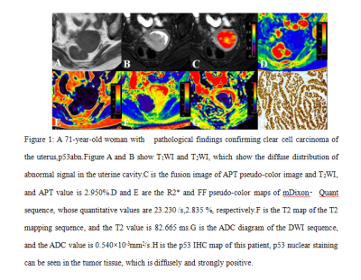 |
30 |
The value of multimodal functional magnetic resonance imaging in
differentiating p53abn from p53wt endometrial carcinoma
Xie Zongyuan1,
Tian Shifeng1,
and Liu Ailian1
1Department of Radiology, the First Affiliated Hospital of Dalian Medical University, Dalian, China Keywords: Cancer, Uterus p53 genotyping is important for EC lymph node metastasis and prognosis determination.Multimodal MRI techniques APTw, T2 mapping, mDIXON-Quant and DWI are new tools to determine p53 genotyping.Results of this study indicate the APT value and R2* value in the p53abn group were higher than those in the p53wt group, while the ADC value was lower than those in the p53wt group.Therefore, APTw, mDIXON-Quant and DWI techniques can quantitatively identify p53abn and p53wt EC, it provides a new way for preoperative quantitative evaluation of EC molecular typing. |
|
3263.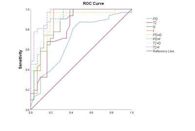 |
31 |
Diagnostic performance of synthetic MRI and reduced
filed-of-view DWI-based IVIM on cervical intraepithelial
neoplasis
Qian Tang1,
Weiyin Vivian Liu2,
Qiqi Zhou3,
Wen Chen3,
and Lin Xu3
1Department of Biomedical Engineering, Hubei University of Medicine, Hubei, China, 2GE Healthcare, Beijing, China, 3Department of Radiology,Taihe Hospital, Hubei University of Medicine, Hubei, China Keywords: Uterus, Cancer CIN III is currently reckoned as a high-risk precancerous lesion for highly-possible transformation into cervical cancers. To improve low diagnostic efficiency of traditional MRI on precancerous CIN III for its subtle changes at the cellular level, this study built a combination model with MAGiC and IVIM derived parameters. In particular, a combined model with T2 and f values showed good performance on differentiation CIN III from cervical cancer with the sensitivity, specificity and AUC of 0.0.968, 0.839, 956, and it indeed elevated diagnostic performance especially specificity. In our study, alteration of heterogeneity at the cellular level was suspected to occur as cervix gradually progresses to CIN III stage despite no obvious morphological changes on structural images. |
|
3264.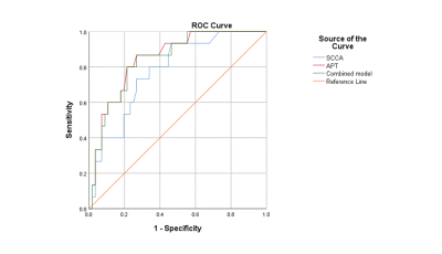 |
32 |
The value of APT combined with squamous cell carcinoma antigen
in predicting parametrial invasion of stage IA to IIB cervical
cancer
Mengdi Zhang1,
Qinghe Han1,
Qinghai Yuan1,
Rui Ma1,
and Jianxiu Lian2
1The Second Hospital of Jilin University, Jilin, China, 2Philips Healthcare, Beijing, China Keywords: Uterus, Cancer, parametrial invasion、squamous cell carcinoma antigen Cervical cancer is one of the most common gynecological malignancies. The accurate diagnosis of cervical cancer is of great significance for the selection of treatment plan. This study is to explore the diagnostic value of the new magnetic resonance technology of amide proton transfer-weighted(APTw) imaging combined with squamous cell carcinoma antigen (SCCA) for parametrial invasion(PMI).The results showed amide proton transfer-weighted(APTw) imaging could provide more valuable for diagnosis efficiency, which maybe an invasive alternative method for evaluating parametrial invasion. |
|
3265. |
33 |
The value of amide proton transfer and glucose-chemical exchange
saturation transfer in predicting tumor grading and staging in
cervical cancer
Han Jiang1,
Nan Meng2,
Ziqiang Li1,
Bo Dai2,
Pengyang Feng3,
Yu Luo2,
Zhiwei Shen4,
and Meiyun Wang*2
1Department of Medical Imaging, Xinxiang Medical University & Henan Provincial People's Hospital, Zhengzhou, China, 2Department of Medical Imaging, Zhengzhou University People's Hospital & Henan Province People's Hospital, Zhengzhou, China, 3Department of Medical Imaging, Henan University People’s Hospital & Henan Provincial People’s Hospital, Zhengzhou, China, 4Philips healthcare, Beijing, China Keywords: Pelvis, CEST & MT, cervical cancer What are the values of three-dimensional amide proton transfer (APT) imaging and glucose-chemical exchange saturation transfer (glucoCEST) in WHO grading and staging of cervical cancer? We performed 3D-APT, 3D-glucoCEST, and DWI scans in 19 cervical cancer cases. APT signal intensity(SI), and GlucoCEST SI could distinguish high-grade from low-grade; GlucoCEST SI can differentiate early (<IIB) from advanced (≥IIB) cervical cancer. Both APT and glucoCEST can be used for the preliminary evaluation of cervical cancer, but glucose CEST has advantages in the evaluation of cervical cancer grading and staging. |
|
3266.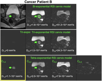 |
34 |
Multi-exponential model of diffusion signal with fixed ADCs in
healthy and cancerous cervix tissues
Ana E Rodríguez-Soto1,
Elin M Lundström1,2,3,
Alexandra Besser1,
Stephane Loubrie1,
Christopher C Conlin1,
Stephan Jordan1,
Summer Batasin1,
Sheida Ebrahimi1,
Alexandra Schlein1,
Michael Hahn1,
Tyler Seibert4,
Anders M Dale1,5,
Marianne Tom-Hedla6,
Cheryl Saenz6,
Shira Varon6,
Michael McHale6,
and Rebecca Rakow-Penner1
1Radiology, University of California San Diego, La Jolla, CA, United States, 2Surgical Sciences, Uppsala University, Uppsala, Sweden, 3Radiology, Uppsala University, Uppsala, Sweden, 4Radiation Medicine, University of California San Diego, La Jolla, CA, United States, 5Neurosciences, University of California San Diego, La Jolla, CA, United States, 6Ob/Gyn & Reproductive Sciences, University of California San Diego, La Jolla, CA, United States Keywords: Pelvis, Cancer Restriction spectrum imaging (RSI) has shown potential to become a DWI-based, contrast-free diagnostic tool for cancer as it separates the different water pools in tissues. Here, we describe the diffusion signal of cancerous and healthy cervical tissues using RSI. Multi-exponential models with fixed ADCs were determined and the signal contribution of each component estimated. Initial results indicate that the slowest diffusion compartment of a tetra-exponential RSI model has the potential to isolate cervical cancer signal from that of surrounding tissues. This model may increase the sensitivity and specificity of DWI to evaluate response to treatment of cervical cancer patients. |
|
3267.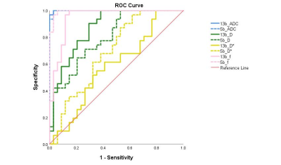 |
35 |
An optimal multiple b-value scheme for applications in female
cervical cancer
Qian Tang1,
Weiyin Vivian Liu2,
Qiqi Zhou3,
Wen Chen3,
Ling Sang3,
and Lin Xu3
1Department of Biomedical Engineering, Hubei University of Medicine, Hubei, China, 2GE Healthcare, Beijing, China, 3Department of Radiology,Taihe Hospital, Hubei University of Medicine, Hubei, China Keywords: Uterus, Cancer Incoherent motion in voxel (IVIM) imaging is widely utilized in grading tumor, cancer, treatment efficacy and prognosis. It is believed utility of at least 8 b-values image sets brings a more stable and accurate results. However, long acquisition time hamper clinical applications. High consistent diagnostic performance of manual ROI-based IVIM-derived measurements including ADC, D, D* and f on distinguishing patients with cervical cancer from healthy controls was found between 5b-value and 13b-value IVIM imaging protocol and no significant difference of all four values was found. We proposed a IVIM diffusion weighted imaging with a 5b-value protocol in cervical cancer. |
|
3268.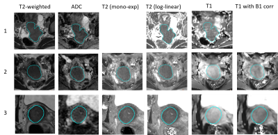 |
36 |
Multicenter consistency of quantitative MRI in cervical cancer
Petra J van Houdt1,
Abel Bregman1,
Kari Tanderup2,
Robert Hudej3,
Marko Zaletelj3,
Barbara Šegedin3,4,
Eva E Schaake1,
Ellen M Kerkhof5,
Laura A Velema5,
Remi A Nout6,
and Uulke A van der Heide1
1Radiation Oncology, The Netherlands Cancer Institute, Amsterdam, Netherlands, 2Clinical Medicine, Aarhus University Hospital, Aarhus, Denmark, 3Radiotherapy, Institute of Oncology Ljubljana, Ljubljana, Slovenia, 4Faculty of Medicine, University of Ljubljana, Ljubljana, Slovenia, 5Radiotherapy, Leiden University Medical Center, Leiden, Netherlands, 6Radiotherapy, Erasmus MC Cancer Institute, University Medical Center Rotterdam, Rotterdam, Netherlands Keywords: Cancer, Quantitative Imaging, cervical cancer The reproducibility of quantitative MRI parameters in a multicenter setting is not trivial. In this study we evaluated the in-vivo multicenter consistency of quantitative MRI parameters in cervical cancer patients. DWI, T2 mapping and T1 mapping were available from 66 patients from 3 institutes. ADC values were measured consistently across institutes despite differences in acquisition protocols. For T2 and T1 values differences between institutes were observed, which need to be accounted in future evaluation of the quantitative MRI parameters in relation to treatment outcome. |
|
3269. |
37 |
Amide Proton Transfer Weighted Combined with Diffusion Kurtosis
Imaging for Predicting Lymph Node Metastasis in Cervical Cancer
Yue Wang1,
Shifeng Tian1,
and Ailian Liu1
1the First Affiliated Hospital of Dalian Medical University, Dalian, China Keywords: Pelvis, Cancer Cervical cancer (CC) is one of the most common gynecological malignancies worldwide, and its incidence is gradually increasing. Acurate identification of lymph node metastases (LNM) is crucial for predicting prognosis and choosing the best available treatment. Amide proton transfer weighted (APTw) imaging can detect the chemical exchange rate of between water and endogenous mobile proteins, peptides or polypeptides[1]. Diffusion kurtosis imaging (DKI) can reflect the limited diffusion movement of water molecules in tissue and the complexity of tissue microstructure [2]. This study is aimed to investigate the quantitative prediction of LNM in CC by APTw combined with DKI. |
|
3270.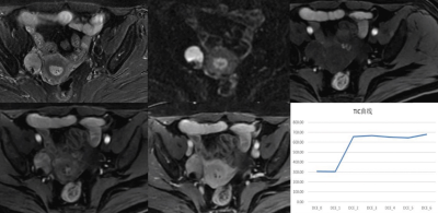 |
38 |
The Value of dynamic contrast-enhanced MRI in differential
diagnosis of solid ovarian tumors with diffusion restriction
Ying Meng1,
Yuting Liang1,
Xinlian Wang1,
Keyang Wang1,
and Lizhi Xie2
1Beijing Obstetrics and Gynecology Hospital, Capital Medical University, Beijing, China, 2GE healthcare, Beijing, Beijing, China Keywords: Pelvis, Pelvis DWI can differentiate benign and malignant lesions by reflecting the diffusion of water molecules in lesions, which has become a routine sequence of MRI examination of gynecological tumors. Generally, DWI signal is high, which will alert to the possibility of malignancy, while benign tumors also show high DWI signal. For solid ovarian tumors with high signal intensity on DWI, the differential diagnosis of ovarian tumors is still a difficult point in routine MRI sequence diagnosis. The results of this study showed that DCE-MRI quantitative parameters of solid ovarian masses were helpful for differential diagnosis of benign and malignant. |
|
3271.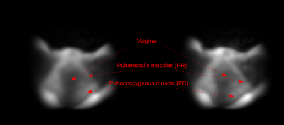 |
39 |
Imaging female pelvic anatomy with a portable, low field
Magnetic Resonance Imager.
Sourajit Mitra Mustafi1,
Meredith Sadinski1,
Muller Gomes1,
Alexander Nacev1,
Hugo Davila2,
and Srirama S Venkataram1
1Promaxo, Oakland, CA, United States, 2Cleveland Clinic Indian River Hospital, Vero Beach, FL, United States Keywords: Pelvis, Body, Pelvic Imaging, Low field MRI Promaxo's single sided, low-field MRI is used for female pelvic imaging. Five healthy female subjects were scanned. The butterfly shape of vagina and normal anatomy of the puborectails regions were observed. Delineation between puborectails and pubococcygneus muscles are clearly seen. MRI’s improved soft tissue contrast provides better visualization of anatomical regions and is less invasive than transvaginal ultrasound. |
|
 Back to Meeting Home
 Back to the Program-at-a-Glance
The International Society for Magnetic Resonance in Medicine is accredited by the Accreditation Council for Continuing Medical Education to provide continuing medical education for physicians.
 Back to Meeting Home
 Back to the Program-at-a-Glance
 View Presentation Video