Digital Poster
More Advances in Prostate Imaging
ISMRM & ISMRT Annual Meeting & Exhibition • 03-08 June 2023 • Toronto, ON, Canada

Digital Poster
More Advances in Prostate Imaging
| Computer # | |||
|---|---|---|---|
2056.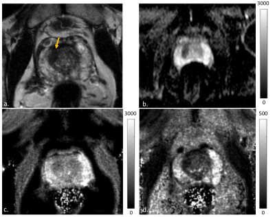 |
61 | Feasibility of Prostate MR Fingerprinting and ADC Mapping in the quantitative characterization of malignant transition zone lesions
Anna Lavrova1, Ananya Panda2, Sarah E. Wido3, Lee Ponsky3, Maria Masotti4, Thomas Chenevert1, Yun Jiang1, Leonardo Kayat Bittencourt5, Anupama Ramachandran1, Katherine Wright1, Nicole Seiberlich1, Hero Hussain1, and Vikas Gulani1
1Department of Radiology, University of Michigan, Ann Arbor, MI, United States, 2Department of Diagnostic and Interventional Radiology, All Indi Institute of Medical Sciences, Jodhpur, India, 3Department of Urology, Case Western Reserve University, Cleveland, OH, United States, 4Department of Biostatistics, University of Michigan, Ann Arbor, MI, United States, 5Department of Radiology, Case Western Reserve University, Cleveland, OH, United States Keywords: Prostate, Cancer The purpose of this study is to assess the feasibility of the use of MR Fingerprinting combined with ADC mapping for characterizing prostate transition zone (TZ) cancers. This quantitative approach has shown promising results in differentiating prostate cancer from normal prostate tissue in peripheral and transition zones, and in separating low from intermediate/high grade tumors in the PZ. Our study aimed to confirm previous observations of quantitative differences in T1, T2 and ADC between cancers and normal appearing TZ, and to assess the utility of MRF-derived T1 and T2 mapping in distinguishing low grade lesions from high/intermediate-grade cancers in TZ. |
|
2057.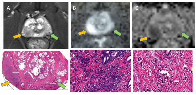 |
62 | Detection of sparse and dense prostate cancers using bi-parametric MRI combined with radiomics features
Bingni Zhou 1,2, Zhangzhe Chen1,2, Rucuan Chen1,2, Wei Liu1,2, Hualei Gan2,3, Yong Zhang4, Liangping Zhou1,2, and Xiaohang Liu1,2
1Radiology, Fudan University Shanghai Cancer Center, Shanghai, China, 2Oncology, Shanghai Medical College of Fudan University, Shanghai, China, 3Pathology, Fudan University Shanghai Cancer Center, Shanghai, China, 4GE Healthcare, Shanghai, China Keywords: Prostate, Radiomics This is a preliminary study which combined bi-parametric MRI with radiomics feature to detect sparse and dense prostate cancers. Fifty-five patients underwent diffusion weighted and T2 weighted imaging. One hundred and nine peripheral zone (PZ) tumors were reviewed using whole-mount histologic findings. The total number of 381 radiomics features were extracted to construct the models for differentiation. Dense tumors showed ADC values significantly lower than sparse tumors and normal PZ tissues. ADC alone should provide sufficient diagnosis efficiency. However, radiomics features can significantly improve the detection of sparse tumors which showed the similar ADC values as compared to normal tissues. |
|
2058.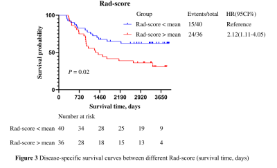 |
63 | Kaplan-Meier survival analysis of the DCE-MRI-based radiomics in the prognosis of prostate cancer
Ting Huang1, Zhiqiang Chen2, Shaoru Zhang1, Zhuo Wang1, Xiaohua Chen1, Yunshu Zhou1, Xiaocheng Wei3, and Aijun Wang4
1Clinical medicine school of Ningxia Medical University, Yinchuan, China, 2Department of Radiology, the First Hospital Affiliated to Hainan Medical College, Haikou, China, 3GE Healthcare, Beijing, China, 4Department of Radiology, General Hospital of Ningxia Medical University, Yinchuan, China Keywords: Prostate, DSC & DCE Perfusion In this study, we aim to investigate whether DCE-MRI-based radiomics can be used in the prognosis of prostate cancer. Rad-score in which DEC-MRI-based radiomics labels were established by screening radiomics features GLCM_Correlation and shape_Flatness through Lasso regression was significantly different between the death group and the censor group. It was concluded that DCE-MRI-based radiomics can be used as prognostic factors for prostate cancer. |
|
2059.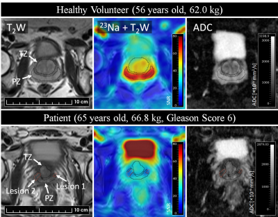 |
64 | Development of an external butterfly coil for sodium (23Na) MRI of human prostate cancer
Josephine L Tan1,2,3, Vibhuti Kalia4,5, Jonathan D Thiessen1,2, Timothy J Scholl1,3,6, and Alireza Akbari1
1Medical Biophysics, Western University, London, ON, Canada, 2Lawson Imaging, Lawson Health Research Institute, London, ON, Canada, 3Robarts Research Institute, London, ON, Canada, 4Medical Imaging, Western University, London, ON, Canada, 5Medical Imaging, St. Joseph's Health Care, London, ON, Canada, 6Ontario Institute for Cancer Research, Toronto, ON, Canada Keywords: Prostate, Cancer This abstract focuses on the development of an external, non-invasive butterfly coil for sodium MRI of human prostate cancer. Sodium MRI of healthy male volunteers and patients revealed higher sodium signal and ADC in the peripheral zone compared to the transition zone of the prostate, and a lower sodium signal and ADC in tumour lesions relative to healthy tissue. Further work will aim to characterize the sensitivity of this coil to detect tumours, which may enhance the workflow of sodium MRI in prostate cancer studies compared to conventional endorectal coils. |
|
2060.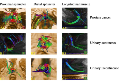 |
65 | Evaluation of urethral sphincter complex after LRP based on 3.0T pelvic floor magnetic resonance imaging combined with DTI and fiber tractography
Zhiheng Zhao1, Yingyu Che1, Jing Zhang1, Yue Wu1, Zitao Yang1, Qinyong Zhang1, Qingwei Wang1, Yifeng Sheng1, Ruilin Fan1, and Jingliang Cheng1
1the First Affiliated Hospital of Zhengzhou University, Zhengzhou, China Keywords: Prostate, Diffusion Tensor Imaging, Radical prostatectomy; Urethral sphincters complex; Urinary incontinence The anatomical-morphological and functional characteristics of the urethral sphincter complex, a key structure in the mechanism of urinary continence, have been studied mostly based on cadaveric dissection or animal experiments. In this study,we qualitatively and quantitatively evaluated the morphology and function of the urethral sphincter complex in patients after laparoscopic radical prostatectomy (LRP) using pelvic floor MRI combined with DTI and fiber tractography, and to investigate the anatomical factors affecting PPI. |
|
2061.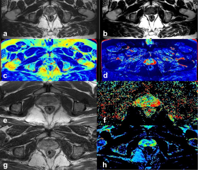 |
66 | Evaluation of the clinical value of Synthetic MRI in the prostate cancer diagnosis: a comparison of quantitative relaxation with conventional MRI
Jun Tian1, Dmytro Pylypenko2, and Haige Li1
12nd Affiliated Hospital of Nanjing Medical University, Nanjing, China, 2MR Research China, GE Healthcare, Beijing, China Keywords: Prostate, Prostate This study aimed to investigate the feasibility of synthetic MRI in distinguishing PCa from benign hyperplasia, and quantitative relaxation was compared with conventional MRI to evaluate the precision of obtained values with synthetic MRI. 38 patients (54 lesions) with cancer and/or hyperplasia were recruited. Quantitative MRI sequences, including T1, T2 mapping and MAGIC, were used. Significantly different T1 and T2 values were shown between cancer and noncancerous tissue, located in peripheral and center zones, as well as between cancer and glandular hyperplasia and stromal hyperplasia. These findings suggest synthetic-MRI may be effective in differentiating PCa from benign hyperplasia. |
|
2062.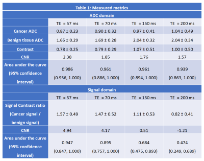 |
67 | Effect of Echo Times on Prostate Cancer Detection on Diffusion Weighted Images at high b-value and Apparent Diffusion Coefficient maps
Aritrick Chatterjee1,2, Audrey Kim1, Batuhan Gundogdu1,2, Milica Medved1,2, Tatjana Antic1, Gregory Karczmar1,2, and Aytekin Oto1,2
1University of Chicago, CHICAGO, IL, United States, 2Sanford J. Grossman Center of Excellence in Prostate Imaging and Image Guided Therapy, Chicago, IL, United States Keywords: Prostate, Diffusion/other diffusion imaging techniques This study compared the effect of echo times on the detection of prostate cancer on DWI at high b-value and ADC maps. Contrast between benign tissue and cancer increased significantly with higher TE on ADC maps. However, due to increased noise at higher TEs, CNR (study endpoint) was highest at TE 57 ms; higher by 23, 26, 33% than that at TE 70, 150 and 200 ms on ADC maps. CNR, contrast ratio AND auc are higher at lower TE on DWI high-b image. Therefore, the use of lower echo times for DWI and ADC mapping may improve prostate cancer. |
|
2063.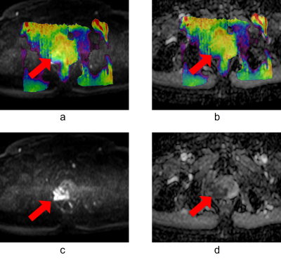 |
68 | The application of Amide proton transfer MR imaging in prostate cancer with different risk
Jianghong Man1, Lina Wang1, Tao Yu1, Yi Zhu2, and Zhiwei Shen2
1Liaoning Cancer Hospital&Institute, Shenyang, China, 2Philips Healthcare, Beijing, China Keywords: Prostate, Cancer, APT, Gleason score Amide proton transfer (APT) imaging is a novel nonintrusive technique for the diagnosis of PCa. In this study, we evaluate the distinction between APT value for prostate cancer and benign region in different Gleason scores. The result shows that APT is a potential tool for evaluating the risk of prostate cancer. |
|
2064.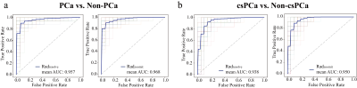 |
69 | Radiomics of MRI Based on Zoomit DWI in Predicting Clinically Significant Prostate Cancer: A Towards Step of a Non-invasive Auxiliary Tool
Xiaomeng Qiao1, Jie Bao1, Yang Song2, Yunzhu Wu2, and Ximing Wang1
1Department of radiology, First Affiliated Hospital of Soochow University, Suzhou, China, 2MR Scientific Marketing, Siemens Healthineers Ltd, Shanghai, China Keywords: Prostate, Diffusion/other diffusion imaging techniques, Zoomit DWI; Resolve DWI; Radiomics models based on ZOOMit DWI had better accuracy in the diagnosis of PCa and csPCa compared with those based on RESOLVE DWI technology, and was promising as a powerful non-invasive auxiliary tool to improve the diagnostic performance of PI-RADS of radiologists with different clinical experience. |
|
2065.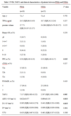 |
70 | The value of the combination of Intravoxel incoherent motion and TAF11 in predicting positive margins of radical prostatectomy
Wanting Gan1, Shuang Meng1, Lihua Chen1, Nan Wang1, Yunsong Liu1, and Ailian Liu1
1the First Affiliated Hospital of Dalian Medical University, Dalian, China Keywords: Prostate, Diffusion/other diffusion imaging techniques Positive surgical margins (PSMs) mean incomplete tumor excision which related with prostate cancer-specific mortality and may lead to additional treatments. As a new technique, Intravoxel incoherent motion can simultaneously demonstrate information about diffusion and perfusion. The result of this study indicate the combination of ADC, D, PPC and TAF11 can be used as a potential method in PSMs of radical prostatectomy. |
|
2066. |
71 | PCa Risk Prediction Model:Combined Clinical Characteristics AND mpMRI Parameters for Prediction of Risk of PCa
Aiqi Chen1, Xiang Li1, Jingxu Xu2, Xiuzheng Yue3, Shoukang Chen1, and Yichuan Ma1
1The First Affiliated Hospital of Bengbu Medical College, Bengbu, China, 2Beijing Deepwise & League, Beijing, China, 3Philips Healthcare, Beijing, China Keywords: Prostate, Prostate It’s still a challenge to accurately diagnose prostate cancer through MRI before operation. PI-RADS was capable of assessing the value of risk, but it did not combine clinical characteristics. we combined clinical characteristics and mpMRI parameters for the prediction of the risk of prostate cancer and compared them with PI-RADS. We found that the model was significantly better than PI-RADS (P=0.01976). The AUC of the model is higher than that of PI-RADS (0.99>0.90, p=0.019). This study demonstrated the feasibility of the model to predict the risk of prostate cancers early so that low-risk patients can avoid unnecessary needle biopsies. |
|
2067.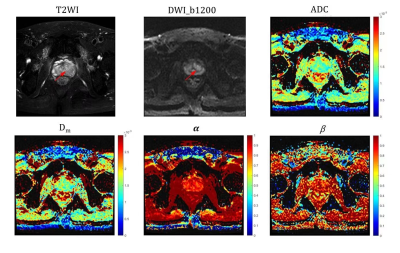 |
72 | Differentiating Chronic Prostatitis and Prostate Cancer Using a Continuous-Time Random-Walk Diffusion Model at High b-Values
Yurui Sheng1, Ke Xue2, Yongming Dai2, and Qingshi Zeng1
1Department of Radiology, The First Affiliated Hospital of Shandong First Medical University & Shandong Provincial Qianfoshan Hospital, Jinan, Jinan, China, 2MR Collaboration, Central Research Institute, United Imaging Healthcare, Shanghai, China Keywords: Prostate, Diffusion/other diffusion imaging techniques In clinical practice, distinguishing between Prostate cancer (PCa) and chronic prostatitis (CP) is difficult but necessary. PCa and CP are heterogeneous at the tissue and cell level, and the continuous time random-walk (CTRW) model can provide information on tissue heterogeneity at the microscopic level. We used the CTRW model to characterize tissue heterogeneity and complexity in CP and PCa. Significant differences were found for the CTRW parameters (α, Dm) between CP and PCa. Moreover, CTRW parameters (α, β, Dm) combined with ADC showed optimal diagnostic efficacy for diagnosis, and this combination would be benefit for the clinical diagnostic work. |
|
2068.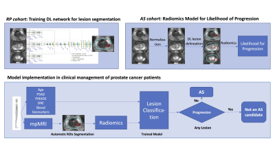 |
73 | mpMRI Radiomic Features Predict the Likelihood for Progression to Treatment of Prostate Cancer Patients on Active Surveillance
Veronica Wallaengen1,2, Evangelia I. Zacharaki1, Mohammad Alhusseini1, Nachiketh Soodana-Prakash2, Ahmad Algohary1, Adrian L. Breto1, Isaac L. Xu1, Sandra M. Gaston1, Rosa P. Castillo Acosta3, Oleksandr N. Kryvenko4, Bruno Nahar2, Dipen J. Parekh2, Alan Pollack1, Sanoj Punnen2, and Radka Stoyanova1
1Department of Radiation Oncology, University of Miami Miller School of Medicine, Miami, FL, United States, 2Department of Urology, University of Miami Miller School of Medicine, Miami, FL, United States, 3Department of Radiology, University of Miami Miller School of Medicine, Miami, FL, United States, 4Department of Pathology, University of Miami Miller School of Medicine, Miami, FL, United States Keywords: Quantitative Imaging, Prostate, Active Surveillance, mpMRI, Prostate Cancer Active surveillance (AS) for prostate cancer has emerged as a safe and attractive alternative to immediate treatment. Here we present an integrated method for baseline mpMRI analysis enabling early detection of patients harboring lesions with a high potential for progression. The approach consists of three steps: (i) Training a deep learning network for automatic segmentation of prostate and lesions, suspicious for cancer; (ii) Application of the network to identify lesions on mpMRI images for patients, enrolled in an AS trial; and (iii) Development of a progression risk stratification model by incorporating radiomic and clinical variables. |
|
2069.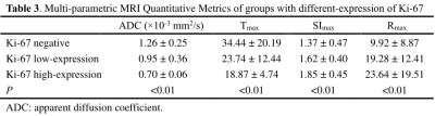 |
74 | Using Multi-parametric MRI Quantitative Metrics as Image Biomarkers to Predict the Tumor Malignant degree of Prostate Cancer
Shili Liu1, Zhiqiang Chen2, Shaoru Zhang3, Xiaohua Chen3, Zhuo Wang3, Yuhui Xiong4, and Aijun Wang5
1Clinical medicine school of Ningxia Medical University, YinChuan, China, 2Department of Radiology ,the First Hospital Affiliated to Hainan Medical College, Haikou, China, 3Clinical medicine school of Ningxia Medical University, Yinchuan, China, 4GE Healthcare,MR Reseaich, Beijing, Beijing, China, 5Department of Radiology, General Hospital of Ningxia Medical University,, Yinchuan, China Keywords: Prostate, Data Processing This study aims to investigate whether the quantitative metrics derived from diffusion weighted imaging (DWI) and dynamic contrast-enhanced (DCE) MRI can be used to predict the tumor malignancy of prostate cancer. The results showed that the apparent diffusion coefficient (ADC) value, Tmax (s), SImax%, Rmax% were correlated with Ki-67 to a certain extent, DWI and DCE-MRI parameters are expected to be a non-invasive examination method to measure the ability of prostatic malignant tumor cells to expand, and provide an important theoretical basis for evaluating the efficacy of prostate cancer and monitoring the prognosis. |
|
2070. |
75 | Distinguishing prostate cancer from prostate hyperplasia nodules using histograms based on amide proton transfer MRI and DWI
You Yun1, Zhiwei Shen2, and Xuejun Chen1
1The Affiliated Cancer Hospital of Zhengzhou University & Henan Cancer Hospital, Zhengzhou, China, 2Philips Healthcare, Beijing, China Keywords: Prostate, CEST & MT The diagnostic value of histograms based on 3D APT MRI and ADC in the central glandular region for the differentiation of prostate cancer and prostate hyperplastic nodules was evaluated in this study. The results showed that the 3D APT MRI and ADC histograms had better diagnostic efficacy in the central glandular region for the differentiation of prostate cancer and prostate hyperplastic nodules, but the diagnostic efficacy of the 3D APT. |
|
2071.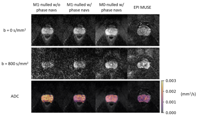 |
76 | 3D Distortionless Diffusion Weighted Imaging in the Prostate using a Diffusion Prepared Fast Spin-Echo Sequence
Jeremiah Joseph Hess1, Philip Kenneth Lee2, Xuetong Zhou1, Andreas Markus Loening2, and Brian Andrew Hargreaves1,2,3
1Bioengineering, Stanford University, Stanford, CA, United States, 2Radiology, Stanford University, Stanford, CA, United States, 3Electrical Engineering, Stanford University, Stanford, CA, United States Keywords: Prostate, Diffusion/other diffusion imaging techniques Diffusion-Weighted Imaging (DWI) of the prostate is commonly used for tumor detection and characterization. However, the echo planar imaging (EPI) based DWI methods commonly used in prostate imaging fail in the setting of field inhomogeneities most related to hip prosthesis and rectal gas. Cartesian Fast Spin-Echo (FSE) is an alternative to EPI that is more robust to off-resonance. In this study, we compare in vivo 3D FSE to multi-shot EPI (MUSE) for DWI prostate imaging, demonstrating that an FSE DWI sequence achieves high image quality while avoiding geometric distortion seen in EPI based DWI. |
|
2072.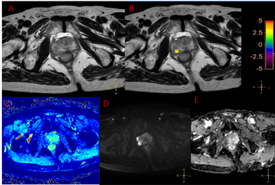 |
77 | Comparison of 3D amide proton transfer imaging and intravoxel incoherent motion imaging in the diagnosis of prostate cancer
Guorui Hou1 and Kai Ai2
1Department of Radiology, Xijing Hospital, Xi'an, China, 2Philips Healthcare, Xi‘an, China Keywords: Prostate, Tumor, amide proton transfer imaging Prostate cancer (PCA), which seriously hazard the life and health, is one of the most common malignant tumors in middle-aged and elderly men, and the incidence population tends to be younger. Amide proton transfer imaging (APT) and intravoxel incoherent motion imaging (IVIM) are two technologies that can be used for prostate cancer diagnostic. This study aims to compare and analyze the diagnostic performance of APT and IVIM in prostate diseases. Besides, we also test the relationship between APT and IVIM derived parameters and Gleason score (GS). |
|
2073.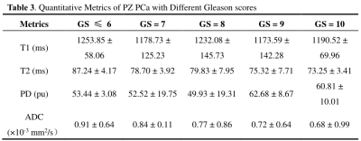 |
78 | Application of Synthetic MRI in the Diagnosis and Invasion Assessment of Peripheral Zone Prostate Cancer
Yunshu Zhou1, Zhiqiang Chen2, Na Song1, Zhuo Wang1, Shaoru Zhang1, Xiaohua Chen1, Xiaocheng Wei3, and Aijun Wang4
1Clinical medicine school of Ningxia Medical University, Yinchuan, China, 2Department of Radiology, the First Hospital Affiliated to Hainan Medical College, Haikou, China, 3GE Healthcare, Beijing, China, 4Department of Radiology, General Hospital of Ningxia Medical University, Yinchuan, China Keywords: Prostate, Data Processing This retrospective study aims to investigate the application value of the quantitative relaxation maps derived from synthetic MRI in the diagnosis and invasion assessment of peripheral zone (PZ) prostate cancer (PCa). 88 patients (45 experimental & 43 control) were scanned using the magnetic resonance image compilation (MAGiC) sequence and the quantitative relaxation metrics were calculated and compared. The results demonstrate that quantitative T1 and T2 values are significant indicators for distinguishing PZ PCa from non-cancerous PZ. T2 value has a similar diagnostic performance to apparent diffusion coefficient value and has potential clinical value in evaluating the invasiveness of PCa. |
|
2074.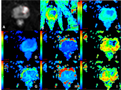 |
79 | Intravoxel incoherent motion (IVIM) imaging: an alternative method to differentiate the prostate diseases
Chen Lihua1, Song Qingwei1, Zhigang Wu2, Jiazhegn Wang3, and Ailian Liu1
1The First Affiliated Hospital of Dalian Medical University, Dalian, China, 2Clinical and Technical Support, Philips Healthcare, Shenzhen, China, 3Clinical and Technical Support, Philips Healthcare, Beijing, China Keywords: Prostate, Prostate Differentiation of prostate hyperplasia, prostatitis and prostate cancerremains challenge using conventional MR techniques, especially for prostatitis and prostate cancer. This study evaluates the value of Intravoxel incoherent motion (IVIM) imaging to differentiate these diseases. Results of this study indicate that ADC value has no significant difference, but Fraction of Fast ADC-Mono and Fraction of Fast ADC-Bi shows significant difference between prostate hyperplasia vs prostate cancer. Slow ADC-Bi values shows difference between prostatitis vs prostate cancer. In summary, IVIM combined with different fitting scheme is potentially a promising and valuable method for differentiation of prostate hyperplasia, prostatitis and prostate cancer. |
|
2075.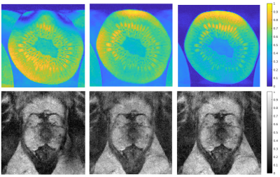 |
80 | Shortening T2-weighted TSE for prostate MRI by reducing the number of echo-trains and using CS reconstruction
Nida Mir1, Saskia Damen1, Jelmer M. Wolterink1, Frank F.J. Simonis1, and Jurgen J. Fütterer1,2
1TechMed Centre, University of Twente, Enschede, Netherlands, 2Radboud UMC, Nijmegen, Netherlands Keywords: Prostate, Cancer The aim of this study was to speed up the turbo spin echo (TSE) sequence for prostate MRI by undersampling the k-space, while aiming to retain PSNR/SSIM. We conducted undersampling retrospectively by omitting echo-trains in the TSE sequence based on the proximity of the echoes (within the echo-trains) to the centre of the K-space. The proposed method speeds up the TSE acquisition time by 20%, while retaining the PSNR/SSIM for a kiwi phantom. Optimizing an undersampling pattern for prostate therefore can lead to a substantial decrease in the total scan time. |
|
 Back to Meeting Home
 Back to the Program-at-a-Glance
The International Society for Magnetic Resonance in Medicine is accredited by the Accreditation Council for Continuing Medical Education to provide continuing medical education for physicians.
 Back to Meeting Home
 Back to the Program-at-a-Glance
 View Presentation Video