Digital Poster
Updates in Body Imaging I
ISMRM & ISMRT Annual Meeting & Exhibition • 03-08 June 2023 • Toronto, ON, Canada

| Computer # | |||
|---|---|---|---|
3974.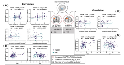 |
41 |
Abnormal functional connectivity of the hippocampus and amygdala
in plateau Tibetan with type 2 diabetes mellitus
Jinli Meng1,2,3,
Yingxue Gao2,
Hailong Li2,
Lingxiao Cao2,
Wanlin He1,
Li Feng1,
Yongyue Guo1,
Xin Hu1,
Hengyan Li1,
Chenghui Zhang4,
Yunhong Wu4,
and Xiaoqi Huang2,3
1Department of Radiology, Hospital of Chengdu Office of People’s Government of Tibetan Autonomous Region (Hospital. C.T.), Chengdu, Sichuan, China, 2Huaxi MR Research Center (HMRRC), Functional and Molecular Imaging Key Laboratory of Sichuan Province, Department of Radiology, West China Hospital, Sichuan University, Chengdu, Sichuan, China, 3Psychoradiology Research Unit of the Chinese Academy of Medical Sciences, West China Hospital of Sichuan University, Chengdu, Sichuan, China, 4Department of Endocrinology and Metabolism, Hospital of Chengdu Office of People’s Government of Tibetan Autonomous Region (Hospital. C.T.), Chengdu, Sichuan, China Keywords: Endocrine, Diabetes We performed a seed-based rsFC study to explore the hippocampus and the amygdala FC patterns in plateau Tibetan patients with T2DM. We found that, compared to plateau Tibetan HC, T2DM showed both increased rsFC between the right hippocampus and left DLPFC and left SFG, and between the left amygdala and right OFC as well as right DLPFC.Moreover, these rsFC abnormalities were significantly correlated with age, RBC and changes of visuospatial memory function in plateau Tibetan patients with T2DM. These findings of altered FC may provide important insights into the neural basis of diabetes-related cognitive decline in plateau Tibetan. |
|
3975. |
42 |
Unpaired Super-Resolution for Anisotropic MRI using a Gradient
Mapping Adversarial Loss
Michele Pascale1,
Vivek Muthurangu1,
Javier Montalt Tordera1,
and Jennifer Steeden1
1University College London, London, United Kingdom Keywords: Digestive, Body We present a novel approach to synthesise high-resolution isotropic 3D abdominal MR images, from anisotropic 3D images in an unpaired fashion. Using a modified CycleGAN architecture with a novel gradient mapping loss, we leverage disjoint patches from the high-resolution (in-plane) data of an anisotropic volume to enforce the network generator to increase the resolution of the low-resolution (through-plane) slices. This will enable accelerated whole-abdomen scanning with high-resolution isotropic images within short breath-hold times. |
|
3976.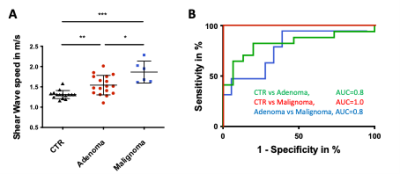 |
43 |
MR Elastography Distinguishes Between Adrenal Adenomas and
Malignomas by Increased Stiffness
Alexandra Webster1,
Agata Dukaczewska2,
Martina Mogl2,
Tom Meyer1,
Bernd Hamm1,
Ingolf Sack1,
and Stephan Marticorena Garcia1
1Department of Radiology, Charité - Universitaetsmedizin Berlin, Germany, Berlin, Germany, 2Department of Surgery, Charité - Universitaetsmedizin Berlin, Germany, Berlin, Germany Keywords: Endocrine, Elastography, Adrenal Gland, Multifrequency magnetic resonance elastography, Adrenal stiffness, Shear wave speed, Viscoelasticity The ability to distinguish a benign from a malignant adrenal mass based on magnetic resonance imaging relaxation times is severely limited. For the first time at all, this study adrenal gland stiffness was investigated by using magnetic resonance elastography in healthy controls and participants with adrenal adenoma and malignoma. In healthy controls MRE was highly reproducible. Adrenal adenoma and malignoma are stiffer than healthy controls and both could be distinguished by an excellent diagnostic performance (AUC 0.8–1.0). MRE is promising non-invasive biomarker for clinically difficult differentiation of a malignancy and could prevent surgical overtreatment in the future. |
|
3977.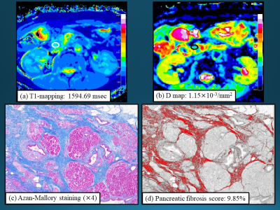 |
44 |
Usefulness of quantitative MRI in the evaluation of pancreatic
fibrosis
Hidemitsu Sotozono1,
Akihiko Kanki1,
Yoshihiko Fukukura1,
Kiyoka Maeba1,
Akira Yamamoto1,
and Tsutomu Tamada1
1Department of Radiology, Kawasaki Medical School, Kurashiki, Japan Keywords: Pancreas, Pancreas We sought to clarify the value of quantitative MRI for evaluating pancreatic fibrosis. Longer T1 relaxation time and lower D within the pancreas parenchyma were significantly related to pancreatic fibrosis score based on histopathological evaluation. T1 relaxation time and D may be sensitive indicators of fibrosis within the pancreas parenchyma. |
|
3978.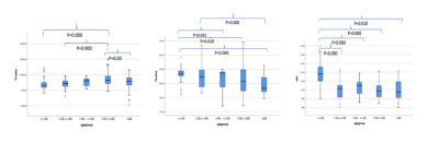 |
45 |
Multiparametric quantitative MRI of Healthy Adult Pancreas:
Correlations with Gender and Age
Lixia Wang1,
Lu Liang2,
Jiyang Zhang2,
Yang Zhou2,
Yang Yu2,
Chen Zhang3,
Tao Jiang2,
Qi Yang2,
Christie Jeon4,
Srinivas Gaddam5,
Stephen J Pandol5,
Yibin Xie1,
and Debiao Li1
1Biomedical Imaging Research Institute, Cedars-Sinai Medical Center, Los Angeles, CA, United States, 2Radiologic department, Beijing Chaoyang Hospital, Captial Medical University, Beijing, China, 3MR Scientific Marketing, Siemens Healthcare, Beijing, China, 4Department of Medicine, Cedars-Sinai Medical Center, Los Angeles, CA, United States, 5Division of Digestive and Liver Diseases, Cedars-Sinai Medical Center, Los Angeles, CA, United States Keywords: Pancreas, Quantitative Imaging, T1 value, T2 value, ADC value The pancreas plays an important role in nutrition and metabolism of the whole body. Changes of pancreatic morphology and function are related to many diseases. Multiparametric MR provides quantitative information about pancreatic morphology and function. In this work, we measured the normal range of T1, T2, and ADC values for different gender and age groups, and found significant differences between age groups. Gender difference was only found in T2. There are negative correlations between T2/ADC and age, and positive correlation between T1 and age. |
|
3979.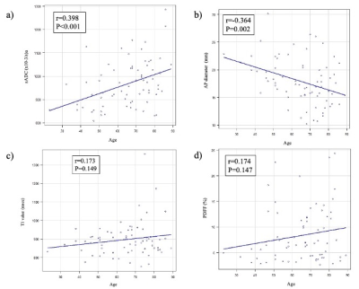 |
46 |
Age-related changes in normal adult pancreatic parenchyma:
multiparametric MR imaging evaluation
Akihiko Kanki1,
Yoshihiko Fukukura1,
Hidemitsu Sotozono1,
Kiyoka Maeba1,
Atsushi Higaki1,
Akira Yamamoto1,
and Tsutomu Tamada1
1radiology, Kawasaki Medical School, kurashiki, Japan Keywords: Pancreas, Pancreas We evaluated relationships of age and gender with shifted apparent diffusion coefficient (sADC), proton density fat fraction (PDFF), T1 relaxation time, and pancreas size using 3-T MRI. sADC increased and anterior-posterior diameter decreased with age, while T1 relaxation time and PDFF showed no correlations with age. These results would offer beneficial information regarding pancreatic imaging. |
|
3980.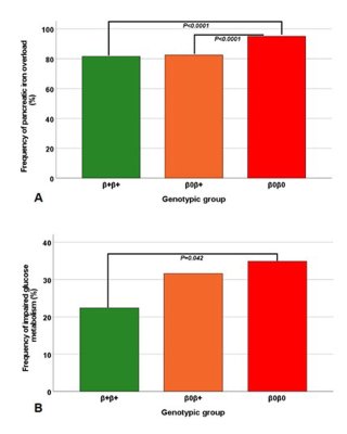 |
47 |
Impact of genotype on pancreatic iron overload and impaired
glucose metabolism in thalassemia major.
Antonella Meloni1,
Laura Pistoia1,
Vincenzo Positano1,
Nicolò Schicchi2,
Gennaro Restaino3,
Luigi Barbuto4,
Ada Riva5,
Giuseppe Peritore6,
Letizia Tedesco7,
Angela Ermini8,
Domenico Visceglie9,
Costanza Bosi10,
Paola Maria Grazia Sanna11,
and Filippo Cademartiri1
1Fondazione G. Monasterio CNR-Regione Toscana, Pisa, Italy, 2Azienda Ospedaliero-Universitaria Ospedali Riuniti "Umberto I-Lancisi-Salesi", Ancona, Italy, 3Gemelli Molise SpA, Fondazione di Ricerca e Cura "Giovanni Paolo II", Campobasso, Italy, 4Azienda Ospedaliera di Rilievo Nazionale Antonio Cardarelli, Napoli, Italy, 5Ospedale “SS. Annunziata” ASL Taranto, Taranto, Italy, 6"ARNAS" Civico, Di Cristina Benfratelli, Palermo, Italy, 7Presidio Ospedaliero Locri - A.S.P di Reggio Calabria, Locri (RC), Italy, 8Ospedale S. Maria Annunziata, Bagno a Ripoli (FI), Italy, 9Ospedale “Di Venere”, Bari, Italy, 10Ospedale “G. Da Saliceto”, Piacenza, Italy, 11Azienda Ospedaliero-Universitaria di Sassari, Sassari, Italy Keywords: Pancreas, Tissue Characterization On the basis of the type of gene mutation, three groups of patients with thalassemia major were identified: homozygotes β+, compound heterozygotes β+/β° and homozygotes β°. β0β0 patients were more likely to have pancreatic iron overload than both β+β+ patients and β0β+patients and had a double risk of alterations of glucose metabolism compared to β+β+ patients. Our findings support the knowledge of the different genotypic groups in the clinical management of β-TM patients. |
|
3981.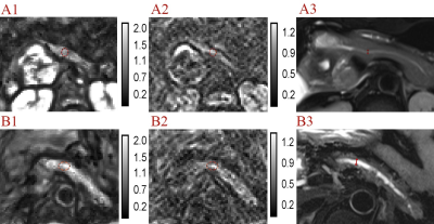 |
48 |
Role of tomoelastography in evaluation for pancreatic fistula
after pancreaticoenteric anastomosis
Siya Shi1,
Liqin Wang1,
Jiaxin Yuan1,
Xuefang Hu1,
Xingyan Xie1,
Tingting Wen1,
Jinhui Yu1,
and Yanji Luo1
1The First Affiliated Hospital, Sun Yat-Sen University, Guangzhou, China Keywords: Pancreas, Surgery, Magnetic resonance elastographies, Pancreatectomy, Pancreatic fistula This prospective study enrolled the patients who underwent both preoperative tomoelastography and pancreaticoenteric anastomosis. Eighty-two patients were included (median age: 59.5 years, 40 men, 18 patients with POPF). Main pancreatic duct diameter (MPDD) (P=0.012), c (P<0.001) and φ (P=0.001) were relevant factors for POPF. The area under the curve (AUC) of c, φ and MPDD for predicting POPF was 0.880, 0.816 and 0.747. Fibrosis was the relevant factor of POPF (P=0.001). There was positive correlation between fibrosis and stiffness (r=0.681, P<0.001). Tomoelastography is a novel and robust multi-frequency MRE technique that can facilitate the prediction of POPF. |
|
3982.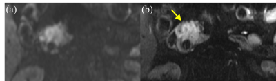 |
49 |
Modified reduced FOV diffusion-weighted MRI using tilted 2D RF
excitation pulses in patients with pancreatic ductal
adenocarcinoma
Masahiro Tanabe1,
Mayumi Higashi2,
Keiko Hideura2,
Kenichiro Ihara2,
Thomas Benkert3,
Hiroshi Imai4,
Masatoshi Yamane5,
Takahiro Yamaguchi6,
Atsuo Inoue2,
and Katsuyoshi Ito2
1Department of Radiology, Yamaguchi University Graduate School of Medicine, Ube, Japan, 2Radiology, Yamaguchi University Graduate School of Medicine, Ube, Japan, 3MR Application Predevelopment, Siemens Healthcare GmbH, Erlangen, Germany, 4MR Research and Collaboration, Siemens Healthcare K.K., Tokyo, Japan, 5Department of Radiological Technology, Yamaguchi University Hospital, Ube, Japan, 6Yamaguchi University Hospital, Ube, Japan Keywords: Pancreas, Diffusion/other diffusion imaging techniques This study evaluated the image quality of modified reduced field-of-view (FOV) DWI using 2D spatially-selective RF pulses with tilted excitation plane (tilted r-DWI) for pancreatic ductal adenocarcinomas (PDACs) compared with conventional full-FOV DWI (f-DWI). Two radiologists evaluated the MR image quality, and SNR, CNR, and ADC values of the pancreatic lesions were quantitatively assessed. Image quality scores and CNR of tilted r-DWI were significantly higher than that of f-DWI. The ADC values of PDACs showed no significant deference between them. Tilted r-DWI provides better image quality with less artifacts and higher pancreatic lesion conspicuity than f-DWI. |
|
3983.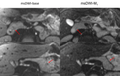 |
50 |
Convex Optimized Diffusion Encoding (CODE) with M1-Nulling for
Pancreatic Diffusion Weighted Imaging
Kang Wang1,
Matthew J. Middione1,
Andreas Markus Loening1,
Daniel Ennis1,
and Ryan Lennex Brunsing1
1Radiology, Stanford University, Palo Alto, CA, United States Keywords: Pancreas, Diffusion/other diffusion imaging techniques, Diffusion Weighted Imaging, M1-nulled, ADC Multi-shot DWI reduces image distortion while increasing SNR. More recently, motion-compensated diffusion encoding has been shown to reduce artifacts and improve ADC consistency in single-shot DWI. Motion-compensated multi-shot pancreatic DWI may also mitigate artifacts arising from shot-to-shot incoherence. Herein, we conducted a pilot study of multi-shot DWI of the pancreas with and without motion-compensated diffusion encoding. Image quality and artifacts were scored by expert radiologists. ADC values of different pancreatic anatomical segments were computed. Multi-shot DWI with motion-compensated gradient encoding increased homogeneity of signal intensity and ADC values across the pancreas. |
|
3984. |
51 |
MR Elastography of the affected mesenteric fat in active Crohn’s
Disease: a feasibility study
Anne-Sophie van Schelt1,2,
Kim Johanna Beek1,3,
Nienke Petronella Maria Wassenaar1,
Eric M. Schrauben1,
Jurgen H. Runge1,
Aart J. Nederveen1,
and Jaap Stoker1,2,3
1Radiology and Nuclear Medicine, AmsterdamUMC, location AMC, Amsterdam, Netherlands, 2Imaging and Biomarkers, Cancer Center Amsterdam, Amsterdam, Netherlands, 3Endocrinology, Metabolism, Amsterdam Gastroenterology, Amsterdam, Netherlands Keywords: Digestive, Quantitative Imaging Crohn’s Disease (CD) is a chronic inflammatory bowel disease. Mesentery of afflicted bowel loops often is involved. MR-Elastography allows assessment of its underlying mechanical properties and gives more insight in the role of the mesentery . Feasibility was first shown in 15 healthy volunteers. Seven patients with active CD (aCD) scheduled for surgical intervention underwent pre-operative MR-Elastography acquisition and histopathological analysis. Seven age- and sex matched volunteers were also scanned. Significantly higher shear wave speed, shear stiffness and phase angle were found in aCD patients, possibly related to increased fibrotic tissue in or inflammation of the mesentery. |
|
3985.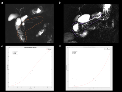 |
52 |
Novel method for quantifying pancreatic exocrine function from
Secretin-MRCP
Alexander R Guimaraes1,
Joyce Kim2,
Kaveh Sharzehi3,
Gregory Cote3,
Lisa Wang1,
Bryan Foster1,
and Cory Wyatt1
1Radiology, OHSU, Portland, OR, United States, 2OHSU, Portland, OR, United States, 3Medicine, OHSU, Portland, OR, United States Keywords: Pancreas, Data Analysis Chronic pancreatitis is a chronic fibro-inflammatory syndrome of the pancreas that often leads to exocrine pancreatic insufficiency EPI. Secretin MRCP provides an approach for quantifying function. Present interpretation of sMRCP lends itself to variability in interpretation . Further, previous approaches for quantitative assessment of sMRCP have been difficult to reproduce. We have developed a mathematical model for quantifying various features of dynamic sMRCP and have shown in a retrospective cohort, significant differences in one feature that correlates to delay of pancreatic production of fluid, to separate and correlate to ePFT and FE. 9 |
|
3986.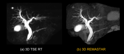 |
53 |
Accelerated 3D free-breathing MRCP using Relaxation-Enhanced
MAgnetization-prepared radial stack-of-STARs (REMASTAR) and
Compressed SENSE
Hiroshi Hamano1,
Masami Yoneyama1,
Masahiro Enzaki2,
Minako Azuma3,
Nobuyuki Toyonari4,
and Takashi Namiki1
1Philips Japan, Tokyo, Japan, 2Division of Radiology, Miyazaki University Hospital, Miyazaki, Japan, 3Department of Radiology, Faculty of Medicine, University of Miyazaki, Miyazaki, Japan, 4Department of Radiology, Kumamoto Chuo Hospital, Kumamoto, Japan Keywords: Pancreas, Pancreas, MRCP 3D TSE RT is commonly used to MRCP imaging. But the image qualities of them are unstable especially in patients with irregular respiratory motion and tachypnea. A T2-prepared pulse is often used to emphasize long T2 component such as CSF, bile, and pancreatic duct. A 3D VANE has been applied in abdominal imaging as a useful free-breathing technique. We attempted to combine bTFE based on 3D VANE with magnetization T2prep pulses (3D REMASTAR) for MRCP instead of, otherwise in addition to 3D TSE RT. We have demonstrated the feasibility of 3D REMASTAR MRCP particularly in subject with unstable respiratory motion. |
|
3987.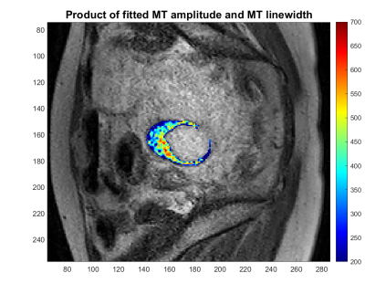 |
54 |
Z-spectrum Imaging of Bowel Wall Fibrosis in Crohn's Disease
Nabeelah Jinnah1,
Olivier Mougin1,
Penny Gowland1,
Caroline Hoad1,
Lauren Gascoyne1,
Christopher Clarke2,
and Gordon Moran1
1University of Nottingham, Nottingham, United Kingdom, 2Nottingham University Hospitals NHS Trust, Nottingham, United Kingdom Keywords: Digestive, CEST & MT Z-spectrum imaging was performed on bowel wall from Crohn's disease patients, to determine it's potential as a method of determining the fibrotic component of scarred bowel wall. Results show that there is a difference between the MT amplitude and linewidth of healthy compared with strictured bowel wall. Future work will involve optimising a protocol to use as a imaging biomarker of the fibrosis within bowel wall in Crohn's disease. |
|
3988.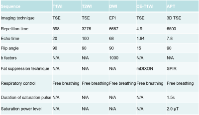 |
55 |
Amide Proton Transfer MRI for Treatment Response of Locally
Advanced Rectal Cancer Following Neoadjuvant Chemotherapy and
Radiation Therapy
Lan Zhang1,
Xin Li1,
Peng Sun2,
and Fan Yang1
1Department of Radiology, Union Hospital, Tongji Medical College, Huazhong University of Science and Techonology, Wuhan, China, 2Philips Healthcare, Beijing, China, Beijing, China Keywords: Digestive, Cancer We conducted this prospective study to determine whether APTw imaging is precise in evaluating treatment response of LARC after NCRT, by comparing the accuracy of evaluation using APTw imaging to that of T2-weighted imaging and DWI, which is value for the selection of organ preservation candidates. |
|
3989.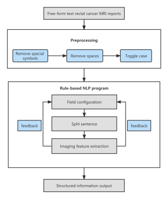 |
56 |
Application of natural language processing to post-structuring
of rectal cancer MRI reports
Wenjuan Liu1
1Beijing Aviation General Hospital, Beijing, China Keywords: Pelvis, Cancer We applied natural language processing (NLP) to the extraction of relevant information from MRI reports of rectal cancer written in Chinese. We used 358 MRI reports written between 2015 and 2021 to develop a rule-based NLP model to extract 11 key image features. The accuracy, precision, recall, and F1 score of our NLP model for correct extraction of values from reports were 93.82%, 95.63%, 87.06%, and 91.15% for pre-2015 reports, and 92.55%, 98.53%, 94.15%, and 96.29% for post-2021 reports. Our NLP system with rule-based pattern matching realized the rapid and accurate structured processing of rectal cancer MRI reports. |
|
3990.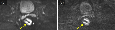 |
57 |
Diffusion-weighted MRI of rectal cancers: Utility of reduced FOV
single-shot EPI with tilted 2D RF excitation pulses
Atsuo Inoue1,
Masahiro Tanabe1,
Kenichiro Ihara1,
Keiko Hideura1,
Thomas Benkert2,
Hiroshi Imai3,
Masatoshi Yamane4,
Takahiro Yamaguchi4,
Mayumi Higashi1,
and Katsuyoshi Ito1
1Department of Radiology, Yamaguchi University Graduate School of Medicine, Yamaguchi, Japan, 2MR Application Predevelopment, Siemens Healthcare GmbH, Erlangen, Germany, 3MR Research and Collaboration, Siemens Healthcare K.K., Tokyo, Japan, 4Department of Radiological Technology, Yamaguchi University Hospital, Yamaguchi, Japan Keywords: Pelvis, Diffusion/other diffusion imaging techniques, Rectum This study evaluated the image quality of reduced FOV DWI using 2D spatially-selective RF pulses with tilted excitation plane (tilted r-DWI) for rectal cancer compared with full-FOV DWI (f-DWI) using readout segmented echo-planar imaging (RS-EPI). Two radiologists evaluated the MR images, and SNR, CNR, and ADC values of the rectal lesions were quantitatively evaluated. All image quality scores and CNR were significantly higher in tilted r-DWI than in f-DWI with RS-EPI. SNR and ADC values showed no significant deference between them. Tilted r-DWI provides better image quality with less artifacts and higher rectal lesion conspicuity than f-DWI with RS-EPI. |
|
3991.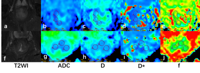 |
58 |
Prediction for P53 Status Using Intravoxel Incoherent Motion
Imaging in Rectal Cancer
Deshuo Dong1,
Anliang Chen1,
Changjun Ma1,
Ailian Liu1,
and Qingwei Song1
1radiology, The First Affiliated Hospital of Dalian Medical University, Dalian, China Keywords: Pelvis, fMRI Mutant P53 promotes tumor cell proliferation, invasion, and resistance to chemotherapy. The purpose of this study was to evaluate the value of intravoxel incoherent motion (IVIM) imaging for prediction of P53 expression status in rectal cancer. The results indicate that D and f values for positive P53 status were significantly lower than those for negative P53 status, while D* was higher. The combination of D, D* and f improved diagnostic efficiency than single parameters. IVIM imaging allows non-invasive visualization and quantification of tissue composition for prediction of P53 status in rectal cancer. |
|
3992.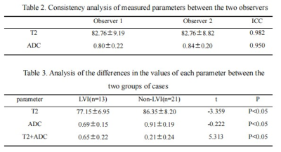 |
59 |
The value of DWI and T2 mapping in evaluating lymphatic vascular
invasion of rectal cancer
Xiwei Li1,
Anliang Chen1,
Nan Wang1,
Jiazheng Wang2,
Liangjie Lin2,
and Ailian Liu1
1The First Affiliated Hospital of Dalian Medical University, Dalian, China, 2Philips Healthcare,Beijing, Beijing, China Keywords: Digestive, Cancer Lymphatic vascular invasion(LVI)can help clinicians choose the best treatment strategy for patients. Pathological examination is the gold standard for the diagnosis of LVI in rectal cancer, but it is an invasive method. Multiparametric magnetic resonance imaging is valuable for the prediction of LVI in rectal cancer. In this study, we quantitatively evaluated the value of LVI in rectal cancer using T2 mapping and DWI sequences. |
|
3993.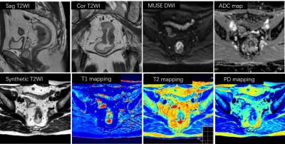 |
60 |
Synthetic magnetic resonance quantitative relaxation maps for
preoperative staging of rectal cancer
Wenguang Liu1,
Yigang Pei1,
Weiyin Vivian Liu2,
Xiao Wang1,
Yu Bai1,
Yijing Luo1,
Gaofeng Zhou1,
and Wenzheng Li1
1Radiology, XiangYa Hospital, Central South University, Changsha, China, 2MR research, GE Healthcare, Beijing, China Keywords: Cancer, Quantitative Imaging To preoperative assessment of tumor and node staging for rectal cancer patients is very important. This study aimed to investigate the feasibility of predicting pathological TN staging with synthetic MRI quantitative relaxation maps for rectal cancer patients. A total of 123 patients were identified in this study. Our results showed that the T1 and T2 values from synthetic MRI can distinguish effectively between pT1-2 and pT3-4 group, pN- and pN+ group, which is vital to selection of therapy strategy for rectal cancer patients. |
|
 Back to Meeting Home
 Back to the Program-at-a-Glance
The International Society for Magnetic Resonance in Medicine is accredited by the Accreditation Council for Continuing Medical Education to provide continuing medical education for physicians.
 Back to Meeting Home
 Back to the Program-at-a-Glance
 View Presentation Video