Digital Poster
Cutting-Edge Breast Imaging
ISMRM & ISMRT Annual Meeting & Exhibition • 03-08 June 2023 • Toronto, ON, Canada

| Computer # | |||
|---|---|---|---|
2725.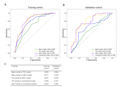 |
21 |
Contrasts between DWI and DCE curve in diagnosing malignancies
of breast non-mass enhancement lesions based on morphology
Yan Li1
1Tongji Hospital, Wuhan, China Keywords: Breast, Cancer The diagnosis of NMEs is a challenge for most radiologists based only on morphological information. The value of DWI or DCE curves for discriminating malignant lesions from benign lesions was controversial. We estabished and compared the ADC model (ADC+morphology) and the TIC (TIC+morphology) for detecting malignancy with a relatively large sample size of NME lesions. We found that the TIC model was superior than the ADC model for differentiating between benign and malignant NME lesions. A whole DCE-MRI scan for NMEs is recommended without the need to acquire additional DWI data. |
|
2726.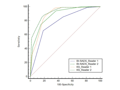 |
22 |
A comparative study of Kaiser score and BI-RADS in the diagnosis
of non-mass enhancement lesions on breast MRI
Jiejie Zhou1,
Haiwei Miao1,
Huiru Liu1,
Zhongwei Chen1,
Youfan Zhao1,
Meihao Wang1,
and Min-Ying Su2
1First affiliated hospital of Wenzhou Medical University, Wenzhou, China, 2University of California, Irvine, California, CA, United States Keywords: Breast, Breast A total of 102 malignant and 60 benign non-mass enhancement lesions were analyzed. Two radiologists with 6 (Reader1) and 14 (Reader2) years of experience assigned the BI-RADS category and Kaiser score (KS). KS decreased biopsy or surgery by 20% for Reader 1 and 16.7% for Reader 2 compared with BI-RADS 4-5. The sensitivity, specificity and accuracy were 87.2%, 78.3% and 84.0% for Reader 1, and 85.2%, 83.3% and 84.6% for Reader 2. Using cutoff threshold of KS>4, the specificity of modified KS increased by 6.7% for both readers, increasing the accuracy slightly by 1.7%-2.5% and keeping the same sensitivity. |
|
2727. |
23 |
Value of Native T1 Mapping for Differentiating Benign and
Malignant Breast Lesions on MRI: A Pilot Study
Chun Lian1,
Lulu Zhuang1,
Zehao Wang 1,
Zhigang Wu2,
Yi Dai1,
and Rong Huang1
1Radiology, Peking University Shenzhen Hospital, Shen Zhen, China, 2Clinical & Technical Support, Philips Healthcare (Shenzhen) Ltd, Shen Zhen, China Keywords: Breast, Breast Breast cancer is the most prevalent cancer among women ,early detection and timely treatment are the key to imprve survival rate. In this preliminary study, we aimed to differentiation lesions using native T1mapping. The mean T1 relaxation time was higher in the malignant group compared to the benign group by both observers (p<0.05). Results indicated that native T1mapping could be used to differentiate benign and malignant lesions, which might be promising in clinical applications. However, the combination of T1mapping and DWI dose not improve the AUC statistically significantly.
|
|
2728.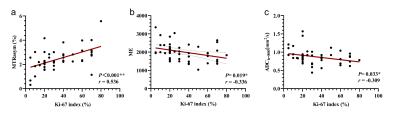 |
24 |
Diagnostic value of APT, DWI, and DCE-MRI in molecular
classification and prognosis of breast cancer
Xiaoyan Liu1,2,
Baojian Wang2,
Litao Zhang1,
Zhenbo Ma1,
Yanlei Wang2,
Yishi Wang3,
Xiujuan Li1,
and Yuanzhong Xie1
1Taian City Central Hospital, Taian, China, 2Shandong First Medical University, Taian, China, 3Philips Healthcare, Beijing, China Keywords: Breast, CEST & MT, Tumor;Amide proton transfer imaging; According to the molecular classification of breast cancer, the clinical therapeutic strategy is formulated. Currently, the diagnostic value of amide proton transfer (APT) imaging in breast cancer is still unclear. This study aims to explore the diagnostic value of APT, DWI, and DCE-MRI in the molecular classification and prognosis of breast cancer. Our results showed that there were differences in TTP and BE values between luminal and non-luminal subtypes of breast cancer. The combination of APT, DWI, and DCE-MRI had the highest diagnostic efficiency. APT, DWI, and DCE-MRI parameters were correlated with different prognostic factors of breast cancer. |
|
2729.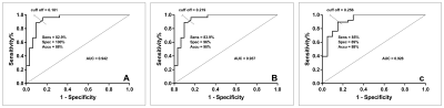 |
25 |
Diagnostic performance of DCE-MRI for the axillary sentinel
lymph nodes status in breast cancer patients
Fang Xiao1,
Mingli Jin2,
Peng wang1,
Zhihua Pan2,
Qi Zhao2,
Ying Jiang2,
Miaoqi Zhang3,
and Yong Zhang3
1Department of Radiology,The First Affiliated Hospital of USTC, Division of Life Sciences and Medicine, University of Science and Technology of China, Hefei, China, 2The Second Affiliated Hospital of Chengdu Medical College, Nuclear Corportation 416 Hosptital, chengdu, China, 3GE Healthcare, MR Research, Beijin, China Keywords: Breast, Cancer, contrast-enhanced (DCE) MRI, sentinel lymph nodes Sentinel lymph nodes(SLN) status is one of the most important indicators affecting breast cancer patient treatment and prognostic. Thus, a non-invasive diagnostic method capable of accurately predicting of the SLN status is of great value in selecting breast cancer patients who would benefit from therapy. In this study, we investigate the diagnostic performance of different SLN status [pN0, pN0 (i+), pN1mi, and pN1] using dynamic contrast-enhanced (DCE) MRI.We found that (1) Krans, Kep, and Ve are significantly different in different SLN status, (2) and the Ktrans is the best single parameter for the detection of SLN status. |
|
2730.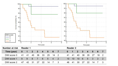 |
26 |
Comparison of DWI score and Kinetic score as a Prognostic Marker
in Breast Cancer Patients receiving Neoadjuvant Systemic
Treatment.
Rie Ota1,
Masako Kataoka2,
Mami Iima2,
Maya Honda3,
Aika Okazawa2,
Yosuke Yamada4,
Yasuhide Takeuchi4,
Masakazu Toi5,
Takeshi Kubo1,
and Yuji Nakamoto2
1Department of Radiology, Tenri Hospital, Nara, Japan, 2Department of Diagnostic Imaging and Nuclear Medicine, Kyoto University graduate school of medicine, Kyoto, Japan, 3Department of Diagnostic Radiology, Kansai Electric Power Hospital, Osaka, Japan, 4Department of Diagnostic Pathology, Kyoto University Hospital, Kyoto, Japan, 5Department of Breast Surgery, Kyoto University Hospital, Kyoto, Japan Keywords: Breast, Cancer, disease-free survival This study aimed to investigate the association of DWI score of breast cancer derived from MRI obtained after neoadjuvant systemic treatment (NST) with disease-free survival (DFS). Kinetic score from standard DCE-MRI at the same timing was analyzed for comparison. Kaplan-Meier analysis showed that patients with DWI score of 2 showed significantly shorter DFS than those with DWI score 0 or 1. Patients with kinetic score of 2 or 3 showed significantly shorter DFS than those with kinetic score of 0 or 1. DWI score demonstrated excellent inter-reader agreement and is suitable for predicting prognosis of breast cancer patients after NST. |
|
2731.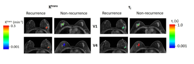 |
27 |
Added Value of Quantitative DCE-MRI in Prediction of Breast
Cancer Recurrence Following Neoadjuvant Chemotherapy?
Rajat Thawani1,
Lina Gao2,
Ajay Mohinani3,
Alina Tudorica4,
Xin Li5,
Zahi Mitri1,
and Wei Huang5
1Hematology and Oncology, Oregon Health and Science University, Portland, OR, United States, 2Biostatistics Shared Resource, Oregon Health and Science University, Portland, OR, United States, 3Medicine, Oregon Health and Science University, Portland, OR, United States, 4Radiology, Oregon Health and Science University, Portland, OR, United States, 5Advanced Imaging Research Center, Oregon Health and Science University, Portland, OR, United States Keywords: Breast, Cancer Breast cancer patients treated with neoadjuvant chemotherapy (NACT) are at risk of recurrence depending on clinicopathological characteristics. This preliminary study aimed to investigate the predictive performances of quantitative dynamic contrast-enhanced (DCE) MRI parameters, alone and in combination with clinicopathological variables, for prediction of recurrence in patients treated with NACT. Pre- and post-NACT DCE-MRI parameters performed better than tumor size measurement in prediction of recurrence, whether alone or in combination with clinicopathological variables. Combining post-NACT Ktrans with residual cancer burden and age showed the best improvement in predictive performance with ROC AUC = 0.965. |
|
2732.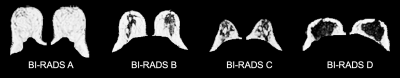 |
28 |
Quantitative calculation of breast density with proton density
fat fraction enables distinction between latest subjective
BI-RADS categories
Isobel Gordon1,2,
George Ralli2,
Amy Herlihy2,
Carolina Fernandes2,
Amy O'Brien3,
Sally Collins1,2,
and Michael Brady2,4
1Nuffield Department of Women's and Reproductive Health, University of Oxford, Oxford, United Kingdom, 2Perspectum Ltd., Oxford, United Kingdom, 3Stoke Mandeville Hospital, Buckinghamshire Healthcare NHS Trust, Aylesbury, United Kingdom, 4Department of Oncology, University of Oxford, Oxford, United Kingdom Keywords: Breast, Fat, Breast density Breast density is a well-established risk factor for breast cancer but conventional visual assessment using mammography has high operator variability. Proton density fat fraction (PDFF) derived from chemical shift encoded (CSE)-MRI has recently been proposed as a quantitative, non-ionising, operator-independent tool for the calculation of breast density. This study demonstrates that breast density derived from PDFF maps which account for a breast-specific fat spectrum can distinguish between the latest density categorisations used in clinical practise and thereby may have use in cancer risk assessment and as a determinant of clinical pathways. |
|
2733.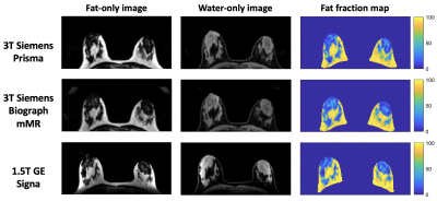 |
29 |
MR-derived breast density (MagDensity) enables reliable density
measures across field strengths and vendors
Jia Ying1,
Renee Cattell1,2,
and Chuan Huang1,3
1Biomedical Engineering, Stony Brook University, Stony Brook, NY, United States, 2Radiation Oncology, Stony Brook Medicine, Stony Brook, NY, United States, 3Radiology, Stony Brook Medicine, Stony Brook, NY, United States Keywords: Breast, Breast The current standard of care for breast density assessment using mammography according to the Breast Imaging Reporting and Data System (BI-RADS) is subjective and can also suffer from intra- and inter-reader variability. Quantitative measurement of breast density is crucial for personalized risk assessment and longitudinal monitoring. Our team has previously developed a quantitative, MR-derived BD (MagDensity) measurement based on fat-water decomposition, which has shown high accuracy and test-retest reproducibility within a single scanner. In order for this measure to be practical for clinical adoption, this study aims to determine the reliability of MagDensity across scanners, field strengths, and vendors. |
|
2734.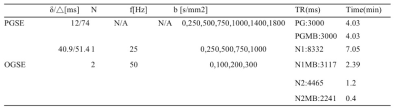 |
30 |
The application of multiband acceleration technique in temporal
diffusion spectroscopy for breast tumor imaging
Jie Ding1,2,3,
Zhen Zhang3,
Yajia Gu1,2,
Yishi Wang4,
Xiuzheng Yue4,
Dazhi Chen3,
Rongrong Zhu3,
and Ruoshui Ha3
1Department of Radiology, Fudan University Shanghai Cancer Center, Shanghai, China, 2Department of Oncology, Shanghai Medical College, Fudan University, Shanghai, China, 3People's Hospital of Ningxia Hui Autonomous Region, Yinchuan, China, 4Philips Healthcare, Beijing, China Keywords: Breast, Diffusion/other diffusion imaging techniques Both the IMPULSED and MRI-cytometry approaches are based on temporal diffusion spectroscopy (TDS), which connotes the acquisitionof dMRI data with a range of diffusion times expressed in terms of components of a diffusion spectrum.MR cell size imaging is possible with IMPULSED and MRI-cytometry using the theory of TDS. In this study, we proposed to use multiband to accelerate the data acquisition for TDS and compared the IMPULSED quantitative parameters and derivative ADC spectrum for two protocols. Our results showed the consistency in IMPULSED quantitative parameters and ADC calculation for the multiband accelerated protocol with the results without using multiband. |
|
2735.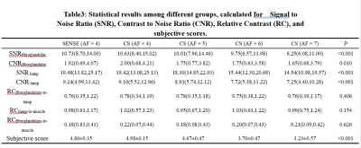 |
31 |
Optimizing Breast Isotropic DCE-MRI Imaging Based on Compressed
SENSE Technique
Ning Ning1,
Lina ZHANG1,
Zhuo WANG1,
Qi WU1,
Hongbing LIANG1,
Qingwei SONG1,
Ailian LIU1,
and Yiming Wang2
1First Affiliated Hospital of Dalian Medical University, Dalian, China, 2Clinical and Technical Support,PhilipsHealthcare, Shanghai, China Keywords: Breast, Breast Dynamic contrast-enhanced (DCE) MRI is a multi-phase sequence that is limited by long scan times, which makes acquisition acceleration necessary to accomplish clinical feasibility. The aim of this study was to investigate the effect of different Acceleration Factors (AFs) on the image quality of isotropic DCE-MRI acquired with the Compressed SENSE (CS) technique, and to find the optimal AF for the isotropic DCE-MRI.CS factors of 4 to 7 were compared against the conventional SENSE technique in 43 patients. Preliminary results show a CS factor of 5 is the optimal AF with optimized image and velocity data quality. |
|
2736.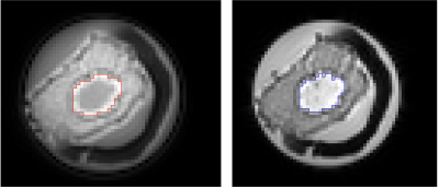 |
32 |
Ultrashort Echo Time (UTE) MRI in the Detection and
Classification of Microcalcification in Breast Cancer
Yazan Ayoub1,
Sai Man Cheung1,
Boddor Maglan1,
Ehab Husain2,
Yazan Masannat3,
and Jiabao He1,4
1Aberdeen Biomedical Imaging Centre, Institute of Medical Sciences, School of Medicine, Medical Sciences and Nutrition, University of Aberdeen, Aberdeen, United Kingdom, 2Department of Pathology, Aberdeen Royal Infirmary, Aberdeen, United Kingdom, 3Breast Unit, Aberdeen Royal Infirmary, Aberdeen, United Kingdom, 4Newcastle Magnetic Resonance Centre, Translational and Clinical Research Institute, Faculty of Medical Sciences, Newcastle University, Newcastle, United Kingdom Keywords: Breast, Cancer Micro-calcification is a central feature of breast cancer, and clinically revealed on mammography. Mammography, a two dimensional imaging approach, is inadequate to provide refined classification. Conventional MRI cannot capture the rapid signal decay from micro-calcification, with Ultra Short Echo Time (UTE) developed to image short T2* species. UTE demands precise control of the hardware to minimise the time delay between radiofrequency transmission and image acquisition, while substantial effort is required to optimise image quality and contrast. We set out to examine the degree of calcification in breast tumour specimens freshly excised from patients using UTE, with comparison against histopathological findings. |
|
2737.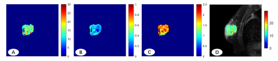 |
33 |
Imaging Microstructural Parameters of Breast Tumor in Patient
Using Temporal Diffusion Spectroscopy
Shuyi Peng1,
Peng Sun2,
Zhigang Wu2,
and Fan Yang1
1Department of Radiology, Union Hospital, Tongji Medical College, Huazhong University of Science and Technology, Wuhan, China, 2Philips Healthcare, Beijing, China Keywords: Breast, Diffusion/other diffusion imaging techniques, OGSE; IMPULSED; Breast tumor This study aimed to investigate the feasibility of temporal diffusion spectroscopy (the IMPULSED technique) for breast imaging. The result shows that the diffusion MRI–based microstructural mapping can evaluate the cell size in breast tumors and may demonstrate pathologic findings promise for characterizing breast cancer. |
|
2738.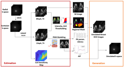 |
34 |
Realistic Digital Reference Object (DRO) toolkit for
quantitative breast Ultra-Fast Dynamic Contrast-Enhanced
(UF-DCE) MRI
Jonghyun Bae1,2,3,4,
Zhengguo Tan5,
Zhengnan Huang1,
Laura Heacock2,3,
Linda Moy2,3,
Florian Knoll5,
and Sungheon Gene Kim4
1Vilcek Institute of Graduate Biomedical Science, NYU School of Medicine, New York, NY, United States, 2Center for Biomedical Imaging, Radiology, NYU School of Medicine, New York, NY, United States, 3Center for Advanced Imaging Innovation and Research, Radiology, NYU School of Medicine, New York, NY, United States, 4Radiology, Weill Cornell Medical College, New York, NY, United States, 5Biomedical Engineering, Friedrich-Alexander-Universitat Erlangen-Nurnberg, Erlangen, Germany Keywords: Breast, DSC & DCE Perfusion Reconstruction of highly accelerated dynamic images is challenging and requires rigorous validation of reconstruction methods. We proposed a digital reference object (DRO) toolkit that provides realistic morphology and contrast dynamics in breast cancer. We acquired various base images containing real breast cancer lesions and simulated realistic contrast dynamics using the estimated kinetic parameters. Our toolkit provides a large number of reference objects with wide ranges of the dynamic images, kinetic parameters, segmentation masks, and k-space data. These realistic DROs with known ground-truth values can be used for different studies, including validation of reconstruction and training deep neural network. |
|
2739.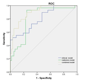 |
35 |
A novel nomogram built from mDixon-Quant MRI-based radiomics
features and clinical characteristics for grading invasive
ductal breast cancer
Zhuo Wang1,
Lina Zhang1,
Qi Wu1,
Ning Ning1,
Hongbing Liang1,
Qingwei Song1,
Qinhe Zhang1,
Ailian Liu1,
and Peng Sun2
1First Affiliated Hospital of Dalian Medical University, Dalian, China, 2Clinical and Technical Support, Philips Healthcare, WuHan, China Keywords: Breast, MR Value The histological grade of invasive breast cancer should be obtained by invasive needle biopsy , and the extracted tissue might not represent the heterogeneity of the whole tumor. Therefore, a novel nomogram was built from mDixon-Quant MRI-based radiomics features and traditional clinical image characteristics for grading invasive ductal breast cancer. The AUC of the combined model reached 0.907, which was significantly higher than that of the model only using traditional clinical image characteristics (0.782) or radiomics features (0.834). The proposed method can be an effective supplement to conventional breast MRI in predicting the histological grade of invasive ductal breast cancer. |
|
2740.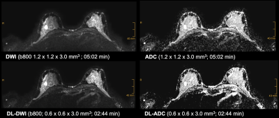 |
36 |
Accelerated DWI with deep learning reconstruction in 3T breast
MRI: initial clinical experiences and image quality
Caroline Wilpert1,
Hannah Schneider1,
Claudia Neubauer1,
Thomas Benkert2,
Elisabeth Weiland2,
Ralph Strecker3,
Marco Reisert4,
Jakob Weiß1,
Matthias Benndorf1,
Fabian Bamberg1,
Marisa Windfuhr-Blum1,
and Jakob Neubauer1
1Departement of Diagnostic and Interventional Radiology, University Medical Center Freiburg, Faculty of Medicine, University of Freiburg, Freiburg im Breisgau, Germany, 2MR Application Predevelopment, Siemens Healthcare GmbH, Erlangen, Germany, 3EMEA Scientific Partnerships, Siemens Healthcare GmbH, Erlangen, Germany, 4MR Physics, University Hospital Freiburg, Freiburg im Breisgau, Germany Keywords: Breast, Diffusion/other diffusion imaging techniques This prospective study evaluated image quality features of a novel accelerated DWI with deep learning image reconstruction in 3T breast MRI in a clinical setting in direct comparison to conventional DWI. Deep learning DWI (DL-DWI) shows a drastically shortened acquisition time of 46% compared to standard DWI, while maintaining a high image quality. Even though some image quality features were rated superior in standard DWI, image quality remained good for DL-DWI and lesion conspicuity scores were rated superior for DL-DWI compared to conventional breast DWI. Therefore, DL-DWI seems a feasible technique for accelerated breast DWI. |
|
2741.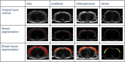 |
37 |
Feasibility study for developing a reproducible AI-driven breast
segmentation and composition algorithm from axial T2-weighted
sequences
Saurabh Garg1,
Saqib Basar1,
Nasrin Akbari1,
Thanh-Duc Nguyen1,
Sean London2,
Yosef Chodakiewitz2,
Rajpaul Attariwala1,
and Sam Hashemi1
1Voxelwise Imaging Technology Inc, Vancouver, BC, Canada, 2Prenuvo, Vancouver, BC, Canada Keywords: Breast, Machine Learning/Artificial Intelligence, Fatty tissue, Screening BI-RADS breast tissue composition defines which imaging modality is best suited for tissue examination. However it is subjective and varies between readers whereas AI techniques have been shown to remove subjectivity. We evaluate the use of state-of-the-art AI algorithms on a general whole-body noncontrast MRI to quantify the amount of fat versus nonfat tissue and compare with radiologists reports. Our results show significant correlation between the AI and radiologists' decisions. Further, we show on large dataset that the rate of replacement of nonfat fibroglandular tissue with fatty tissue is almost triple the rate in premenopausal women than postmenopausal women. |
|
2742.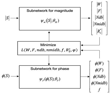 |
38 |
Estimation of Fatty Acid Composition in Mammary Adipose Tissue
Using Unsupervised Approach of Deep Learning
Suneeta Chaudhary1,
Elizabeth Lane2,
Eileen Chang3,
Anika McGrath2,
Eralda Mema2,
Allison Levy4,
Melissa Reichman2,
Katerina Dodelzon4,
Marcel Dominik Nickel5,
Linda Moy6,
Michele Drotman2,
and Sungheon Gene Kim1
1Radiology, Weill Cornell Medical College, New York, NY, United States, 2Weill Cornell Medical College, New York, NY, United States, 3Weill Cornell Medical college, New York, NY, United States, 4Weill Cornell Medical College, New york, NY, United States, 5MR Application Predevelopment, Siemens Healthcare GmbH, Erlangen, Germany, 6New York University School of Medicine, New York, NY, United States Keywords: Breast, Cancer The purpose of this study is to develop a non-invasive imaging method to measure the fatty acid composition (FAC) of mammary adipose tissue (MAT) and to investigate its role in breast cancer. A novel unsupervised deep learning approach has been developed using the MRI signal equation of fat peaks in the loss function to generate the FAC maps without using any training data. It takes less computational efforts than conventional voxel-wise analysis techniques. The repeatability and reproducibility of the proposed method have been examined on six subjects, which showed no statistically significant difference between repeated analyses and scans. |
|
2743.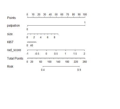 |
39 |
Deep Learning Radiomics of Preoperative Breast MRI for
Prediction of Axillary Lymph Node Metastasis in Breast Cancer
yanhong chen1,
lijun wang1,
ran luo1,
huanhuan liu1,
and dengbin Wang1
1Xinhua Hospital Affiliated to Shanghai Jiao Tong University School Of Medicine, shanghai, China Keywords: Breast, Radiomics, Deep Learning This study described the application of MRI-based deep learning radiomics in patients with breast cancer, presenting a novel individualized clinical decision nomogram that could be used to predict axillary lymph node metastasis providing a noninvasive approach to assist clinicians in clinical decision-making. |
|
 Back to Meeting Home
 Back to the Program-at-a-Glance
The International Society for Magnetic Resonance in Medicine is accredited by the Accreditation Council for Continuing Medical Education to provide continuing medical education for physicians.
 Back to Meeting Home
 Back to the Program-at-a-Glance
 View Presentation Video