Digital Poster
What's New in Liver Imaging
ISMRM & ISMRT Annual Meeting & Exhibition • 03-08 June 2023 • Toronto, ON, Canada

| Computer # | |||
|---|---|---|---|
3136.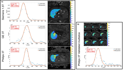 |
81 |
Translation of a non-contrast quantitative MRI protocol for
portal pressure prediction
Chris R Bradley1,2,
Eleanor F Cox1,2,
and Susan T Francis1,2
1University of Nottingham, Nottingham, United Kingdom, 2NIHR Biomedical Research Centre, Nottingham, United Kingdom Keywords: Liver, Liver Most complications in liver cirrhosis arise from portal hypertension. Using a vendor-specific fat suppressed spin-echo echo planar imaging T1 mapping method along with measures of flow within the superior mesenteric artery, we previously validated MRI as a surrogate measure of portal pressure at 1.5 and 3T. Here we translate this work to three MR vendors (GE, Philips and Siemens) using commercially available sequences allowing multi-site studies of assessment of portal pressure. Importantly, this could provide a non-invasive measure of portal pressure for clinical use to replace current invasive gold standard measures. |
|
3137.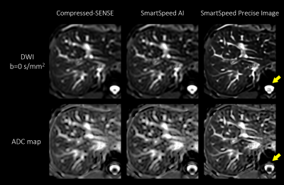 |
82 |
Deep learning-based image quality and spatial resolution
improvement for Diffusion Weighted Imaging in liver
Jihun Kwon1,
Kohei Yuda2,
Masami Yoneyama1,
Yasutomo Katsumata3,
and Marc Van Cauteren3
1Philips Japan, Tokyo, Japan, 2Tokyo Metropolitan Police Hospital, Nakano, Japan, 3Philips Healthcare, Best, Netherlands Keywords: Liver, Diffusion/other diffusion imaging techniques Diffusion-weighted imaging (DWI) in liver plays a significant role for lesion characterization and staging of fibrosis. Single-shot echo-planar imaging (ssh-EPI) readout is typically used; however, spatial resolution of ssh-EPI-DWI is limited by acquisition time. In this study, we investigated the use of prototype AI-based reconstruction technique (SmartSpeed Precise Image) to improve the image quality of liver ssh-EPI-DWI images. The image quality was compared between conventional Compressed-SENSE (C-SENSE), SmartSpeed AI, and SmartSpeed Precise Image. Volunteer data demonstrated a significant improvement of sharpness in DWI images and ADC map, and reduction of ringing artifact compared with C-SENSE and SmartSpeed AI reconstruction. |
|
3138.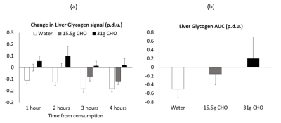 |
83 |
Using 13C MRS to Assess Fasted and Postprandial Glycogen Stores
in Healthy Children: A Randomized Controlled Study
Stephen Bawden1,2,
Astrid Horstman3,
Abi Spicer2,
Noura Darwish3,
Denis Breuille3,
Penny Gowland2,
Ian Macdonald4,
and Liz Simpson4
1NIHR Nottingham Biomedical Research Centre, Nottingham University Hospitals NHS Trust and the University of Nottingham, Nottingham, UK, University of Nottingham, Nottingham, United Kingdom, 2Sir Peter Mansfield Imaging Centre, School of Physics and Astronomy, University of Nottingham, Nottingham, United Kingdom, 3Nestle Institute of Health Sciences, Lausanne, Switzerland, 4David Greenfield Human Physiology Unit, MRC/ARUK Centre for Musculoskeletal Ageing Research, University of Nottingham School of Life Sciences, University of Nottingham, Nottingham, United Kingdom Keywords: Liver, Metabolism The aim of this study was to use 13C MRS to compare hepatic glycogen levels before and after an overnight fast and following a small breakfast of varying carbohydrate quantities (0, 15.5 and 31g CHO) in children . Liver glycogen concentration decreased overnight from 378 ± 141 to 277 ± 107 mmol l-1 with a between visit coefficient of variation of 21 ± 15 %. There was also a significant difference in the postprandial AUC (P < 0.005) with a linear CHO ‘dose’ response (R=0.51, P < 0.001). This study expands knowledge of normative glycogen storage in children. |
|
3139.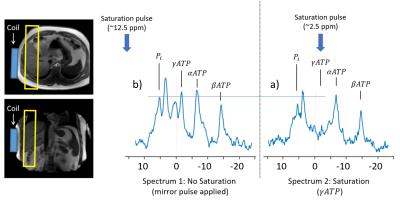 |
84 |
Change in ATP flux after propionate ingestion in healthy
volunteers Using 31P MRS Saturation Transfer.
Abi Spicer1,
Stephen Bawden1,2,
Penny Anne Gowland1,
Susan Francis1,
Douglas Morrison3,
Guruprasad Aithal4,
and Gary Frost5
1University of Nottingham, Nottingham, United Kingdom, 2NIHR Nottingham Biomedical Research Centre, Nottingham University hospitals NHS trust, Nottingham, United Kingdom, 3Scottish Universities Environmental Research Centre (SUERC), University of Glasgow, Glasgow, United Kingdom, 4Nottingham Biomedical Research Centre NUH, University of Nottingham, Nottingham, United Kingdom, 5Section for Nutrition, Faculty of Medicine, Imperial College London, London, United Kingdom Keywords: Liver, Spectroscopy, Multinuclear Propionate is a short chain fatty-acid absorbed in the colon and metabolised in the liver with some evidence showing that consumption can increase energy homeostasis. Three healthy participants have been studied to date with 31P saturation transfer experiments performed pre and 180 minutes post consumption of an inorganic-propionate ester using 1D-slice-selective-ISIS. The exchange rate constant (k) was calculated to be higher than previous studies despite similar methodologies. Whilst at this time no conclusions can be drawn about the effect of consuming Inorganic Propionate Ester thus far, this study shows the applicability of using 31P saturation transfer in simpler intervention studies. |
|
3140.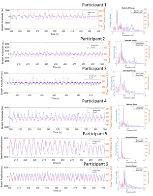 |
85 |
Characterising the effect of free breathing on abdominal MR
Spectroscopy and impact on X-nuclei spectra.
Abi Spicer1,
Stephen Bawden1,2,
Zachary Peggs1,
Alexander J Daniel1,
Andrew Peters1,
Susan Francis1,
and Penny Anne Gowland1
1University of Nottingham, Nottingham, United Kingdom, 2NIHR Nottingham Biomedical Research Centre, Nottingham University hospitals NHS trust, Nottingham, United Kingdom Keywords: Liver, Spectroscopy Multinuclear spectroscopy requires the use of free breathing acquisitions to achieve acceptable SNR. Here proton spectroscopy is used as an analogue for multinuclear MRS along with dynamic phase imaging to characterise the effect of respiration. Consistent agreement was seen between the respiratory bellows trace and the timeseries of the water peak frequency and imaging field offset, with overlapping primary frequencies on respective Fourier transforms. On free breathing a maximum frequency shift of 31Hz was measured, this equates to a 0.24ppm shift which could cause significant line broadening in the spectra of multinuclear 31P and 13C liver experiments. |
|
3141.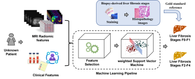 |
86 |
Stratification of Liver Histologic Fibrosis using Machine
Learning on MRI Radiomic Data and Clinical Features
Hailong Li1,
Jinzhao Qian1,
Ziang Chen1,
Wen Pan1,
Scott B. Reeder2,
David T. Harris2,
William R. Masch3,
Anum Alsam3,
Krishna P. Shanbhogue4,
Anas Bernieh1,
Sarangarajan Ranganathan1,
Nehal A. Parikh1,
Jonathan R. Dillman1,
and Lili He1
1Cincinnati Children's Hospital Medical Center, Cincinnati, OH, United States, 2University of Wisconsin-Madison, Madison, WI, United States, 3Michigan Medicine, University of Michigan, Ann Arbor, MI, United States, 4NYU Langone Health, New York, NY, United States Keywords: Liver, Liver, Liver fibrosis, biopsy Chronic liver diseases can lead to variable amounts of liver fibrosis, which impacts patient management and outcomes. Percutaneous liver biopsy is the clinical reference standard for assessment of liver fibrosis. However, biopsy is subject to sampling errors and poor patient acceptance. The aim of this study is to develop machine learning models to stratify the severity of biopsy-derived liver fibrosis using MR radiomic data and clinical data. Using clinical, routinely collected MRI and clinical data, our machine learning was able to stratify the severity of liver fibrosis with an AUROC of 0.71, demonstrating the feasibility of the machine learning approaches. |
|
3142.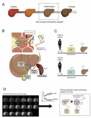 |
87 |
Pharmacokinetic Modelling of Gd-EOB-DPTA Uptake: Early
Progression of NASH in A Clinically Relevant Cohort, at 1.5 and
3 T
Christian Simonsson1,2,3,
Nils Dahlström1,3,
Markus Karlsson1,
Shan Cai1,
Simone Ignatova4,
Patrik Nasr5,
Mattias Ekstedt3,5,
Stergios Kechagias3,5,
and Peter Lundberg1,3
1Department of Radiation Physics, Radiology, Department of Medical and Health Sciences, Linköping University, Linköping, Sweden, 2Department of Biomedical Engineering, Linköping University, Linköping, Sweden, 3Center for Medical Image Science and Visualization (CMIV), Linköping University, Linköping, Sweden, 4Department of Clinical Pathology and Clinical Genetics, Department of Biomedical and Clinical Sciences, Linköping University, Linköping, Sweden, 5Department of Gastroenterology and Hepatology, Department of Health, Medicine and Caring Sciences, Linköping University, Linköping, Sweden Keywords: Liver, Contrast Agent, NAFLD,NASH, Gd-DPTA-EOB, Pharmacokinetic Modeling Due to the increased global prevalence of non-alcoholic fatty liver disease (NAFLD), there is a significant need for precise and non-invasive clinical methods to detect early stages of non-alcoholic steatohepatitis (NASH), which can progress to cirrhosis. We investigate the possibility of using the hepatocyte specific contrast agent Gd-EOB-DPTA based DCE-MRI in combination with mathematical modelling to assess hepatobiliary influx, as a possible biomarker for early NASH detection. We show a tentative correlation between increased portal inflammation and decreased hepatic Gd-EOB-DPTA uptake in a cohort of prospectively included patients with suspected chronic liver disease. |
|
3143.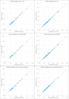 |
88 |
A statistical analysis of ROI weighting by area for MRE derived
liver stiffness measurements
Justin Yu1,
Alvin C Silva1,
Annelise Silva2,
Matthew Doan1,
and Anshuman Panda1
1Mayo Clinic Arizona, Phoenix, AZ, United States, 2Wright State University, Fairborn, OH, United States Keywords: Liver, Elastography MR elastography (MRE) derived liver stiffness measurements (LSMs) require manual drawing of ROIs on elastogram images. These ROIs may differ in size, and some clinicians suggest that reporting mean stiffness weighted by ROI area is more accurate than the unweighted mean. In this study the unweighted and weighted mean liver stiffness were calculated for 161 patients undergoing liver MRE exams using various scanning hardware/field strengths, and the statistical significance of the difference between the two sets of LSMs was tested using a paired two-tailed t-test. No statistically significant differences were found between weighted and unweighted LSMs. |
|
3144.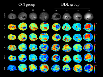 |
89 |
Diffusion Metrics for Staging Liver Fibrosis: An Experimental
Study in Rats with Bile Duct Ligation and Carbon Tetrachloride
at 11.7 T MRI
yimei Lu1,
qianfeng Wang2,
and dengbin Wang3
1Xinhua Hospital affiliated to Shanghai Jiao Tong University School of Medicine, Shanghai, China, 2Institute of Science and Technology for Brain-Inspired Intelligence, Fudan University, shanghai, China, 3Xinhua Hospital affiliated to Shanghai Jiao Tong University School of Medicine, shanghai, China Keywords: Liver, Diffusion/other diffusion imaging techniques As a common pathological feature of chronic liver diseases, liver fibrosis is a significant cause of global morbidity and mortality. An accurate assessment of the fibrosis stage and early detection of hepatic fibrosis are essential for preventing further adverse consequences. Our study suggests diffusion metrics are useful tools for noninvasively staging liver fibrosis. Among them, MK is a more valuable imaging biomarker for evaluating the degree of liver fibrosis, and f is less consistent in our two fibrosis models. |
|
3145.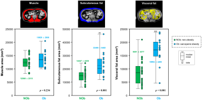 |
90 |
Assessment of association between body composition contents and
liver fibrosis in the patients with sarcopenic obesity
Tae-Hoon Kim1,
Chang-Won Jeong1,
ChungSub Lee1,
SiHyeong Noh1,
DongWook Lim1,
Youe Ree Kim2,
and Young Hwan Lee2
1Medical Convergence Research Center, Wonkwang University, Iksan, Korea, Republic of, 2Radiology, Wonkwang University School of Medicine and Hospital, Iksan, Korea, Republic of Keywords: Liver, Quantitative Imaging Sarcopenic obesity is a disease which associates both sarcopenia and obesity and may trigger worse clinical outcomes including hepatic manifestation of fat accumulation and musculoskeletal disabilities. It is likely that sarcopenia and obesity share common physiological pathways and are interconnected through the muscle-liver-adipose tissue axis. Assessment of muscle mass is a key for the whole-body insulin-mediated glucose metabolism and energy homeostasis. However, it is little known the interconnection between hepatic fibrosis and sarcopenic obesity. This study compared body composition contents in non-obese and sarcopenic obese patients using abdominal MRIs and investigated the relationship between hepatic fibrosis and sarcopenic obesity factors. |
|
3146.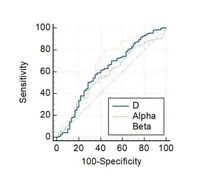 |
91 |
Evaluating chronic hepatitis B-related liver fibrosis with a
continuous-time random-walk diffusion model
Jing Zhang1,
Zhiyang Lu1,
Kaifan Yang1,
Liyun Zheng2,
Yongming Dai3,
and Yikai Xu1
1Department of Medical Imaging Center, Nanfang Hospital, Southern Medical University, Guangzhou, China, 2Shenzhen United Imaging Research Institute of Innovative Medical Equipment, Shenzhen, China, 3MR Collaboration, Central Research Institute, United Imaging Healthcare, Shanghai, China Keywords: Liver, Diffusion/other diffusion imaging techniques Early characterization of liver fibrosis has great clinical significance as it allows monitoring of treatment response to antifibrotic agents in patients with chronic liver disease. Most recently, high b-values diffusion-weighted imaging (DWI) based on the continuous-time random-walk (CTRW) model has been introduced. Compared to the healthy control group, the mean D, α, and β values were significantly lower in the hepatic fibrosis group. Spatial diffusion heterogeneity β showed the best diagnostic performance. This non-invasive method has the potential to realize efficient evaluation and improve clinical treatment decision-making. |
|
3147.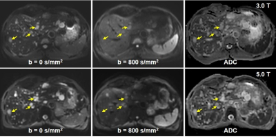 |
92 |
Preliminary Experience of 5.0 Tesla Higher Field Abdominal DWI:
Agreement of Apparent Diffusion Coefficient With 3.0 Tesla
Imaging
Yunfei Zhang1,
Yongming Dai2,
and Mengsu Zeng3
1MR Collaboration, Central Research Institute, United Imaging Healthcare, Shanghai, China, 2MR Collaboration, Central Research Institute, United Imaging Healthcare, Shanghai, China, 3Zhongshan Hospital of Fudan University, Shanghai, China Keywords: New Devices, Diffusion/other diffusion imaging techniques Recently, the 5.0 T whole-body MRI scanner was developed. This study aims to evaluate the feasibility of 5.0 T high field DWI and assess the agreement of the apparent diffusion coefficient (ADC) with that from 3.0 T DWI. The results showed that 5.0 T DWI displayed an increase in subjective image quality score. Both the inter-field and inter-observer agreements of ADC values were substantial to excellent. The substantial to excellent agreements between the ADC values measured with 3.0 T and 5.0 T DWI for liver, pancreas, spleen and kidney suggested that 5.0 T DWI can be applied for abdominal imaging. |
|
3148.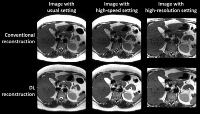 |
93 |
Deep Learning Reconstruction for Abdomen Diagnosis: Improvement
of Diagnostic Performance with higher Spatial or Temporal
Resolution
Bo-Ting Chen1,2,
Cheng-Ya Yeh2,
Yi-Chen Chen2,
Chia-Wei Li3,
Charng-Chyi Shieh3,
Chien-Yuan Lin3,
and Kao-Lang Liu1,2
1Department of Medical Imaging, National Taiwan University Hospital and National Taiwan University College of Medicine, Taipei, Taiwan, 2Department of Medical Imaging, National Taiwan University Cancer Center and National Taiwan University College of Medicine, Taipei, Taiwan, 3GE Healthcare, Taipei, Taiwan Keywords: Liver, Machine Learning/Artificial Intelligence, Deep learning reconstruction, Abdomen diagnosis We have previously validated the SNR improvement of abdominal MRI by Deep Learning Reconstruction (DLRecon). In this study, we further investigate the improvement of image quality and diagnostic performance when trading the SNR with higher spatial and temporal resolution imaging setting. In the result, the clinical scoring of images with high-speed or high-resolution settings in DLRecon was superior to that of the images with conventional setting and reconstruction. |
|
3149.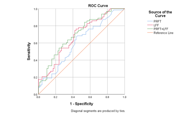 |
94 |
Efficacy of Perirenal Fat and Intra-organ Fat in differentiation
Metabolic Syndrome from Adults with Overweight and Obesity of
Suspected NAFLD
Li Wang1,
Xinzhong Ruan1,
and Mengxiao Liu2
1Department of Radiology, Ningbo First Hospital, Ningbo, China, 2Siemens healthineers, Shanghai, China Keywords: Liver, Fat This study investigated the fat quantitative Q-DIXON sequence to evaluate intra-organ fat content and visceral adipose tissue (VAT) to find the association with metabolic syndrome (MetS) in adults with overweight and obesity suspected with NAFLD. The results showed that the cut-off level of 9.15mm of perirenal fat thickness (PRFT) and 14.68% of liver fat fraction (LFF) can effectively predict MetS, and the combination of PRFT and LFF predict MetS better. Moreover, ectopic fat levels in pancreas and lumbar spines are positively associated with PRFT. |
|
3150.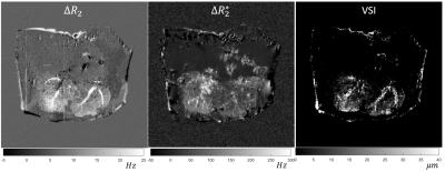 |
95 |
Building a Liver Perfusion Phantom for Vessel Size Imaging
Dominick Jon Romano1,
Mert Şişman2,3,
Qihao Zhang1,3,
Thanh Nguyen3,
Pascal Spincemaille3,
Martin Prince3,4,
and Yi Wang1,3
1Biomedical Engineering, Cornell University, Ithaca, NY, United States, 2Electrical & Computer Engineering, Cornell University, Ithaca, NY, United States, 3Radiology, Weill Cornell Medical College, New York, NY, United States, 4Radiology, Columbia University Vagelos College of Physicians and Surgeons, New York, NY, United States Keywords: Liver, Vessels We have developed a methodology of preparing explant livers for perfusion and vessel size imaging. Off-label use of ferumoxytol is known to provide high quality vessel size maps in vivo; however, known adverse reactions to ferumoxytol may hinder adoption into the clinical workflow. In this case, gadolinium may be more attractive. VSI experiments on a gadolinium perfused liver provide a proof of concept that Gadolinium may be used as an alternative contrast for vessel size imaging. |
|
3151.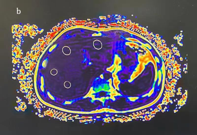 |
96 |
The feasibility of fat quantification by MRI-PDFF maps for
assessing grading of NAFLD
Hongyan Qi1,2,
Jianxiu Lian3,
Jiang Nan2,
Xuan Wang2,
and Junqiang Lei2
1First School of Clinical Medicine ,Lanzhou University, Lanzhou, China, 2Department of Radiology, the First Hospital of Lanzhou University, Lanzhou, China, 3Philips Healthcare, Beijin, China Keywords: Liver, Quantitative Imaging, pdff-mapping Non-alcoholic fatty liver disease (NAFLD) is a serious threat to human health, and the investigation for non-invasive biomarkers to quantify hepatic fat content appears to be crucial. In the study, PDFF was used to quantify liver fat content, assess the grade and degree of hepatic steatosis. PDFF was found to be effective in assessing fat content in NAFLD, which is expected to be used as a biomarker to assess the grading of steatosis in patients with NAFLD. |
|
3152.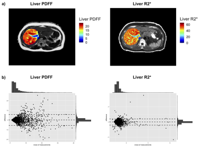 |
97 |
Evaluating Spatial Heterogeneity for Liver PDFF and R2* using
Radiomics Features
Marjola Thanaj1,
Nicolas Basty1,
Ramprakash Srinivasan2,
Madeleine Cule2,
Elena Sorokin2,
Jimmy Bell1,
Elizabeth Louise Thomas1,
and Brandon Witcher1
1Life Sciences, University of Westminster, London, United Kingdom, 2Calico Life Sciences LLC, South San Francisco, CA, United States Keywords: Liver, Data Analysis, PDFF, R2* MRI measures specific to the liver such as proton density fat fraction (PDFF) and R2* are proven biomarkers for assessing hepatic fat and iron content. There is an interest in using radiomics to extract additional information relating to spatial heterogeneity from images and apply these to clinical data. Here, we extracted radiomics features from liver PDFF and R2* and selected reproducible features and features that provide independent information to that derived from median liver PDFF and R2*. Additionally, we show that most radiomics features are negatively correlated with age and positively correlated with body mass index (BMI). |
|
3153.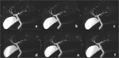 |
98 |
Influence of Different uCS Acceleration Factors on
Three-dimensional Magnetic Resonance Cholangiopancreatography
Image Quality
Chen Lihua1,
Song Qingwei1,
Nan Wang1,
Yongming Dai2,
Dan Yu2,
Guobin Li2,
and Ailian Liu1
1The First Affiliated Hospital of Dalian Medical University, Dalian, China, 2MR Collaboration,Central Research Institute, United Imaging Healthcare, Shanghai, China Keywords: Liver, MR Value To investigate the effects of different united compressed sensing (uCS) acceleration factors (AFs) on three-dimensional magnetic resonance cholangiopancreatography (3D-MRCP) image quality and to compare the quality with that using conventional parallel imaging (PI). |
|
3154.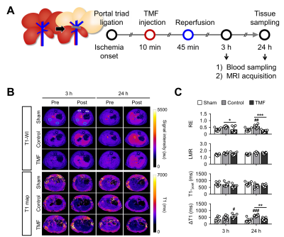 |
99 |
Is Aryl Hydrocarbon Receptor Antagonism after Ischemia Effective
in Alleviating Hepatic Ischemia-Reperfusion Injury in Rats?
Dong Cheol Woo1,
Hwon Heo1,
Yeon Ji Chae1,
Joongkee Min1,
Do-Wan Lee1,
Sang Tae Kim1,
Monica Young Choi1,
Yu Sub Sung1,
Kyung Won Kim1,
Yoonseok Choi2,
and Chul-Woong Woo1
1Asan Medical Center, Seoul, Korea, Republic of, 2Gangneung Asan Hospital, Gangneung, Korea, Republic of Keywords: Liver, Ischemia, reperfusion, aryl hydrocarbon receptor, L-kynurenine Recent studies suggest that aryl hydrocarbon receptor (AhR) act as an important mediator of ischemic injury in brain. In particular, pharmacological inhibition of AhR activation after ischemia has been shown to attenuate cerebral ischemia-reperfusion (IR) injury. In this study, we investigated whether AhR antagonist administration after ischemia was also effective in ameliorating hepatic IR injury. Our results indicate that treatment with the AhR antagonist after ischemia alleviate liver damage, as evidenced by serum ALT and AST levels, MRI-based liver function indices, and histologic and molecular analysis. Thus, adequate AhR antagonist activity is a potential therapeutic approach for hepatic IR injury. |
|
3155.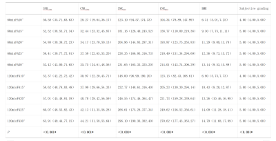 |
100 |
Optimal flip angle and delay time of Gd-BOPTA-enhanced MRI in
the normal liver and biliary imaging
Xue Ren1,
Ying Zhao1,
Qingwei Song1,
Geli Hu2,
Jiazheng Wang2,
and Ailian Liu1
1The First Affiliated Hospital of Dalian Medical University, Dalian, China, 2Clinical and Technical Support, Philips Healthcare, Beijing, China Keywords: Liver, Liver Gadobenate dimeglumine (Gd-BOPTA) is a novel contrast agent for hepatobiliary specific magnetic resonance imaging.At present, there is no exact and recognized turning Angle and delay time for Gd-BOPTA enhanced MRI scanning, and no relevant research reports have been reported at home and abroad. This study intends to analyze the image quality of the liver and biliary system in the hepatobiliary stage of Gd-BOPTA enhanced MRI by using the combination of different rotation angles and delay time. |
|
 Back to Meeting Home
 Back to the Program-at-a-Glance
The International Society for Magnetic Resonance in Medicine is accredited by the Accreditation Council for Continuing Medical Education to provide continuing medical education for physicians.
 Back to Meeting Home
 Back to the Program-at-a-Glance
 View Presentation Video