Digital Poster
Updates in Body Imaging II
ISMRM & ISMRT Annual Meeting & Exhibition • 03-08 June 2023 • Toronto, ON, Canada

| Computer # | |||
|---|---|---|---|
4110.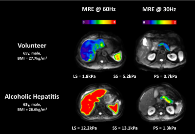 |
21 |
Assessment of Alcoholic Impact on Multiple Organs by
Dual-frequency Dual-driver Abdominal MR Elastography (MRE)
Jiahui Li1,
Nana K. Owusu1,
Kevin J. Glaser1,
Yi Sui1,
Douglas A. Simonetto2,
Armando Manduca1,
Sudhakar K. Venkatesh1,
Vijay H. Shah2,
Richard L. Ehman1,
and Meng Yin1
1Radiology, Mayo Clinic, Rochester, MN, United States, 2Gastroenterology and Hepatology, Mayo Clinic, Rochester, MN, United States Keywords: Digestive, Digestive We performed dual-frequency dual-driver abdominal MR Elastography (MRE) on 11 patients with alcoholic hepatitis (AH), and 6 healthy volunteers without known histories of chronic alcohol consumption. Two flexible drivers were placed over the anterior abdominal wall to deliver sufficient shear wave propagation in multiple abdominal organs at two different mechanical frequencies. The liver stiffness (LS) and spleen stiffness (SS) calculated from MRE at 60Hz, and pancreas stiffness derived from 30Hz, all significantly increased in patients with AH, compared with controls. This pilot study indicates the feasibility of dual-frequency dual-driver abdominal MRE to assess the alcoholic impact on multiple organs. |
|
4111.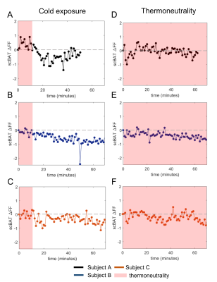 |
22 |
Comparing supraclavicular brown adipose tissue fat fraction
changes between mild cold and thermoneutrality in healthy adults
Robin van Eenige1,
Aashley S.D. Sardjoe Mishre1,
Maaike E. Straat1,
Borja M. Martinez-Tellez1,
Mariëtte R. Boon1,
Oleh Dzyubachyk1,
Andrew G. Webb1,
Patrick C.N. Rensen1,
and Hermien E. Kan1
1Leiden University Medical Center, Leiden, Netherlands Keywords: Endocrine, Fat, Supraclavicular brown adipose tissue Brown adipose tissue (BAT) is considered a therapeutic target in cardiometabolic health for its capacity to combust triglyceride-derived fatty acids into heat. Previous MRI studies consistently reported reductions in fat fraction (FF) in supraclavicular BAT (scBAT) after cold exposure. However, there is limited research on the validity of this outcome against thermoneutral conditions. We compared scBAT FF dynamics and thermal perception scores between mild-cold and thermoneutrality. Cold exposure decreased scBAT ΔFF (-0.73±0.19%), accompanied with colder thermal perceptions during cooling. At thermoneutrality, two out of three participants also showed a scBAT ΔFF decrease (-0.39±0.06%), yet normal thermal perceptions were reported. |
|
4112.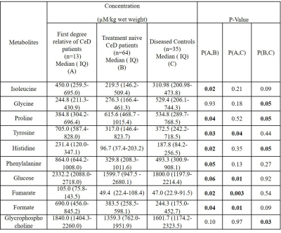 |
23 |
Metabolic Profiling of Small Intestinal Mucosa of patients with
Celiac Disease and their Family members by NMR Spectroscopy
Uma Sharma1,
Deepti Upadhyay1,
Prasenjit Das 2,
and Govind K Makharia3
1Department of Nuclear Magnetic Resonance, All India Institute of Medical Sciences, New Delhi, NEW DELHI, India, 2Department of Pathology, All India Institute of Medical Sciences, New Delhi, NEW DELHI, India, 3Department of Gastroenterology & Human Nutrition, All India Institute of Medical Sciences, New Delhi, NEW DELHI, India Keywords: Digestive, Body, Celiac disease, Metabolomics, First Degree Relative Celiac Disease, Biomarker, Pathophysiology, NMR Spectroscopy The present study demonstrated distinct metabolic features of intestinal mucosa biopsies in first-degree relatives (FDRs) of patients with celiac disease (CeD) patients and disease controls (DC) using proton NMR spectroscopy. FDRs had significantly lower levels of proline and allantoin while higher levels of 7 metabolites compared to CeD patients. The results showed a higher concentration of glucose, fumarate, tyrosine, and formate in the FDRs as compared to CeD and DC indicating altered energy metabolism and gut microbiome of FDRs. The obtained results may provide insight into the underlying mechanism involved in the pathophysiology of CeD patients. |
|
4113.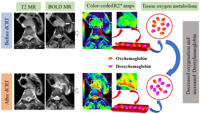 |
24 |
Value of BOLD MR in early evaluating the response and prognosis
of esophageal squamous cell carcinoma treated with definitive
chemoradiotherapy
Huanhuan Zheng1,
Hailong Zhang1,
Yan Zhu1,
Xiaolei Wei1,
Song Liu1,
and Wei Ren2
1Department of Radiology, Nanjing Drum Tower Hospital, The Affiliated Hospital of Nanjing University Medical School, Nanjing, China, 2The Comprehensive Cancer Center of Drum Tower Hospital, Medical School of Nanjing University and Clinical Cancer Institute of Nanjing University, Nanjing, China Keywords: Digestive, fMRI To confirm a quantitative imaging predictor for evaluation of early treatment response and prognosis to definitive chemoradiotherapy (dCRT) in patients with esophageal squamous cell carcinoma , using BOLD MR images. R2* values were obtained pre- and post-dCRT in 28 patients using BOLD MR. Independent samples t-test (normality) or Mann-Whitney U test (non-normality) was used to compare differences of R2*-related parameters between CR and non-CR groups. Diagnostic performance of parameters in predicting response was tested with receiver operating characteristic curve analysis. 3-years overall survival (OS) was evaluated using by Kaplan Meier curve, log rank test, and Cox proportional hazards regression analysis. |
|
4114.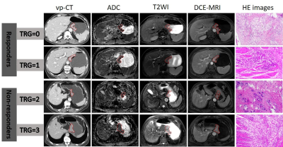 |
25 |
Comparison and combination of CT and MRI radiomics for early
prediction of pathological response to neoadjuvant chemotherapy
in gastric cancer
Jing Li1,
Hongkun Yin2,
Jinxia Guo3,
and Jinrong Qu4
1Radiology, the Affiliated Cancer Hospital of Zhengzhou University (Henan Cancer Hospital), Zhengzhou, China, 2Institute of Advanced Research, Infervision Medical Technology Co., Ltd, Beijing, China, 3GE Healthcare, MR Research China, Beijing, Beijing, China, 4the Affiliated Cancer Hospital of Zhengzhou University (Henan Cancer Hospital), Zhengzhou, China Keywords: Digestive, Radiomics, Gastric cancer; Computed tomography, X-rayed; Neoadjuvant chemotherapy To compare and combine CT and multi-parametric MRI (mp-MRI) radiomics for early prediction of pathologic response to neoadjuvant chemotherapy (NAC) in locally advanced gastric cancer (LAGC). The CT radiomics model, mp-MRI radiomics model and the combined nomogram were all associated with pathologic response. The multi-modal nomogram containing both CT and MRI radiomics scores exhibited added predictive ability and was linked to patients’ outcome. The CT and MRI radiomics model exhibited equivalent capability. This study proposed a multi-modal radiomics nomogram by incroperating concurrent CT and MRI images, which presents favorable efficacy in predicting treatment response to NAC in LAGC. |
|
4115.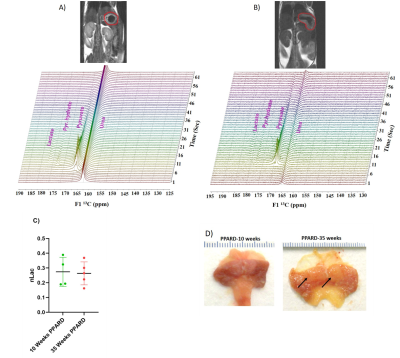 |
26 |
Identifying the Metabolic Signatures of PPARD-Overexpressing
Gastric Tumors by HP MRI and Metabolomics
Shivanand Pudakalakatti1,
Mark Titus2,
Imad Shureiqi3,
Xiangsheng Zuo4,
and Pratip Bhattacharya1
1Cancer Systems Imaging, University of Texas MD Anderson Cancer Center, Houston, TX, United States, 2Genitourinary Medical Oncology, University of Texas MD Anderson Cancer Center, Houston, TX, United States, 3University of Michigan, Ann Arbor, MI, United States, 4Gastrointestinal Medical Oncology, University of Texas MD Anderson Cancer Center, Houston, TX, United States Keywords: Digestive, Hyperpolarized MR (Non-Gas), 13C Pyruvate, Metabolomics Peroxisome proliferator-activated receptor delta (PPARD) is a ligand-dependent nuclear transcription factor that regulates a multiplicity of pathophysiological processes vital to cell metabolism. Recent discovery of PPARD overexpressed in villin-positive gastric progenitor cells, demonstrated spontaneous development of large, invasive gastric tumors as the mice aged. These unique animal models allowed us to address the knowledge gap of PPARD-regulated downstream metabolic changes and determine the significance of changes in gastric tumorigenesis by hyperpolarized MRI, NMR spectroscopy and LC-MS. Unlike many cancer systems, we found these gastric cancer tumors are not primarily dependent on aerobic glycolysis but on fatty acid oxidation for energy. |
|
4116.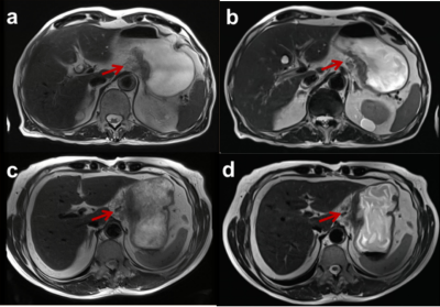 |
27 |
Deep Learning-accelerated, Single-breath-hold T2-weighted
Imaging for Tumor Invasion Assessment in Gastric Cancer
Wei-Yue Xu1,
Qiong Li1,
Ya-Jun Hou1,
Yi-Cheng Hsu2,
Dominik Nickel3,
Yu-Dong Zhang1,
and Xi-Sheng Liu1
1Department of Radiology, the First Affiliated Hospital with Nanjing Medical University, Nanjing, China, 2MR Collaboration, Siemens Healthineers Ltd, Shanghai, China, 3MR Applications Predevelopment, Siemens Healthcare GmbH, Erlangen, Germany Keywords: Contrast Mechanisms, Contrast Mechanisms, deep learning-accelerated T2WI T2-weighted imaging (T2WI) is an indispensable sequence of gastric magnetic resonance imaging (MRI) for tumor assessment. This study compared the deep learning-accelerated T2WI sequence (DL-T2WI) with BLADE T2WI in image quality assessment and tumor invasion evaluation of gastric cancer (GC). It revealed that DL-T2WI, acquired within a single-breath-hold, performed better than BLADE T2WI in terms of image quality for both quantitative and qualitative analyses. Furthermore, DL-T2WI images displayed comparable accuracy to BLADE T2WI images for serous invasion evaluation. The study suggests that DL-T2WI might be superior to BLADE T2WI for GC. |
|
4117.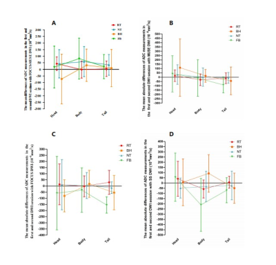 |
28 |
Reproducibility and image quality of diffusion weighted imaging
in volunteers with pancreas using different respiratory schemes
Yigang Pei1,
Yu Bai1,
Weiyin Vivian Liu2,
Wenguang Liu1,
and Wenzheng Li1
1Xiangya Hospital of the Central South University, China, China, 2GE Healthcare, MR Research China, Beijing, China, China Keywords: Pancreas, Diffusion/other diffusion imaging techniques DWI is essential in clinical diagnosis of pancreas. Image quality highly influence ADC measurements. Pancreases in abdomen easily suffers from artifact induced by abdominal and chest respiratory motion. A new diffusion-weighted imaging (DWI) sequence was explored to better delineate pancreas in our previous study (named FOCUS-MUSE DWI). In this study, we compared the clinical utility of FOCUS-MUSE DWI, MUSE DWI, FOCUS DWI and SS DWI using four different breathing schemes (RT, NT, BH and FB) for the repeatability of apparent diffusion coefficient (ADC) measurements and image quality. Our results suggested that RT‑DWI provided the best ADC reproducibility and image quality among four breathing schemes on 3.0 T MRI, making it as the recommended protocol for clinical DWI of the pancreas. |
|
4118.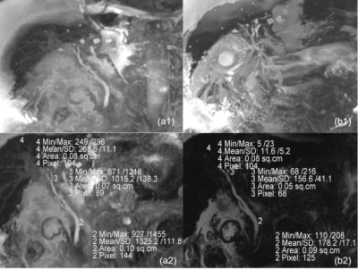 |
29 |
Comparison of NATIVE-TrueFISP and 3D-SPACE in the magnetic
resonance cholangiopancreatography of patients with ascites at
3T scanner
Lanbin Huang1,
Jinglian Zhong2,
Liping Liao1,
Yingying Huang1,
Zeping Liu3,
Qingchun Li1,
Qiceng Ruan1,
Mingxia Tan1,
Mengzhu Wang4,
Shasha Liao5,
Zehe Huang1,
and Song Chen1
1Department of Radiology, The First People's Hospital of QinZhou, Qinzhou, China, 2Department of Radiology, Sun Yat-Sen Memorial Hospital, Sun Yat-Sen University, Guangzhou, China, 3School of Biomedical Engineering, Guangzhou Medical University, Guangzhou, China, 4MR Scientific Marketing, Siemens Healthineers Ltd, Guangzhou, China, 5Department of Clinical Pharmacy, Maternity and Child Health care of Guangxi Zhuang Autonomous Region, Nanning, China Keywords: Digestive, Biliary, MRCP To compare the image quality of NATIVE-TrueFISP and 3D-SPACE in the MRCP of patients with ascites at 3T scanner. 5 patients with ascites and another 5 patients without underwent conventional upper abdominal MRI and MRCP with two kinds of sequences: NATIVE-TrueFISP and 3D-SPACE. The image quality of the two MRCP methods were independently assessed by two readers. Image quality of 3D-SPACE was superior to TrueFISP in patients without ascites while the TrueFISP sequence achieved better duct visualization in patients with ascites. Native TrueFISP can be used as an optional fast imaging sequence for MRCP of patients with ascites. |
|
4119.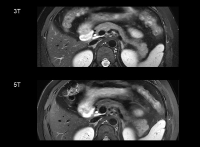 |
30 |
5T MRI of the pancreas in comparison to 3T: An initial study
Liyun Zheng1,2,3,
Chun Yang1,2,
Yongming Dai4,
and Mengsu Zeng1,2
1Shanghai Institute of Medical Imaging, Shanghai, China, 2Department of Radiology, Zhongshan Hospital, Fudan University, Shanghai, China, 3Shenzhen United Imaging Research Institute of Innovative Medical Equipment, Shenzhen, China, 4MR Collaboration, Central Research Institute, United Imaging Healthcare, Shanghai, China Keywords: High-Field MRI, Pancreas Despite the growing clinical acceptability of magnetic resonance imaging (MRI) evaluation, continued efforts are required to increase the resolution and robustness of pancreatic imaging. The feasibility of pancreatic MRI at 5T was studied in this study, which used a brand-new 5T MR scanner. 5T pancreatic MRI exhibited greater signal-to-noise ratio (SNR) and sufficient image quality as compared to the 3T examination. Diffusion-weighted imaging (DWI) at 5T was sufficient for measuring the longitudinal pancreatic apparent diffusion coefficient (ADC). As a result, pancreatic MRI at 5T may be effective for diagnosing and managing pancreatic diseases. |
|
4120.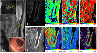 |
31 |
Multiparametric MRI for Staging of Bowel Inflammatory Activity
in Crohn's Disease with IVIM and DCE-MRI: A Preliminary Study
Jianguo Zhu1,
Liangqiang Mao1,
Yan Li1,
Weiqiang Dou2,
and Dmytro Pylypenko2
1Radiology, the Second Affiliated Hospital of Nanjing Medical University, Nanjing, China, 2MR Research China, GE Healthcare, Beijing, China Keywords: Digestive, Diffusion/other diffusion imaging techniques In summary, our findings suggested that the combination of IVIM and DCE-MRI can be used to accurately stage CD lesions activity. However, we must acknowledge that further investigation area is warranted to validate this preliminary finding. |
|
4121.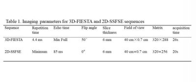 |
32 |
A comparative study of 2D SSFSE with deep learning based
reconstruction and 3D FIESTA in the assessment of active
inflammation in Crohn’s Disease
Yan Wang1,
Jingyun Cheng1,
Weiyin Vivian Liu2,
and Yunfei Zha1
1Department of Radiology, Renmin Hospital of Wuhan University, Wuhan, China, 2MR Research, GE Healthcare, Beijing, China Keywords: Digestive, Image Reconstruction Higher resolution and better image quality of 3D FIESTA sequence is used for evaluating bowel inflammation in CD; however, we found 2D SSFSEDLwith significantly higher SNR and CNR of ileum and terminal ileum than 3D FIESTA and, of course, conventional 2D SSFSE. 2D SSFSEDL shortened scan time and also offered better image quality than 3D FIESTA in assessment of bowel inflammation in CD. The SSFSE sequence alone has been considered sufficient in lesion detection[2]; SSFSE with DLR has great potential in elevation of detection rate without extra imaging time. |
|
4122.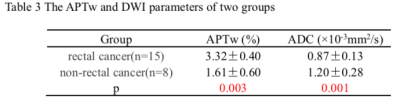 |
33 |
Clinical value of DWI combined with APT in differentiating
rectal cancer from non-rectal cancer
Yaxin Niu1,
Xiaoyan Lei1,
Li Zhang1,
Min Tang1,
and Kai Ai2
1Shaanxi Provincial People's Hospital, XI'AN, China, 2Philips Healthcare, XI'AN, China Keywords: Cancer, Diffusion/other diffusion imaging techniques We aimed to explore the diagnostic value of amide proton transfer-weighted (APTw) imaging combined with diffusion-weighted imaging (DWI) in differentiating rectal cancer from non-rectal cancer. The result showed that, compared with APTw or DWI (AUC=0.846 and 0.854, respectively), the highest diagnostic efficacy (AUC=0.967) was acquired using APTw combined with apparent diffusion coefficient (ADC) values. |
|
4123.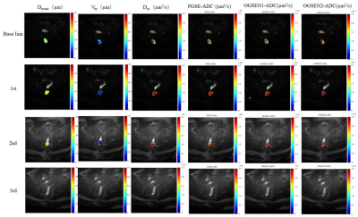 |
34 |
Longitudinally Evaluation of Locally Advanced Rectal Carcinoma
Treated with Preoperative Chemotherapy using Time-Dependent
Diffusion MRI
Xiaoling Gong1,
Yu Shen2,
Xiaoxiao Zhang3,
Zhigang Wu3,
Ziqiang Wang2,
and Bing Wu4
1Department of Radiology, West China Hospital, Sichuan University, Cheng du, China, 2Colorectal Cancer Center,Department of General Surgery, West China Hospital, Sichuan University, Chengdu, China, 3Philips Healthcare, China, Beijing, China, 4Department of Radiology, West China Hospital, Sichuan University, Chengdu, China Keywords: Cancer, Microstructure, rectal cancer The effectiveness of neoadjuvant Chemotherapy (NACT) is most commonly assessed by the change in tumor size based on conventional rectal cancer imaging. However, morphologic changes often occur later during treatment. Time-Dependent Diffusion MRI (TD MRI) is a novel MRI tool for depicting cellular microstructures. This study aimed to investigate the feasibility of Time-Dependent Diffusion MRI–based microstructural mapping for noninvasively characterizing cellular properties of LARC during NACT. |
|
4124.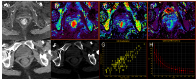 |
35 |
Comparation of DCE-MRI and dual-energy CT imaging in
discriminating tumor deposits from lymph node metastasis in
rectal cancer.
WEN JUN HU1,
Anliang Chen1,
and AIlian Liu1
1The First Affiliated Hospital of Dalian Medical University, Dalian, China Keywords: Digestive, Cancer Tumor deposits (TDs) in rectal cancer have been shown to be an important marker of poor prognosis. Although very similar to lymph node metastasis (LNMs), TDs have unique features in terms of biology and outcome, suggesting that distinguishing between these two entities may be of great importance. Results of this study indicate iodine-based material-decomposition (MD) images and DCE-MRI can effectively differentiate TDs and LNMs in rectal cancer, and iodine-based MD images showed better diagnostic efficiency. |
|
4125. |
36 |
Amide proton transfer-weighted imaging histogram analysis to
predict pathological extramural venous invasion in rectal
adenocarcinoma
Weicui Chen1,
Guoqing Liu1,
Jialiang Chen1,
Qiurong Wei1,
Yongsong Ye1,
Xiaohua Du1,
Jiepin Feng1,
Zhaoxian Yan1,
Kan Deng2,
and Xian Liu1
1The Second Affiliated Hospital of Guangzhou University of Chinese Medicine, Guangzhou, China, 2Philips Healthcare, Guangzhou, China Keywords: Digestive, Cancer, Rectal neoplasms This study evaluate amide proton transfer-weighted (APTw) derived whole-tumour histogram analysis parameters in predicting pathological extramural venous invasion (pEMVI) positive status of rectal adenocarcinoma (RA). The method is to calculate APTw histogram parameters between the pEMVI-negative and positive groups, assess the independent risk factors and diagnosis performance. Our results demonstrated that RA with pEMVI-positive status is associated with higher APTw-SI. The best prediction for EMVI involvement was obtained with a combined model of histopathological factors and APTw histogram parameters. Therefore, the APTw histogram analysis may represent a valuable non-invasive tool in predicting pEMVI positive status of RA. |
|
4126.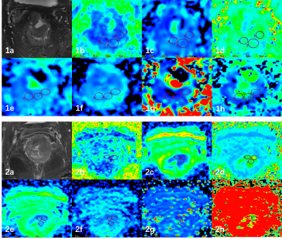 |
37 |
Diffusion Kurtosis Imaging Combined with Intravoxel Incoherent
Motion Imaging in Prediction of P53 Status in Rectal Cancer
Anliang Chen1,
Deshuo Dong1,
Changjun Ma1,
Ailian Liu1,
and Qingwei Song1
1radiology, The First Affiliated Hospital of Dalian Medical University, Dalian, China Keywords: Digestive, Pelvis Mutant P53 promotes tumor cell proliferation, invasion, and resistance to chemotherapy. The purpose of this study was to evaluate the value of combination of DKI and IVIM for prediction of P53 expression status in rectal cancer. The results indicate that the combination of MK and D* improved the diagnostic efficiency than single parameters. DKI and IVIM imaging allows non-invasive visualization and quantification of tissue composition for prediction of P53 status in rectal cancer. |
|
4127.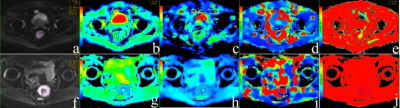 |
38 |
A nomogram based on IVIM-DWI and Clinical parameters to Predict
Survival of Rectal Cancer Patients after Radical Resection
Haodong Jia1
1Anhui Provincial Hospital Affiliated to Anhui Medical University, Hefei, China Keywords: Pelvis, fMRI This is an abstract by Haodong Jia.
|
|
4128.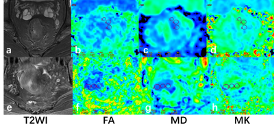 |
39 |
Evaluation of Diffusion Kurtosis Imaging in Prediction of P53
Expression in Rectal Cancer
Anliang Chen1,
Deshuo Dong1,
Changjun Ma1,
Ailian Liu1,
and Qingwei Song1
1radiology, The First Affiliated Hospital of Dalian Medical University, Dalian, China Keywords: Cancer, Cancer Mutant P53 promotes tumor cell proliferation, invasion, and resistance to chemotherapy. The purpose of this study was to evaluate the value of Diffusion Kurtosis Imaging (DKI) for prediction of P53 expression status in rectal cancer. The results indicate that positive P53 status group had significantly lower MD and higher MK values than negative P53 status group. DKI reflect a non-Gaussian phenomenon of water molecules within biologic tissues for prediction of P53 expression status in rectal cancer. |
|
4129.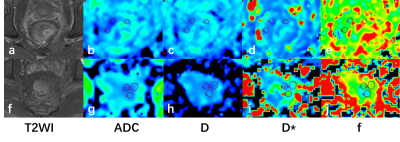 |
40 |
Prediction for Ki-67 Expression Using Intravoxel Incoherent
Motion Imaging in Rectal Cancer
Anliang Chen1,
Deshuo Dong1,
Changjun Ma1,
Ailian Liu1,
and Qingwei Song1
1radiology, The First Affiliated Hospital of Dalian Medical University, Dalian, China Keywords: Cancer, Digestive The Ki-67 expression has been verified as a prognostic indicator in colorectal cancer, correlating with biological behavior of tumor and response to the chemotherapy. The purpose of this study was to evaluate the value of intravoxel incoherent motion (IVIM) imaging for prediction of Ki-67 expression status in rectal cancer. The results indicate that high Ki-67 status group had significantly lower ADC and D values than low Ki-67 status group. IVIM imaging allows non-invasive visualization and quantification of tissue composition for prediction of Ki-67 expression status in rectal cancer. |
|
 Back to Meeting Home
 Back to the Program-at-a-Glance
The International Society for Magnetic Resonance in Medicine is accredited by the Accreditation Council for Continuing Medical Education to provide continuing medical education for physicians.
 Back to Meeting Home
 Back to the Program-at-a-Glance
 View Presentation Video