Digital Poster
Pipes & Tubes
ISMRM & ISMRT Annual Meeting & Exhibition • 03-08 June 2023 • Toronto, ON, Canada

| Computer # | |||
|---|---|---|---|
4811.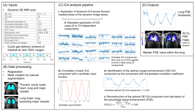 |
41 |
Pipeline for Robust Functional Lung Imaging with Oxygen-Enhanced
MRI (OE-MRI) and Independent Component Analysis (ICA)
Sarah Helen Needleman1,
Mina Kim1,
Jamie R. McClelland1,
and Geoff J. M. Parker1,2
1Centre for Medical Image Computing (CMIC), Department of Medical Physics & Biomedical Engineering, University College London, London, United Kingdom, 2Bioxydyn Limited, Manchester, United Kingdom Keywords: Oxygenation, Lung Analysis of dynamic lung oxygen-enhanced MRI (OE-MRI) is challenging due to the presence of substantial artefacts and poor SNR. Understanding and minimising sources of error is critical for reliable use of the method. We have created a pipeline using independent component analysis (ICA) for the automatic extraction of functional lung information from dynamic lung OE-MRI, for which confounding factors are reduced. The pipeline demonstrated good repeatability when utilised for the analysis of a scan-rescan dynamic lung OE-MRI study at 3.0 T; the algorithmic uncertainty of ICA on the analysis pipeline was found to be minimal. |
|
4812.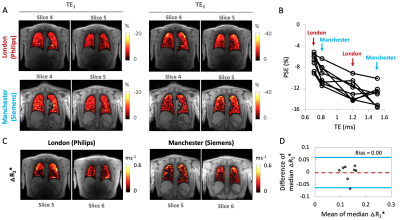 |
42 |
Repeatability and reproducibility of oxygen-enhanced MRI of the
lung at 3 tesla: cross-center, cross-vendor evaluation
Mina Kim1,
Sarah H. Needleman1,
Josephine H. Naish2,3,
Yohn Taylor1,
Marta Tibiletti2,
James P. B. O’Connor4,5,
and Geoff J. M. Parker1,2
1Centre for Medical Image Computing (CMIC), Department of Medical Physics & Biomedical Engineering, University College London, London, United Kingdom, 2Bioxydyn Limited, Manchester, United Kingdom, 3BHF Manchester Centre for Heart and Lung Magnetic Resonance Research (MCMR), Manchester, United Kingdom, 4Division of Cancer Sciences, University of Manchester, Manchester, United Kingdom, 5Division of Radiotherapy and Imaging, Institute of Cancer Research, Manchester, United Kingdom Keywords: Lung, Contrast Mechanisms, oxygen-enhanced MRI A recently developed protocol with a dual echo RF-spoiled gradient echo acquisition enabled simultaneous measurement of dynamic OE signal change and the substantial ∆R2* effect at 3 tesla. To progress towards clinical translation, we evaluated repeatability and reproducibility across two sites, vendors and time points. Our results demonstrate good repeatability for the quantitative and semi-quantitative indices across time points, consistent signal behaviour and repeatable ∆R2* across two sites and vendors, suggesting potential utility in multi-centre clinical studies. However, echo time dependence should be considered when interpreting percentage signal enhancement. |
|
4813.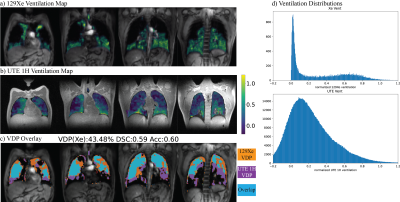 |
43 |
3D Free-Breathing Ultrashort Echo Time (UTE) 1H Ventilation
Compared with Hyperpolarized 129Xe Ventilation
Fei Tan1,
Rachel L. Eddy2,3,
Vanessa M. Diamond2,
Jonathan H. Rayment2,4,
and Peder E. Z. Larson1,5
1UC Berkeley-UCSF Graduate Program in Bioengineering, University of California, Berkeley and University of California, San Francisco, San Francisco, CA, United States, 2BC Children's Hospital Research Institute, Vancouver, BC, Canada, 3Centre for Heart Lung Innovation, University of British Columbia, Vancouver, BC, Canada, 4Department of Pediatrics, University of British Columbia, Vancouver, BC, Canada, 5Department of Radiology and Biomedical Imaging, University of California, San Francisco, San Francisco, CA, United States Keywords: Lung, Lung This abstract compares the 3D UTE 1H ventilation calculated from Motion-Compensated Low-Rank constrained reconstruction (MoCoLoR) with the hyperpolarized 129Xe ventilation for validation. The dataset covers healthy volunteers and pediatric and adult patients with cystic fibrosis (CF). We validated that 3D UTE 1H has a positively correlated VDP with the gold standard 129Xe and a high spatial VDP accuracy. |
|
4814.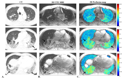 |
44 |
Evaluation of a Patient with Interstitial Lung Disease Using 3D
Perfusion Maps Based on 3D Ultrashort Echo-time Imaging
Hyeonha Kim1,2,
Seokwon Lee1,
Jinil Park2,
Jooae Choe3,
So Hyeon Bak3,
Ho Cheol Kim4,
and Jang-Yeon Park1,2
1Department of Biomedical Engineering, Sungkyunkwan University, Suwon, Korea, Republic of, 2Department of Intelligent Precision Healthcare Convergence, Sungkyunkwan University, Suwon, Korea, Republic of, 3Department of Radiology and Research Institute of Radiology, University of Ulsan College of Medicine, Asan Medical Center, Seoul, Korea, Republic of, 4Department of Pulmonary and Critical Care Medicine, University of Ulsan College of Medicine, Asan Medical Center, Seoul, Korea, Republic of Keywords: Lung, Perfusion, Interstitial lung diseases Since vascular abnormalities are common features of interstitial lung disease, not only structural images but also functional images such as perfusion maps are required to accurately evaluate vascular abnormalities including pulmonary hypertension. In this preliminary study, we proposed a method of evaluating an interstitial lung disease (ILD) patient particularly with idiopathic interstitial pneumonia (IIP) using 3D perfusion maps as well as high-resolution 3D structural images, both of which were obtained from 3D ultrashort echo-time imaging. Fibrotic areas were well identified in the structural UTE images as well as in the perfusion maps showing increased perfusion signals in the corresponding lesions. |
|
4815.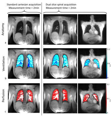 |
45 |
A simultaneous dual slice acquisition with spiral trajectory for
morphological and functional lung assessment
Anne Slawig1,2,
Andreas Max Weng2,
and Herbert Köstler2
1University Clinic and Outpatient Clinic for Radiology, University Hospital Halle (Saale), Halle (Saale), Germany, 2Department of Diagnostic and Interventional Radiology, University Hospital Würzburg, Würzburg, Germany Keywords: Lung, Data Analysis, ventilation, functional lung imaging, senceful SElf-gated Non-Contrast-Enhanced FUnctional Lung imaging (SENCEFUL) is a well-established method to determine functional lung parameters and was already successfully combined with 3D-UTE for robust ventilation quantification. Nevertheless, the evaluation of not only ventilation, but also perfusion, requires a 2D excitation, as it is based on the inflow effect. Additionally, coverage of more than one slice within short measurement times is valuable. Here, it is shown that functional information can be obtained from two slices acquired simultaneously, thus doubling the anatomical coverage of a conventional 2D acquisition. |
|
4816.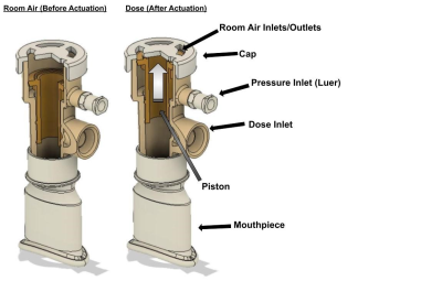 |
46 |
Remote Pneumatic Dose Administration for 129Xe MRI: Effects on
Lung Inflation and Patient Experience
Alexander Church1,
Shou Zhang1,
Junlan Lu1,
Cody Blanton1,
Jennifer Korzekwinski1,
David Mummy1,
and Bastiaan Driehuys1
1Radiology, Duke University, Durham, NC, United States Keywords: Lung, Hyperpolarized MR (Gas), New Devices Hyperpolarized 129Xe MRI requires repeatable lung inflation volumes to ensure consistent and accurate data. Current dose administration techniques require specialized study personnel to not only properly coach the subject through the inhalation maneuver, but also to directly administer the 129Xe dose, often while leaning deep into the MRI bore. Since lung inflation directly affects imaging data, it is important to maximize the consistency of inflation volumes across imaging sessions. We propose a novel 129Xe gas administration device intended to mitigate lung inflation differences as well as allow for remote administration of doses from outside the bore. |
|
4817. |
47 |
Diffusion of 129Xe in the lung: airspace size heterogeneity and
airway connectivity assessed by filter exchange imaging
Agilo L. Kern1,2,
Marcel Gutberlet1,2,
Jens M. Hohlfeld2,3,4,
Frank Wacker1,2,
and Jens Vogel-Claussen1,2
1Institute for Diagnostic and Interventional Radiology, Hannover Medical School, Hannover, Germany, 2Biomedical Research in Endstage and Obstructive Lung Disease Hannover (BREATH), German Center for Lung Research (DZL), Hannover, Germany, 3Clinical Airway Research, Fraunhofer Institute for Toxicology and Experimental Medicine (ITEM), Hannover, Germany, 4Department of Respiratory Medicine, Hannover Medical School, Hannover, Germany Keywords: Lung, Hyperpolarized MR (Gas) A filter exchange imaging (FEXI) sequence was developed for studying dynamic changes of apparent diffusion coefficient (ADC) in hyperpolarized 129Xe MRI, thereby probing heterogeneity and connectivity of lung airspaces. The sequence was tested in healthy volunteers and patients of chronic obstructive pulmonary disease (COPD). An initial reduction of ADC after filter application was observed in all subjects. Relative reduction and recovery rate tended to be increased in COPD. Weak/moderate correlations of initial reduction and recovery rate with ADC before filter application were observed. 129Xe FEXI lung MRI is feasible and derived quantities may be useful for disease phenotyping in COPD. |
|
4818.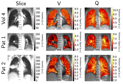 |
48 |
Feasibility and Reproducibility of Ventilation and Perfusion
Imaging at a 0.35 T MR-Linac in Healthy Volunteers and Lung
Cancer Patients
Rabea Klaar1,2,
Moritz Rabe3,
Thomas Gaass1,2,
Moritz J. Schneider1,2,4,
Ilyes Benlala1,2,5,
Stefanie Corradini3,
Chukwuka Eze3,
Claus Belka3,6,
Guillaume Landry3,
Christopher Kurz3,
and Julien Dinkel1,2
1Department of Radiology, University Hospital, LMU Munich, Munich, Germany, 2Comprehensive Pneumology Center (CPC-M), Member of the German Center for Lung Research (DZL), Munich, Germany, 3Department of Radiation Oncology, University Hospital, LMU Munich, Munich, Germany, 4Antaros Medical AB, BioVenture Hub, Mölndal, Sweden, 5Univ. Bordeaux, INSERM, Centre de recherche Cardio-Thoracique de Bordeaux, U1045, F-33000 Bordeaux, France, 6German Cancer Consortium (DKTK), Munich, Germany Keywords: Lung, Cancer, Ventilation, Perfusion Hybrid MR-imaging and linear accelerators for radiotherapy allow highly accurate treatment of lung cancer patients. The potential of Non-uniform Fourier Decomposition (NuFD), a non-contrast enhanced free-breathing technique that allows the assessment of lung ventilation and perfusion, was investigated in 10 healthy subjects and 2 lung cancer patients at a 0.35 T MR-Linac. Due to challenging ventilation quantification and reproducibility, two normalization strategies were proposed. Evaluation on repeated scans demonstrated clear improvement compared to uncorrected cases. First longitudinal ventilation assessment over treatment course shows promising results for the use of NuFD as treatment response monitoring tool in the clinical workflow. |
|
4819.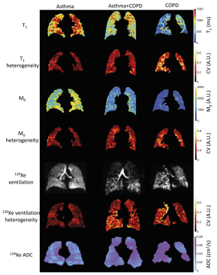 |
49 |
Differentiating patients with asthma and/or chronic obstructive
pulmonary disease using lung T1 and M0
Paul John Clifford Hughes1,
Helen Marshall1,
Laurie Smith1,
Demi Jakymelen1,
Alberto Biancardi1,
Guilhem Collier1,
Ho-Fung Chan1,
Martin Brook1,
Joshua Astley1,
Ryan Munro1,
Smitha Rajaram1,
Andy Swift1,
David Capener1,
Jody Bray1,
James Ball1,
Oliver Rodgers1,
Bilal Tahir1,
Madhwesha Rao1,
Graham Norquay1,
Nicholas Weatherley1,
Leanne Armstrong1,
Latife Hardaker2,
Titti Fihn-Wikander3,
Rod Hughes4,
and Jim Wild1
1POLARIS, University of Sheffield MRI Unit, IICD, The University of Sheffield, Sheffield, United Kingdom, 2Priory Medical Group, York, United Kingdom, 33BioPharmaceuticals Medical, AstraZeneca, Gothenburg, Sweden, 4Early Development Respiratory, AstraZeneca, Cambridge, United Kingdom Keywords: Lung, Lung Directing treatment based on diagnosis is important in patients suffering with respiratory disease. This work investigated the ability of whole-lung mean T1 and M0, and measures of heterogeneity (median coefficient of variation and its interquartile range) to differentiate between patients with asthma and/or COPD and correlated these metrics with 129Xe ventilation and diffusion MRI and pulmonary function test metrics. Measures of heterogeneity of T1 and M0 correlated with lung function test outcomes and sensitive measures of lung function derived from 129Xe MRI and had the ability to separate patients with asthma+COPD and COPD from patients with asthma alone. |
|
4820.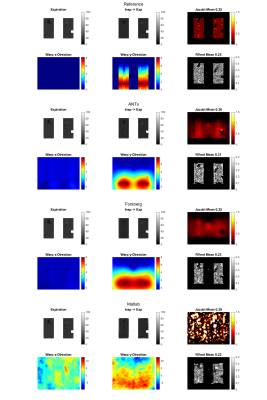 |
50 |
Introduction of a Digital Lung Model for Validation and
Refinement of Functional Lung Imaging Methods
Andreas Voskrebenzev1,2,
Marcel Gutberlet1,2,
Filip Klimeš1,2,
Frank Wacker1,2,
and Jens Vogel-Claussen1,2
1Institute of Diagnostic and Interventional Radiology, Medical School Hannover, Hannover, Germany, 2Biomedical Research in Endstage and Obstructive Lung Disease Hannover (BREATH), German Center for Lung Research (DZL), Hannover, Germany Keywords: Lung, Quantitative Imaging Functional lung imaging with proton MRI based on acquisition of free-breathing image-time-series gained interest during the last years, as it offers potential for ventilation / perfusion measurements without requirement for high patient compliance or administration of contrast agents. Since these methods rely on indirect surrogate measurements, validation with more direct techniques (e.g. hyperpolarized MRI), is required, which is elaborate, costly and limited. In this study a framework for a digital lung model, including artificial pathologies, is introduced and tested for different ventilation and perfusion measurements. Using the proposed method important differences were found between the methods regarding ventilation defect detection.
|
|
4821.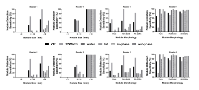 |
51 |
Identification of invasive adenocarcinoma in pulmonary
ground-glass nodules using zero echo time magnetic resonance
imaging (ZTE-MRI)
Qiao Zou1,
Qi Wan1,
Xinchun Li1,
Weiyin Vivian Liu2,
and Yongzhou Xu3
1Department of Radiology, The First Affiliated Hospital of Guangzhou Medical University, Guangzhou, China, 2MR Research China, GE Healthcare, Beijing, China, 3Philips Healthcare, Guangzhou, China Keywords: Lung, Cancer Intra-modality and inter-reader agreements of ground-glass opacity nodule (GGN) size were good between modalities (ZTE-MRI, T2WI-FS, CT). ZTE-MRI was susceptible to motion artifacts caused by breathing and vascular pulsation and had difficulty in displaying GGNs in specific lung locations, but was sensitive to GGNs with CT values of >-455 HU, highly possibly lung invasive adenocarcinomas , giving a hint to treatment strategies. In contrast, T2WI-FS can detect a smaller size of 6 mm for GGNs than ZTE-MRI. Overall, ZTE might play a unique role in differentiating invasive adenocarcinomas from precursor glandular lesions and minimally invasive adenocarcinoma (MIA) in eligible GGNs. |
|
4822.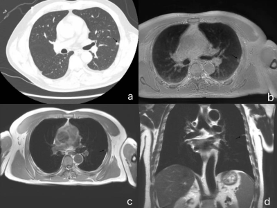 |
52 |
Feasibility study of using Zero TE Sequence to evaluate
pulmonary lesions: compared with CT examination
wang xiaoyan1 and
zhang yan1
1The First Affiliated Hospital of Zhengzhou University, Zhengzhou, China Keywords: Lung, MR Value Pulmonary MRI zero-echo time (ZTE) and MRI conventional sequences were compared with thoracic computed tomography (CT) to evaluate the lesions. All participants in the study underwent chest CT, ZTE, T2-TSE, T2-HASTE, and T1-VIBE scans. ZTE can be used as a supplementary examination of CT imaging. ZTE combined with T2WI can improve the detection rate and display performance of pulmonary nodules |
|
4823.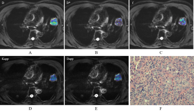 |
53 |
Intravoxel incoherent motion and diffusion kurtosis imaging can
be used to assess the differentiation degree of non-small cell
lung cancer
Yu Zheng1,
Wenjing Huang1,
Jing Zhang1,
and Na Han1
1Lanzhou University Second Hospital, Lanzhou, China Keywords: Lung, Lung, intravoxel incoherent motion, diffusion kurtosis imaging, differentiation degree, lung cancer The purpose of this study was to assess differentiation degree of non-small cell lung cancer (NSCLC) by using intravoxel incoherent motion (IVIM) and diffusion kurtosis imaging (DKI) . There were statistically significant differences in D and Dapp values between the poorly differentiated group and the moderately and highly differentiated group (p>0.05). D value had the best diagnostic efficacy in evaluating the differentiation degree of NSCLC [area under the curve (AUC) =0.773]. Our study demonstrated that D value derived from IVIM and Dapp derived from DKI can distinguish the differentiation degree of NSCLC. |
|
4824.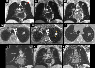 |
54 |
Improved strategy for diagnosing incidental benign and malignant
lung nodules during cardiac MR using volumetric multi-contrast
dark blood SSFP
Robert R Edelman1,2,
Nondas R Leloudas1,
Jianing Pang3,
and Ioannis R Koktzoglou1,4
1Radiology, NorthShore University HealthSystem, Evanston, IL, United States, 2Radiology, Feinberg School of Medicine, Northwestern University, Chicago, IL, United States, 3Siemens Medical Solutions USA, Chicago, IL, United States, 4Pritzker School of Medicine, University of Chicago, Chicago, IL, United States Keywords: Lung, Cancer, unbalanced steady state free precession Cardiovascular MR is an invaluable diagnostic tool for the heart and great vessels. As with cardiac CT, it is imperative to carefully evaluate areas outside of the heart to exclude the presence of clinically relevant lesions such as pulmonary infiltrates or lung cancer. Unfortunately, existing CMR techniques are grossly inadequate for detecting and characterizing extra-cardiac lesions. We hypothesized that prototype volumetric dark blood SSFP techniques could overcome these limitations and tested them in patients undergoing CMR. In this pilot study, dark blood SSFP outperformed standard CMR techniques for the evaluation of incidental pulmonary lesions, including benign nodules and cancer. |
|
4825.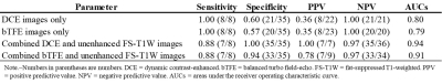 |
55 |
Follow-up non-contrast-enhanced MRA using balanced turbo
field-echo sequence for visceral artery aneurysm after
endovascular therapy
Nobuyuki Kawai1,
Yoshifumi Noda1,
Tetsuro Kaga1,
Kimihiro Kajita2,
Hiroshi Kawada1,
and Masayuki Matsuo1
1Radiology, Gifu University, Gifu, Japan, 2Radiology Services, Gifu University Hospital, Gifu, Japan Keywords: Vessels, Blood vessels Contrast-enhanced MRA has been shown to be a safe and effective way to provide posttreatment visceral artery aneurysm (VAA) follow-up. However, this study cannot be performed for those who have contraindications to MR contrast agents. Balanced turbo field-echo (bTFE) sequence enables static and moving fluids to be shown as high intensity without the use of gadolinium. We assessed the feasibility of follow-up non-contrast-enhanced MRA using bTFE sequence for posttreatment VAA. Combined bTFE and unenhanced fat-suppressed T1-wighted (FS-T1W) images demonstrated excellent performance for the diagnosis of aneurysm reperfusion comparable to combined dynamic contrast-enhanced and unenhanced FS-T1W images. |
|
4826.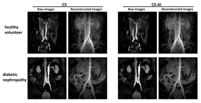 |
56 |
The application of REACT based on CS-AI for abdominal vessel
imaging in kidney-pancreas transplantation and diabetic
nephropathy patients
Hongbin Wang1,
Wenyun Liu1,
Lei Zhang1,
Zhuo Wang1,
Ying Qiu1,
Yuejiao Sun1,
Dandan Guo1,
and Yi Zhu2
1Department of Radiology, the First Hosipital of Jilin University, Changchun, China, 2Philips Healthcare, Beijing, China Keywords: Vessels, Image Reconstruction, REACT Patients with diabetic nephropathy and terminal renal insufficiency and those requiring combined kidney-pancreas transplantation should avoid using gadolinium-based contrast agents MR Angiography due to its potential nephrotoxicity. Compressed SENSE (CS) Artificial Intelligence (CS-AI) reconstructed Relaxation-Enhanced Angiography without Contrast and Triggering (REACT) sequence provides a simultaneous depiction of abdominal arterial and venous vessels. This study aims to improve the image quality of REACT sequence with CS-AI and further quantitatively evaluate the image quality in patients with diabetic nephropathy, and CS-AI reconstructed REACT sequence was first applied to image transplanted renal arteries in patients with combined kidney-pancreas transplantation. |
|
4827.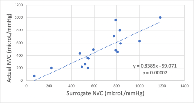 |
57 |
Common Carotid Arteries as Surrogate to the Internal Carotid
Arteries in the MRI Quantification of Neurovascular Compliance
Marianne Nabbout1,
Michael C Langham1,
and Felix W Wehrli1
1Department of Radiology, University of Pennsylvania, Philadelphia, PA, United States Keywords: Vessels, Blood vessels, Neurovascular Compliance Neurovascular compliance (NVC) is a vascular property involving the intracranial vascular arterial tree expressed as the arterial blood volume in response to a transient systolic blood pressure increase. NVC attracted attention due to its association with neurovascular diseases. We computed the NVC at the common carotid arteries with cine phase-contrast MR. This measure was shown to be significantly correlated with the quantification of the actual NVC in N=17 subjects (r= 0.84; p= 0.00002). Aortic pulse wave velocity, marker of aortic stiffness, was not found to be correlated with any of the two NVC computations. |
|
4828.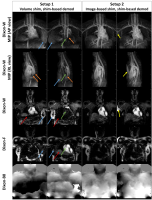 |
58 |
Mitigation of water-fat swap caused by off-resonance effects in
Dixon-based non-contrast-enhanced MR angiography
Teresa Nolte1,
Jihun Kwon2,
Mark Terwolbeck1,
Masami Yoneyama2,
Christiane Kuhl1,
and Shuo Zhang3
1Diagnostic and Interventional Radiology, University Hospital RWTH Aachen, Aachen, Germany, 2Philips Japan, Tokyo, Japan, 3Philips GmbH Market DACH, Hamburg, Germany Keywords: Vessels, Vessels, non-contrast-enhanced angiography, REACT Recently introduced Dixon-based MR angiographic techniques have shown promise for non-contrast enhanced vascular imaging, though off-resonance effects induced by B0 inhomogeneities may result in water-fat separation errors (i.e., swaps) during Dixon water fat separation, typically seen in a large field of view at high field strength. In this work, we investigate possible swaps via numeric simulations and in vivo measurements with various shimming techniques as well as reconstruction options for B0 demodulation. Initial results may suggest practical methods to mitigate water-fat swaps and improve technical robustness even in challenging applications. |
|
4829.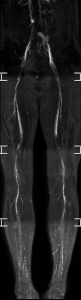 |
59 |
Ultrafast and versatile non-contract-enhanced MRA for
visualization of whole-body dynamic blood flow using
flow-sensitive CINE imaging
Isao Shiina1,
Michinobu Nagao2,
Masami Yoneyama3,
Yasutomo Katsumata3,
Jihun Kwon3,
Yasuhiro Goto1,
Yutaka Hamatani1,
Kazuo Kodaira1,
Takumi Ogawa1,
Mana Kato1,
and Shuji Sakai2
1Department of Radiological Services, Tokyo Women's Medical University, Tokyo, Japan, 2Department of Diagnostic imaging & Nuclear Medicine, Tokyo Women's Medical University, Tokyo, Japan, 3Philips Japan, Tokyo, Japan Keywords: Vessels, Blood vessels Flow-sensitive CINE imaging is non-contrast-enhanced MRA technique based on the Turbo Field Echo Planar Imaging (TFEPI). Because TFEPI is sensitive to blood flow, the MRA acquired with flow-sensitive CINE imaging includes dynamic flow information. In this study, we applied flow-sensitive CINE imaging to a variety of body parts such as trunk and lower extremity. Volunteer data demonstrated its usefulness to a variety of body parts. The combination of anatomical vascular pattern and the dynamic flow information may enable comprehensive assessment of whole-body arteries. |
|
 Back to Meeting Home
 Back to the Program-at-a-Glance
The International Society for Magnetic Resonance in Medicine is accredited by the Accreditation Council for Continuing Medical Education to provide continuing medical education for physicians.
 Back to Meeting Home
 Back to the Program-at-a-Glance
 View Presentation Video