Digital Poster
Body: Miscellaneous II
ISMRM & ISMRT Annual Meeting & Exhibition • 03-08 June 2023 • Toronto, ON, Canada

| Computer # | |||
|---|---|---|---|
4435.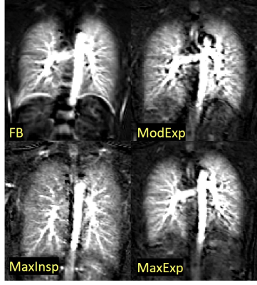 |
1 |
The Effect of Breathing on Pulmonary Blood Flow using
Pseudocontinuous Arterial Spin Labeling (PCASL) in a Test-Retest
Design
Manuel Kolb1,
Ferdinand Seith1,
Isabella Mack1,
Thomas Küstner1,
Rolf Pohmann2,
and Petros Martirosian3
1Department of Diagnostic and Interventional Radiology, University Hospital of Tübingen, Tübingen, Germany, 2High-Field MR Center, Max Planck Institute for Biological Cybernetics, Tübingen, Germany, 3Section of Experimental Radiology, Department of Radiology, University Hospital of Tübingen, Tübingen, Germany Keywords: Lung, Arterial spin labelling, Pulmonary blood flow, Lung perfusion PCASL can quantitively measure pulmonary blood flow and detect pulmonary embolism. However, patients suffering from lung embolisms are usually unable to follow breathing commands required for optimal imaging. In this study, the influence of respiratory states on pulmonary perfusion measured with PCASL was investigated. Nineteen healthy volunteers were studied with an ECG-triggered PCASL sequence in a test-retest design. It was found that mean perfusion was highest during expiration, followed by free breathing and lowest in inspiration. A significant difference was observed between the maximal inspiration and expiration breath-hold conditions. Perfusion values acquired by PCASL in the lung parenchyma showed high reproducibility. |
|
4436.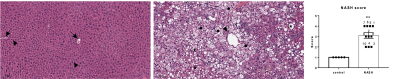 |
2 |
Application of Metabolic Imaging of Hyperpolarized
[1-13C]Pyruvate to a Genetic Mouse Model of Nonalcoholic
Steatohepatitis
Aditya Jhajharia1,
Salaheldeen Elsaid1,
Minjie Zhu1,
Joshua Rogers1,
Youngshim Choi2,
Liqing Yu2,
Sui Seng Tee1,
and Dirk Mayer1
1Diagnostic Radiology and Nuclear Medicine, University of Maryland School of Medicine, Baltimore, MD, United States, 2Department of Medicine, University of Maryland School of Medicine, Baltimore, MD, United States Keywords: Liver, Hyperpolarized MR (Non-Gas), NASH This study applied metabolic imaging of hyperpolarized [1-13C]pyruvate to a genetic mouse model of nonalcoholic steatohepatitis (NASH). Histology of the NASH (livKO) mice showed different levels of liver injury that were categorized into mild, moderate, and severe. NASH mice with severe liver injury had significantly higher Lac-to-Pyr and Ala-to-Pyr ratios in liver ROIs than that in control animals . These results suggest that hyperpolarized MRSI can be a promising method to differentiate liver injury based on severity in NASH. We also discussed several sources for variability of the MR data and how to potentially address them. |
|
4437.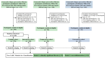 |
3 |
A Fully Automated Hybrid Approach to Assessing Liver Fibrosis
and Necroinflammation on Conventional MRI:A Multi-center Study
Junhao Zha1,
Yang Song2,
and Shenghong Ju1
1Jiangsu Key Laboratory of Molecular and Functional Imaging, Department of Radiology, Zhongda Hospital, School of Medicine, Southeast University, Nanjing, China, 2MR Scientific Marketing, Siemens Healthineers Ltd, Shanghai, China Keywords: Liver, Radiomics, liver fibrosis To our knowledge, this is the first study developing a multi-task hybrid models incorperating conventional MR tissue texture and routine clinical biomarkers with both good accuracy and explainability in detecting fibrosis and necroinflammation. Our study used an interactive deep learning approach to automatedly segment the entire volumetric liver contours more effectively. Our CoRC models outperformed routine clinical fibrotic scores (FIB-4, APRI), and TE-LSM by discrimination, calibration in the large multicenter cohorts. Our CoRC models could be as a potential alternative when biopsy, hepatobiliary phase (HBP) images, liver stiffness measurement (LSM) are unavailable. |
|
4438.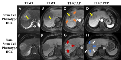 |
4 |
Preoperative Prediction of Hepatocellular Carcinoma with Stem
Cell Phenotype on Enhanced Magnetic Resonance Imaging and Its
Prognostic Value
Yidi Chen1,
Hanyu Jiang1,
and Bin Song1
1Radiology, West China Hospital, Sichuan University, Chengdu, China Keywords: Liver, Tumor Hepatocellular carcinoma (HCC) with stem cell phenotype is characterized by activating classic cell proliferation pathways and altering intrinsic regulators, it can stimulate HCC tumor proliferation, make the tumor more prone to recurrence, metastasis and therapy resistance. In this study, we developed an easy-to-use and noninvasive risk score integrating preoperative clinical indicators and enhancement magnetic resonance imaging (MRI) to predict stem cell phenotype HCC. |
|
4439.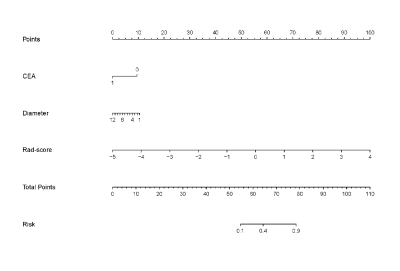 |
5 |
MRI-based radiomics nomogram for preoperatively differentiating
intrahepatic cholangiocarcinoma from colorectal liver metastases
Abstract
Ying Xu1,
Lu Li1,
Yi Yang1,
Feng Ye1,
Sicong Wang2,
Lizhi Xie2,
Yanan Wang3,
and Xinming Zhao1
1Cancer Hospital, Chinese Academy of Medical Sciences, Beijing, China, 2GE Healthcare, China, Beijing, China, 3Affiliated Cancer Hospital of Zhengzhou University, Henan Cancer Hospital, Zheng Zhou, China Keywords: Liver, Radiomics, Cholangiocarcinoma; colorectal liver metastases; nomogram; differential diagnosis. A total of 133 patients in training cohort (64 IMCC and 69 CRLM) and 57 patients in validation cohort (29 IMCC and 28 CRLM) were included. Radiomics features were extracted from the DCE-MR images and selected by the least absolute shrinkage and selection operator algorithm to establish the radiomics model.The radiomics nomogram was constructed combining the radiomics model and clinical model.The radiomics nomogram combining radiomics signatures based on DCE-MRI with clinical factors (serum CEA level and tumor diameter) may provide a reliable and noninvasive tool to discriminate IMCC from CRLM, which could help guide treatment strategies and prognosis prediction preoperatively. |
|
4440. |
6 |
The PRISM clinical trial: Quantitative imaging for personalized
liver radiation therapy
Sirisha Tadimalla1,
Tim Wang2,
Val Gebski3,
Sheryl Foster4,5,
Vincent Lam6,
Jonathan Sykes2,
Robert Finnegan1,7,8,
Jacob George9,
Verity Ahern2,
Steven Sourbron10,
and Annette Haworth1
1Institute of Medical Physics, The University of Sydney, Sydney, Australia, 2Sydney West Radiation Oncology Network, Western Sydney Local Health District, Westmead, Australia, 3NHMRC Clinical Trials Centre, The University of Sydney, Sydney, Australia, 4Faculty of Medicine and Health, The University of Sydney, Sydney, Australia, 5Department of Radiology, Westmead Hospital, Westmead, Australia, 6Acute Surgical Unit, Westmead Hospital, Westmead, Australia, 7Ingham Institute of Applied Medical Research, Liverpool, Australia, 8Northern Sydney Cancer Centre, Royal North Shore Hospital, St Leonards, Australia, 9Department of Gastroenterology and Hepatology, Westmead Hospital, Westmead, Australia, 10Department of Infection, Immunity and Cardiovascular Disease, University of Sheffield, Sheffield, United Kingdom Keywords: Cancer, Radiotherapy Quantitative imaging biomarkers of liver function offer a potential opportunity to optimize radiation dose distributions for personalized treatment of patients with liver cancers using radiation therapy. The PRISM clinical study aims to determine if radiation treatment plans guided by liver function maps from MRI can increase the therapeutic dose to tumours. We have developed a dedicated multi-parametric MRI protocol for the quantitative assessment of liver composition, structure and function. Details of the protocol and pilot data are presented here. |
|
4441. |
7 |
Toward Assessment of Renal Tubule Volume Fraction in Rat Kidney
Using Decomposition of Parametric T2 Mapping
Ehsan Tasbihi1,
Thomas Gladytz1,
Ludger Starke1,2,
Jason M. Millward1,
Erdmann Seeliger3,
and Thoralf Niendorf1,4
1Berlin Ultrahigh Field Facility (B.U.F.F.), Max Delbrück Center for Molecular Medicine in the Helmholtz Association (MDC), Berlin, Germany, 2Hasso Plattner Institute for Digital Engineering, University of Potsdam, Potsdam, Germany, 3Institute of Translational Physiology, Charité – Universitätsmedizin, Berlin, Germany, 4Experimental and Clinical Research Center, a joint cooperation between the Charité Medical Faculty and the MAX Delbrück Center for Molecular Medicine in the Helmholtz Association, Berlin, Germany Keywords: Kidney, Preclinical Increased incidence of kidney diseases is a concern, for which rapid biomarkers are lacking. Changes in tubule volume fraction (TVF) is a potential biomarker for kidney disease. T2-mapping may be useful to asses TVF in noninvasive manner. To study this, we developed a protocol tailored for T2-mapping of the rat kidney, and determined a suitable numerical solution for multi-exponential decomposition of the T2-decay. Evaluation and validation of these algorithms using synthetic data and phantom showed an accuracy of 98%. This demonstrates that our approach is promising for research into quantitative assessment of renal TVF in in vivo applications. |
|
4442.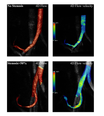 |
8 |
The feasibility of 4D Flow in evaluating hemodynamic changes of
transplant renal artery stenosis
Songlin Guo1,
Peng Wu2,
Lu Han2,
Liang Pan 1,
and Wei Xing1
1Third Affiliated Hospital of Soochow University, Department of Radiology, Changzhou, China, 2Philips Healthcare, Shanghai, China, Shanghai, China Keywords: Kidney, Transplantation Although 4D-Flow MRI is capable of detecting hemodynamic abnormalities, it is rarely employed in transplanted kidneys. Using 4D-Flow MRI, we assessed total volume, net flux, maximum flow, and mean flow rate and performed a statistical analysis of the correlation in this study. Total volume, net flux, maximum flow, and mean flow rate were shown to be inversely linked to the degree of stenosis in the transplanted renal artery. As a result, 4D-Flow MRI may be applied to study the hemodynamic changes of the transplanted renal artery noninvasively. |
|
4443.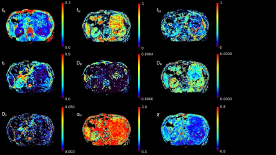 |
9 |
Feasible study of multimodal apparent diffusion (MAD) in kidney
Shuang Wang1,
Tuo Ji2,
Jingdong Zhang 1,
Dan YU3,
Yongming Dai3,
and Lin Liu1
1The Radiology Apartment of China-Japan Union Hospital of Jilin University, Changchun, China, 2The first Urology Apartment of China-Japan Union Hospital of Jilin University, Changchun, China, 3Central Research Institute, United Imaging Healthcare, Shanghai, China Keywords: Kidney, Diffusion/other diffusion imaging techniques The multimodal apparent diffusion (MAD) is a new approach with a predefined number of components associated with microstructure. In this work, the performance of quad-modal apparent diffusion in the kidney was assessed with simulation for different conditions. In-vivo demonstration and comparison with other models showed an explicable parametrization and improved fitting performance. |
|
4444.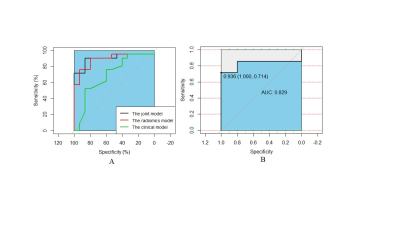 |
10 |
MRI Radiomics Model Preoperatively Predicts Key Gene Mutation in
Pancreatic Ductal Adenocarcinoma Patients
Yan Deng1,
Xiao ming Zhang2,
and Bin Song1
1Radiology, West China Hospital, Sichuan University, Chengdu, China, 2Affiliated Hospital of North Sichuan Medical College, Nanchong, China Keywords: Pancreas, Cancer Individualized management of pancreatic ductal adenocarcinoma (PDAC) is difficult and understanding it underlying biology is important. Radiomics model based on MRI was developed to predict status of KRAS and TP53 in PDAC, univariate analysis, MRMR and LASSO were applied for feature selection and multivariable logistic regression used for modeling. The classification metrics were applied to assess these models discrimination. Results suggested radiomics model based on MRI outperformed clinical model for predicting gene mutations of PDAC. This study confirmed that radiomics model based on MRI had a potentiality to predict the KRAS and TP53 mutation in PDAC. |
|
4445.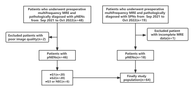 |
11 |
Role of tomoelastography for the differentiation between solid
pseudopapillary neoplasms and pancreatic neuroendocrine
neoplasms
Jiaxin Yuan1,
Siya Shi1,
Liqin Wang1,
Xuefang Hu1,
Jinhui Yu1,
Tingting Wen1,
Shiting Feng1,
Zhenpeng Peng1,
and Yanji Luo2
1The First Affiliated Hospital, Sun Yat-Sen University, Guangzhou, China, 2The First Affiliated Hospital, Sun Yat-Sen University, Guanghzou, China Keywords: Pancreas, MR Value, Magnetic resonance elastographies,SPNs,pNENs This prospective study enrolled patients with pathologically confirmed solid pseudopapillary tumors (SPNs) and pancreatic neuroendocrine tumors (pNENs) who underwent preoperative tomoelastography examinations at our hospital. Sixty-four patients were included (18 patients with SPNs; 46 patients with pNENs). SPNs showed significantly lower stiffness (P<0.001) and fluidity (P=0.001) than pNENs. Both stiffness and fluidity allowed distinguishing SPNs from pNENs. AUCs were 0.899 for stiffness, 0.760 for fluidity. Tomoelastography is a novel multi-frequency MRE technique that can facilitate the differentiation between SPNs and pNENs. |
|
4446. |
12 |
High resolution diffusion weighted imaging on pancreas through
reduced FOV and compressed SENSE: a feasibility study
Yajing Zhang1,
Zhigang Wu2,
Wengu Su3,
Jiazheng Wang4,
Yan Zhao5,
and Marc Van Cauteren6
1MR Clinical Science, Philips Health Technology, Suzhou, China, 2Philips Healthcare (China), Shenzhen, China, 3MR Application, Philips Health Technology, Suzhou, China, 4Philips Healthcare (China), Beijing, China, 5MR R&D, Philips Health Technology, Suzhou, China, 6Philips Healthcare, Tokyo, Japan Keywords: Pancreas, Data Acquisition Pancreas imaging remains challenging because of its small size and motion, and conventional diffusion weighted imaging (DWI) suffers from poor image quality due to low signal-to-noise (SNR) and image blurring. This study performs several schemes to achieve high resolution high image quality pancreas DWI with reduced FOV using Compressed SENSE. |
|
4447.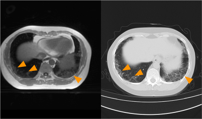 |
13 |
Oxygen-enhanced high-resolution 3D ultrashort echo time MRI for
patients with connective tissue diseases: a preliminary study
Feng Zhang1,
Yu Xin Yang2,
Lei Jiang3,
Liang Zhao1,
Yongming Dai4,
and Jiong Zhu1
1Renji Hospital, School of Medicine, Shanghai Jiao Tong University, Shanghai, China, 2United Imaging Research Institute of Intelligent Imaging, Beijing, China, 3Ningbo Hangzhou Bay Hospital, Ningbo, China, 4Central Research Institute, United Imaging Healthcare, Shanghai, China Keywords: Lung, Lung This preliminary study investigates the utility of high-resolution 3D-UTE to assess patients with CTD-ILD and their pulmonary function, including both pulmonary parenchyma and lesions, using OE-UTE-MRI. Our results suggest that high-resolution 3D UTE and OE-UTE MRI could potentially be as useful as thin-section CT for lesion detection of CTD patients with ILD and provide additional pulmonary functional loss assessment. |
|
4448.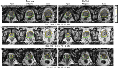 |
14 |
Development and clinical utility analysis of a prostate zonal
segmentation model on T2-weighted imaging: a multicenter study
Lili Xu1,
Gumuyang Zhang1,
Li Mao2,
Xiuli Li2,
Hao Sun1,
and Zhengyu Jin1
1Peking Union Medical College Hospital, Beijing, China, 2Deepwise AI Lab, Beijing, China Keywords: Prostate, Machine Learning/Artificial Intelligence, Multicenter study Zonal segmentation is important in the management of prostatic diseases. Many studies have demonstrated the feasibility of training CNN models for zonal segmentation. However, they lack validation in non-public datasets and consideration of the patients’ characteristics. In this study, we validated the model’s utility for prostate zonal segmentation on T2WI in different external testing datasets. The model yielded good performance regardless of the variations in the patients’ clinicopathological characteristics. The model showed higher performance than the junior radiologist in PZ segmentation. Prostate morphology and MR scanner parameters, especially CG volume and vendor, impact zonal segmentation performance. |
|
4449.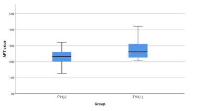 |
15 |
The value of amide proton weighted imaging in predicting
perineural invasion of prostate cancer
Xiwei Li1,
Lihua Chen1,
Nan Wang1,
Jiazheng Wang2,
and Ailian Liu1
1The First Affiliated Hospital of Dalian Medical University, Dalian, China, 2Philips Healthcare,Beijing, Beijing, China Keywords: Prostate, Cancer Perinerval infiltration (PNI) is closely related to the degree of radical operation and postoperative recurrence. Pathological examination is the gold standard for PNI diagnosis, but it is an invasive examination. The APTw imaging can be used for disease diagnosis and therapeutic evaluation by detecting the content of protein and polypeptide in tissues and cells. In this study, APTw was used to evaluate the PNI in prostate cancer. The results showed that the APT values of PNI (+) group was higher than other one group. Therefore, APTw is of great value in differentiation of perinerval invasion from prostate cancer. |
|
4450.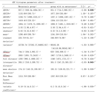 |
16 |
ADC histogram features of rectal cancer before and after
neoadjuvant radiation to predict heterochronic hematogenous
metastasis
Xiaoxian Zhang1,
Chunmiao Xu1,
Zhiwei Shen2,
Ke Jiang2,
and Xuejun Chen1
1Henan tumor hospital, Zhengzhou, China, 2Philips healthcare,Beijing,China, Beijing, China Keywords: Cancer, Diffusion/other diffusion imaging techniques Bloodstream metastasis to the liver, lung, and bone is the most prevalent mechanism of rectal cancer metastasis. However, there are currently no good indications for diagnosing heterochronic hematogenous metastases at the early stage. Therefore, this study aimed to explore whether ADC histograms might predict new heterochronic hematogenous metastases before and after neoadjuvant radiation for rectal cancer. Results showed that the first-order histogram feature ADC5% derived from post-treatment ADC maps might be utilized as an imaging biomarker and an independent predictor of new heterochronic hematogenous metastases after surgery following neoadjuvant chemotherapy for rectal cancer. |
|
4451.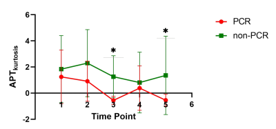 |
17 |
APT histogram parameters for Response Prediction of Neoadjuvant
Chemotherapy in Rectal Cancer: A Prospective Study
Xiaoling Gong1,
Yu Shen2,
Daguang Wen3,
Xiaoxiao Zhang4,
Xiaoyong Zhang5,
Ziqiang Wang2,
and Bing Wu3
1Department of Radiology, West China Hospital, Sichuan University, Cheng du, China, 2Colorectal Cancer Center,Department of General Surgery, West China Hospital, Sichuan University, Chengdu, China, 3Department of Radiology, West China Hospital, Sichuan University, Chengdu, China, 4Philips Healthcare, China, Beijing, China, 5Philips Healthcare, China, Chengdu, China Keywords: Cancer, fMRI, rectal cancer Early imaging prediction of neoadjuvant chemotherapy (NACT) response would enable a personalized treatment approach to improve therapeutic response and avoid treatment morbidity in rectal cancer. Morphological changes base on T2 weighted MRI and free dispersion limitation of water molecules in tumor have limited value in evaluating efficacy after NACT. Amide proton transfer (APT)–weighted MRI, indirectly detecting the concentration of tissue macromolecular proteins, could help to inform us of the proliferation and biological status of tumor cells. The aim of this study is to determine if APT MRI is useful in early assessment of treatment response in persons with rectal cancer. |
|
4452.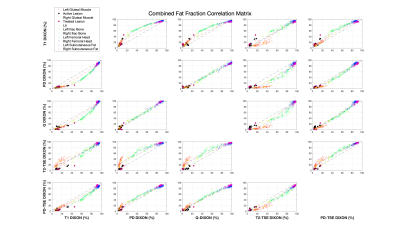 |
18 |
Comparison of fat fraction techniques within the pelvis:
evaluating repeatability and accuracy
Yassine N Azma1,2,
Nada Boci1,
Katarzyna Abramowicz1,
Matthew R Orton1,2,
Nina Tunariu1,2,
Dow-Mu Koh1,2,
Geoff Charles-Edwards1,2,
David J Collins1,2,
and Jessica M Winfield1,2
1MRI Unit, The Royal Marsden NHS Foundation Trust, London, United Kingdom, 2Division of Radiotherapy and Imaging, The Institute of Cancer Research, London, United Kingdom Keywords: Fat, Whole Body, Dixon Dixon-derived fat fraction (FF) has proven to play a pivotal role in whole-body MRI examinations for assessment of bone marrow disease in advanced prostate cancer. However, Dixon sequences are known to be subject to multiple sources of bias compared to the proton-density fat fraction (PDFF) gold standard. In this study, FF within the pelvis was measured for healthy tissues, active lesions and treated lesions with six different Dixon sequences. All sequences demonstrated good repeatability in scan-rescan studies of healthy volunteers. Statistically significant differences were observed between T1w-FF and PDFF for both active and treated lesions which requires further investigation. |
|
4453.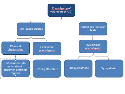 |
19 |
MR-based body composition analysis, resting state fMRI and
autonomic function evaluation for phenotyping biomarkers
CHETNA BANGA1,
DR. PREETI BHOSLE1,
DR. DUSHANT KUMAR1,
PROF. K.K DEEPAK2,
DR. DINU CHANDRAN3,
and PROF. RAMA JAYASUNDAR4
1NUCLEAR MAGNETIC RESONANCE, ALL INDIA INSTITUTE OF MEDICAL SCIENCES, NEW DELHI, NEW DELHI, IN, India, 2PHYSIOLOGY, ALL INDIA INSTITUTE OF MEDICAL SCIENCES, NEW DELHI, NEW DELHI, India, 3PHYSIOLOGY, ALL INDIA INSTITUTE OF MEDICAL SCIENCES, NEW DELHI, NEW DELHI, IN, India, 4NUCLEAR MAGNETIC RESONANCE, ALL INDIA INSTITUTE OF MEDICAL SCIENCES, NEW DELHI, NEW DELHI, India Keywords: Screening, Quantitative Imaging, MR body composition analysis, phenotyping, subcutaneous fat, resting state fMRI Phenotyping individuals are gaining importance in personalized medicine. This MR based body composition analysis and resting state (R-fMRI) along with autonomic functions (AFT) evaluation have determined parameters for physical, functional and physiological phenotyping in healthy volunteers (n=30). MR studies (at 3T) were R-fMRI using EPI sequence, subcutaneous fat evaluation by mDIXON sequence and AFT included parasympathetic (deep breathing, HUT) and sympathetic (HUT, hand grip test, cold pressor test). Increased functional activation, higher abdominal fat and fall in Baroreflex independent sympathetic reactivity were observed in volunteers having BMI ≥25 Kg/m2 compared to those with BMI <25Kg/m2. |
|
4454.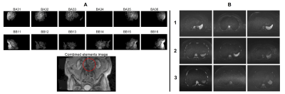 |
20 |
Evaluation of a vendor-agnostic scan-specific quality control
acquisition for clinical whole-body MRI (WB-MRI) protocols
Sam Keaveney1,2,
Georgina Hopkinson1,
Julia Markus3,
Andrew N Priest4,5,
Erica Scurr1,
Julie Hughes1,
Cheryl Richardson1,
Simon J Doran2,
David J Collins1,2,
Christina Messiou1,2,
Dow-Mu Koh1,2,
and Jessica M Winfield1,2
1MRI Unit, Royal Marsden NHS Foundation Trust, Sutton, United Kingdom, 2Division of Radiotherapy and Imaging, The Institute of Cancer Research, London, United Kingdom, 3Centre for Medical Imaging, University College London, London, United Kingdom, 4Department of Imaging, Addenbrookes Hospital, Cambridge University Hospital NHS Foundation Trust, Cambridge, United Kingdom, 5Department of Radiology, University of Cambridge, Cambridge, United Kingdom Keywords: Whole Body, Cancer, Quality Assurance / Quality Control A short quality control (QC) acquisition and analysis process was developed previously to detect faulty RF coil elements and coil positioning errors in whole-body (WB) MRI. This work demonstrates the feasibility of this process on systems from three manufacturers and reports on the evaluation of this approach following its addition to the clinical WB-MRI protocol for all examinations over a period of six months on six scanners, providing on-going monitoring of image quality across 450 examinations. Two previously unidentified broken RF coil elements were detected, which were affecting clinical image quality, demonstrating the value of this active detection approach. |
|
 Back to Meeting Home
 Back to the Program-at-a-Glance
The International Society for Magnetic Resonance in Medicine is accredited by the Accreditation Council for Continuing Medical Education to provide continuing medical education for physicians.
 Back to Meeting Home
 Back to the Program-at-a-Glance
 View Presentation Video