Digital Poster
Cancer: Technical Advances for Clinical Benefit
ISMRM & ISMRT Annual Meeting & Exhibition • 03-08 June 2023 • Toronto, ON, Canada

Digital Poster
Cancer: Technical Advances for Clinical Benefit
| Computer # | |||
|---|---|---|---|
1487.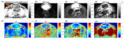 |
21 | Assessment of IVIM-DKI for characterization of benign and malignant lymph nodes in lymphoma
Archana Vadiraj Malagi1,2, Devasenathipathy Kandasamy3, Deepam Pushpam4, Kedar Khare5, Raju Sharma3, Rakesh Kumar6, Sameer Bakhshi4, and Amit Mehndiratta1,7
1Centre for Biomedical Engineering, Indian Institute of Technology Delhi (IIT Delhi), New Delhi, India, 2Biomedical Imaging Research Institute, Cedars-Sinai Medical Center, Los Angeles, CA, United States, 3Department of Radiodiagnosis, All India Institute of Medical Sciences Delhi, New Delhi, India, 4Department of Medical Oncology, Dr. B.R. Ambedkar Institute-Rotary Cancer Hospital (IRCH), All India Institute of Medical Sciences Delhi, New Delhi, India, 5Department of Physics, Indian Institute of Technology Delhi (IIT Delhi), New Delhi, India, 6Department of Nuclear Medicine, All India Institute of Medical Sciences Delhi, New Delhi, India, 7Department of Biomedical Engineering, All India Institute of Medical Sciences Delhi, New Delhi, India Keywords: Cancer, Diffusion/other diffusion imaging techniques Intravoxel Incoherent motion-diffusion kurtosis imaging (IVIM-DKI) was used for evaluation and characterization of malignant lymph nodes in lymphoma. A total of twenty-one (n=21) patients diagnosed with biopsy proven Hodgkin lymphoma(HL: n=13) or non-Hodgkin lymphoma(NHL: n=8) were prospectively evaluated. IVIM-DKI parameters were estimated using standard IVIM-DKI model (standard model) and IVIM-DKI model with total variation(TV) penalty function method(IDTV model). Perfusion fraction (f) and kurtosis (k) estimated using IDTV model, and apparent diffusion coefficient(ADC) were significantly(p<0.05) lower in malignant lymph nodes than benign lymph nodes. f and k showed high AUC of 0.88 and 0.83, respectively for malignant vs. benign lymph nodes. |
|
1488.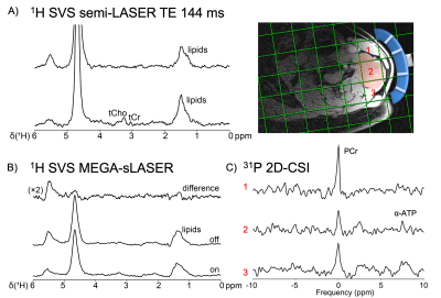 |
22 | 31P and 1H MRS for in vivo metabolic profiling of diffuse large B-cell lymphoma
Katharina J Wenger1,2,3,4, Seyma Alcicek1,2,3, Ulrich Pilatus1, Dennis C Thomas1,2,3, Elke Hattingen1,2,3,4, Florian Buettner4, Andreas Loth5, and Thomas Oellerich2,3,4,6
1Institute of Neuroradiology, University Hospital Frankfurt, Frankfurt am Main, Germany, 2University Cancer Center Frankfurt (UCT), Frankfurt am Main, Germany, 3Frankfurt Cancer Institute (FCI), Frankfurt am Main, Germany, 4German Cancer Research Center (DKFZ) Heidelberg, Germany and German Cancer Consortium (DKTK), Partner Site Frankfurt/Mainz, Frankfurt am Main, Germany, 5Department of Oto-Rhino-Laryngology, University Hospital Frankfurt, Frankfurt am Main, Germany, 6Department of Hematology/Oncology, University Hospital Frankfurt, Frankfurt am Main, Germany Keywords: Cancer, Tumor, lymphoma, multiomics, MR-Spectroscopy With the presented protocol, key metabolites related to different metabolic profiles of aggressive lymphomas (such as lactate, alanine, as a surrogate for pyruvate, as well as choline components) can be detected in vivo in a clinical setting. |
|
1489.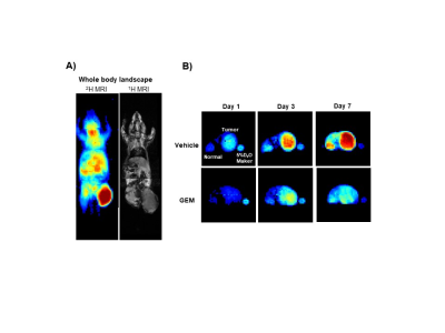 |
23 | Using D2O-Induced 2H labeling, Deuterium MRI at 1.5T Enables In Vivo Visualization of Body Tissues and Early Assessment of Anticancer Therapies
Abdelazim Elsayed Elhelaly1,2, Fuminori Hyodo1, Yoshifumi Noda3, Hiroki Kato3, Koki Ichihashi4, Hiroyuki Tomita4, and Masayuki Matsuo3
1Department of Radiology, Frontier Science for Imaging, Gifu University, Gifu, Japan, 2Department of Food Hygiene and Control, , Faculty of Veterinary Medicine, Suez Canal University, Ismailia, Egypt, 3Department of Radiology, School of Medicine, Gifu University, Gifu, Japan, 4Department of Tumor Pathology, Gifu University Graduate School of Medicine, Gifu University, Gifu, Japan Keywords: Cancer, Deuterium, deuterium MR imaging DMI emerged as a promising alternative in cancer metabolic imaging. We D2O-induced 2H-labeling followed by DMI to study deuterium kinetics and track tumor response to treatment at 1.5T. We succeeded to use dMRI to monitor 2H kinetics in tissues of a pancreatic carcinoma model. Higher 2H build-up was observed in tumor. Treated mice showed a significant decrease in 2H in tumor during 7 days of treatment and before anatomical changes were detectable. Tumor homogenates of treated mice also showed a significant reduction in 13C lactate production. DMI of tumors is a potential imaging method for assessment of early treatment responses |
|
1490.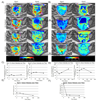 |
24 | Quantification of Acute and Chronic Fibrosis in Gynecologic and Prostate Cancer over the course of Radiotherapy
Khadija Sheikh1, Akila N Viswanathan1, Daniel Y Song1, Junichi Tokuda2, Junghoon Lee1, Michael Jerosch-Herold2, Mindy K Graham1, Ravi Seethamraju3, Thomas Benkert4, Himanshu Bhat5, Bruce L Daniels6, and Ehud J Schmidt1,7
1Radiation Oncology and Molecular Sciences, Johns Hopkins University School of Medicine, Washington, DC, United States, 2Radiology, Brigham and Women’s Hospital, Boston, MA, United States, 3Siemens Medical Solutions, Boston, MA, United States, 4MR Applications Predevelopment, Siemens Healthcare GmbH, Erlangen, Germany, 5Siemens Healthineers, Boston, MA, United States, 6Radiology, Stanford University, Stanford, CA, United States, 7Medicine (Cardiology), Johns Hopkins University School of Medicine, Baltimore, MD, United States Keywords: Pelvis, Radiotherapy, acute and chronic fibrosis We developed means to quantify the volume and voxel-wise fraction of acute (FA) and chronic fibrosis (FC) present during and following radiation therapy (RT), using non-contrast and contrast enhanced stack-of-spirals IR-UTE scans. Imaging was performed in gynecologic and prostate cancer patients at multiple time points in relation to RT (pre-RT, on-RT, post-RT, and post-3 months RT). This study provides mm-resolution quantitative information on fibrosis changes during the course of RT based on MRI. Increases in FA, followed by subsequent-time FC increases were observed in both prostate and gynecologic cancer patients. |
|
1491.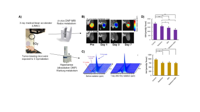 |
25 | Early monitoring of radiation treatment based on tumor redox status using in vivo dynamic nuclear polarization MRI
Fuminori Hyodo1, Norikazu Koyasu1, Ryota Iwasaki1, Hinako Eto2, Abdelazim Elsayed Elhelaly1, Hiroyuki Tomita1, Takashi Elsayed Mori1, Masaharu Murata2, Yoshifumi Noda1, Hiroki Kato1, and Masayuki Matsuo1
1Gifu University, Gifu, Japan, 2Kyushu University, Fukuoka, Japan Keywords: Cancer, Hyperpolarized MR (Non-Gas), redox, DNP MRI, radiation In vivo dynamic nuclear polarization-MRI (DNP-MRI, also called OMRI, PEDRI) using carbamoyl-PROXYL(CmP) as a redox sensitive DNP probe enables the accurate monitoring of the tissue redox status. We found that the redox status decreases 1 day after radiation treatment, and the decay of redox status occurs before any micro- or macroscopic changes in tumor morphology and pyruvate metabolism based on the Warburg effect. This decay of redox status can also be associated with the decreased production of intratumor reducing redox molecules such as GSH and AsA. |
|
1492.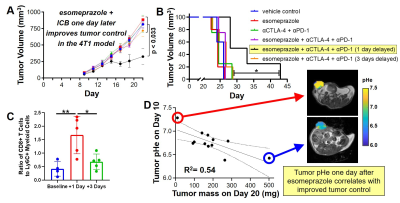 |
26 | A pH-sensitizer can improve cancer immunotherapy treatment as monitored with acidoCEST MRI
Renee L Chin1,2, Jorge de la Cerda1, F William Schuler3, Sanhita Sinharay4, and Mark D Pagel1
1Cancer Systems Imaging, UT MD Anderson Cancer Center, Houston, TX, United States, 2Immunology, MD Anderson Cancer Center, Houston, TX, United States, 3Cancer Systems Imaging, MD Anderson Cancer Center, Houston, TX, United States, 4Center for Biosystems Science and Engineering, Indian Institute of Science, Bangalore, India Keywords: Cancer, CEST & MT, immunotherapy Tumor acidosis causes resistance to immune checkpoint blockade (ICB) immunotherapy. We have identified a “pH-sensitizer” that increases the extracellular pH of the tumor microenvironment, improving immunogenicity and tumor control with ICB. AcidoCEST MRI can measure the extracellular pH of the tumor microenvironment. We used acidoCEST MRI to correlate the increase in tumor pHe immediately caused by the pH-senstizer with the tumor volume at study endpoint, in a pre-clinical model of breast cancer. These results show that acidoCEST MRI can contribute to predicting the outcome of immunotherapy. |
|
1493.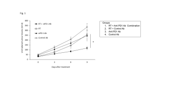 |
27 | MRI assessment of abscopal effect enhanced by immune checkpoint blockade
Kota Yamashita1, Kazumasa Horie1, Kazutoshi Yamamoto1, Jeffery R. Brender1, Nallathamby Devasahayam1, Murali C. Krishna1, and Shun Kishimoto1
1National Cancer Institute, Bethesda, MD, United States Keywords: Cancer, DSC & DCE Perfusion Radiation therapy (RT) on primary tumor rarely induces regression of non-irradiated metastatic lesions (abscopal effect), which is reported to be enhanced by immune-checkpoint blockade. In this study, we examined the physiological changes induced by abscopal effect using MRI-based imaging modalities such as EPR oximetry, DCE MRI, and 13C DNP MRI. Hypoxic fraction < 10 mmHg (HF10), permeability, perfusion, and CD8+ T cell infiltration in metastatic tumor increased after the combination of RT and PD-1 blockade. Interestingly, higher permeability/perfusion and lower HF10 in primary tumor before the treatment were associated with slower growth of the metastatic tumor after the treatment. |
|
1494.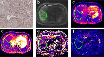 |
28 | Preoperative assessment of vessels encapsulating tumor clusters (VETC) in hepatocellular carcinoma using IVIM diffusion-weighted imaging
Chenhui Li1, Jinhuan Xie1, Liling Long1, Huiting Zhang2, and Yang Song2
1The First Affiliated Hospital of Guangxi Medical University, Nanning, China, 2MR Scientific Marketing, Siemens Healthineers, Shanghai, China Keywords: Cancer, Liver Vessels encapsulating tumor clusters (VETC) is a powerful predictor of a poor prognosis in patients with hepatocellular carcinoma (HCC) .Therefore, preoperative radiologic prediction of these histopathological results can allow optimized management of HCC and help improve long-term survival for patients.However,to our knowledge, no published studies have investigated preoperative VETC patten of HCC using IVIM parameters and comparing with DWI. Our study indicated that IVIM-derived D* provides a non-invasive analytical approach for preoperative predicting VETC of HCC.ADC value were not predictive for VETC. |
|
1495.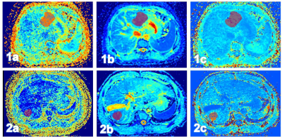 |
29 | Texture Analysis of Diffusion Kurtosis Imaging for Predicting Early Recurrence of Hepatocellular Carcinoma After Hepatectomy
Yue Wang1, Ying Zhao1, and Ailian Liu1
1the First Affiliated Hospital of Dalian Medical University, Dalian, China Keywords: Cancer, fMRI The global incidence and mortality of hepatocellular carcinoma (HCC) are high [1]. Although radical resection of HCC is the most effective treatment, the postoperative recurrence rate is high. Diffusion kurtosis imaging (DKI) is based on the non-Gaussian diffusion motion of water molecules, which can quantitatively reflect the characteristics of lesions from the perspective of diffusion and heterogeneity[2]. Texture analysis provides information about tissue complexity and heterogeneity based on mathematical methods. This study showed that DKI-based entire-tumor texture analysis could assist in prediction of the early recurrence of HCC patients after hepatectomy, which was beneficial to improve the prognosis. |
|
1496.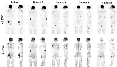 |
30 | Clinical assessment of noise corrected exponentially weighted diffusion weighted MRI (niceDWI) for imaging patients with metastatic melanoma
Annemarie Knill1,2, Matthew Blackledge1, Jessica Winfield1,2, Hannah Barnsley2, Benjamin Malawo2, Georgina Hopkinson2, Dow-Mu Koh1,2, Samuel Withey2, and Christina Messiou1,2
1The Institute of Cancer Research, London, United Kingdom, 2The Royal Marsden NHS Foundation Trust, London, United Kingdom Keywords: Cancer, Diffusion/other diffusion imaging techniques Systematic clinical validation of noise-corrected, exponentially-weighted, diffusion-weighted MRI (niceDWI) is performed in metastatic melanoma including soft tissue disease. Previous assessments of niceDWI demonstrated improvement in the in-plane bias field. No clinical advantage is found over clinical DWI: the signal-to-noise ratio was assessed as significantly worse in niceDWI both quantitatively and by an experienced reader; there was no difference demonstrated in the contrast-to-noise ratio (CNR) of bone or nodal lesions; when soft tissue disease is included CNR is significantly worse on niceDWI. It is recommended that future evaluations of niceDWI assess the maximum intensity projection for use as a survey tool. |
|
1497. |
31 | Detecting cancer immunotherapy response in ccRCC patients from 1H MRS of plasma
Raj Kumar Sharma1, Michael T. McMahon2, Meiyappan Solaiyappan1, Yasser Ged3, and Zaver M Bhujwalla1
1Division of Cancer Imaging Research, The Russell H. Morgan Department of Radiology and Radiological Science, JOHNS HOPKINS UNIVERSITY, SCHOOL OF MEDICINE, BALTIMORE, MD, United States, 2F.M. Kirby Research Center for Functional Brain Imaging, Kennedy Krieger Institute, JOHNS HOPKINS UNIVERSITY, SCHOOL OF MEDICINE, Baltimore, MD, United States, 3Genitourinary Oncology, Sidney Kimmel Comprehensive Cancer Center, JOHNS HOPKINS UNIVERSITY, SCHOOL OF MEDICINE, Baltimore, MD, United States Keywords: Cancer, Metabolism, Clear Cell Renal Cell Carcinoma , Cancer , 1H MR metabolomics A minimally invasive, blood-based assay to predict and detect response to immune-checkpoint inhibitors (ICIs) would be of significant importance. 1H MRS of plasma samples provides a robust reproducible assay that require minimal sample preparation, and convenience of sample storage. Here we evaluated changes in plasma metabolites of clear cell renal cell carcinoma (ccRCC) patients receiving ICIs using 1H MRS. We identified a significant increase of acetate, formate, glutamine, glycine, myoinositol, and lactate following ICI that may reflect response. |
|
1498.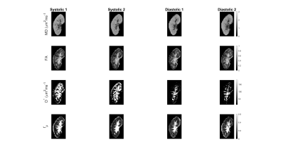 |
32 | The effect of cardiac gating on the repeatability of quantitative renal diffusion MRI
Nima Gilani1, Artem Mikheev1, Inge Manuela Brinkmann2, Dibash Basukala1, Thomas Benkert3, Malika Kumbella1, James S. Babb1, Hersh Chandarana1, and Eric E. Sigmund1
1Department of Radiology, NYU Langone Health, New York, NY, United States, 2Siemens Medical Solutions USA Inc., New York, NY, United States, 3Siemens Healthcare GmbH, Erlangen, Germany Keywords: Kidney, Diffusion/other diffusion imaging techniques The effect of cardiac gating on quantitative diffusion weighted magnetic resonance imaging of the kidney was investigated using an advanced cardiac triggered diffusion-weighted imaging sequence which allows the acquisition and estimation of diffusion tensor imaging and intra-voxel incoherent motion parameters. Cardiac gating significantly influences the repeatability of IVIM parameters that reflect flow of blood and fluids in the kidney and allow interpretations of renal function. |
|
1499.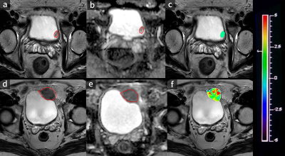 |
33 | Evaluation of Amide Proton Transfer Imaging for Bladder Cancer Histopathologic Features: A Comparative Study with Diffusion- Weighted Imaging
Fang Wang1, Yong-Sheng Xiang1, Peng Wu2, Ai-Jun Shen1, and Pei-Jun Wang1
1Tongji Hospital, School of Medicine, Tongji University, Shanghai, China, 2Philips Healthcare, Shanghai, China Keywords: Urogenital, Bladder Amide proton transfer (APT) imaging is an emerging chemical exchange saturation transfer (CEST)-based MRI technique that is sensitive to mobile proteins and peptides in tissue and has drawn considerable attention in the field of cellular and molecular imaging.In the present work, our result demonstrates that APT imaging can predict tumor grade and muscular invasion in bladder cancer, the diagnostic performance for evaluating muscular invasion is better than that of diffusion-weighted imaging (DWI) and adding APT imaging to DWI significantly improved the diagnostic accuracy for evaluating muscular invasion versus DWI alone, thus opening new research avenues in this field. |
|
1500.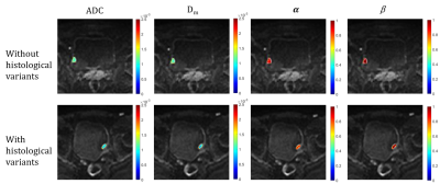 |
34 | Characterizing pathological grade and histological variant of bladder urothelial carcinoma with a continuous-time random walk diffusion model
Wei Wang1, Ke Xue2, Yongming Dai2, and Jianxing Qiu1
1Department of Radiology, Peking University First Hospital, Beijing, China, 2MR Collaboration, Central Research Institute, Shanghai United Imaging Healthcare, Shanghai, China Keywords: Urogenital, Diffusion/other diffusion imaging techniques Accurately determining the histologic grade and variant status is essential. In this study, the diagnostic value of the CTRW model in characterizing histological grade or histological variants was evaluated and compared with ADC. The optimal diagnostic performance of CTRW diffusion model suggested it could provide more diagnostic value than conventional ADC not only for pathological grading but also for histological variant of bladder urothelial carcinoma. |
|
1501.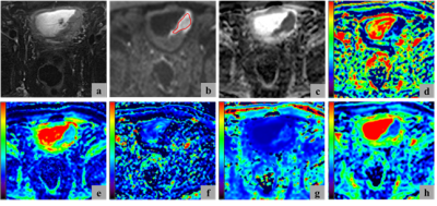 |
35 | Models based on diffusion-weighted imaging in tumor staging and histologic grading of bladder cancer: A comparison study
Cong You1, Yujiao Zhao2, Cheng Zhang1, Mengyao Chen1, Jinxia Zhu3, Feifei Qu3, Thomas Benkert4, Robert Grimm4, and Wen Shen2
1The First Central Clinical School, Tianjin Medical University, Tianjin, China, 2Department of Radiology, Tianjin First Central Hospital, School of Medicine, Nankai University, Tianjin, China, 3MR Collaboration, Siemens Healthineers Ltd., Beijing, China, 4MR Application Predevelopment, Siemens Healthcare GmbH, Erlangen, Germany Keywords: Urogenital, Diffusion/other diffusion imaging techniques, bladder cancer; diffusion-weighted imaging; monoexponential model;intravoxel incoherence motion;diffusion kurtosis imaging This comparative study aimed to investigate the ability of Gaussian distribution models, including monoexponential model (MEM) and intravoxel incoherence motion (IVIM), and non-Gaussian diffusion kurtosis models to differentiate the pathologic stages and histologic grades of bladder cancer. The results indicated that the diffusion kurtosis imaging (DKI) parameters had the highest diagnostic performance. The mean kurtosis (MK) value among individual parameters and the combination of MK and mean diffusivity (MD) among combined values had the largest area under the curves. Also, the MK values were most strongly correlated with the Ki-67 labeling index. |
|
1502.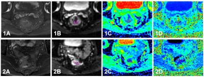 |
36 | Evaluation of Diffusion Kurtosis Imaging in Predicton of Tumor Budding Grade in Rectal Cancer
Yue Wang1, Anliang Chen1, Lizhi Xie2, and Ailian Liu1
1the First Affiliated Hospital of Dalian Medical University, Dalian, China, 2GE Healthcare (China), Beijing, China Keywords: Cancer, Quantitative Imaging, tumor budding This study used diffusion kurtosis imaging (DKI) to predict the tumor budding (TB) grade in rectal cancer. The results showed that the parameters MK, MD and their combination parameter both perform great diagnostic efficiency. DKI could noninvasively predict TB grade of rectal cancer, which is an important prognostic information, and has important guiding significance for clinical treatment. |
|
1503.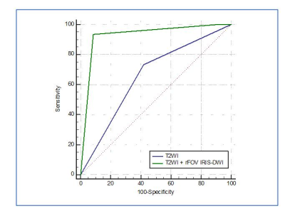 |
37 | The Added Value of Reduced Field-of-view IRIS-DWI Sequence for T2WI Sequence in T Staging of Rectal Cancer
Ziyou Wang1, Chong Wang2, Haini Zhang1, Jianwei Zeng2, Zhongxiao Liu2, Wenbei Xu1, Shenman Qiu2, Yanchun Zhang3, Hao Wang3, Peng Wu4, Yankai Meng2, and Kai Xu2
1Xuzhou medical university, Xuzhou,Jiangsu Province, China, 2The affilliated hospital of Xuzhou Medical University, Xuzhou,Jiangsu Province, China, 3Suining Country People's hospital affiliated to Xuzhou Medical University, Xuzhou,Jiangsu Province, China, 4Philips Healthcare, Shanghai Province, China Keywords: Cancer, Diffusion/other diffusion imaging techniques, diagnosis confidence We aimed to evaluate the added value of reduced field-of-view (rFOV) IRIS-DWI sequence for T2WI sequence in evaluating T staging of rectal cancer. ROC analysis showed the diagnosis performance based on T2WI + rFOV IRIS-DWI imaging was superior to single T2WI sequence. The AUC, sensitivity and specificity were 0.928, 93.3%, 91.7% and 0.658, 73.3%, 58.3% for two diagnosis methods respectively. The result showed significant difference between two methods with P = 0.002. rFOV IRIS-DWI combined T2WI imaging could provide higher diagnosis confidence and higher diagnosis accuracy for rectal cancer T staging. |
|
1504. |
38 | Discriminating Locally advanced and Metastatic Lung Cancer using Restriction Spectrum Imaging and Intravoxel Incoherent Motion Model
Zhongyan Xiong1, Yu Luo2, Yang Yang3, Meiyun Wang2, Xin Liu4,5, Hairong Zheng1, and Chao Zou4,5
1Institute of Biomedical and Health Engineering, Shenzhen Institutes of Advanced Technology, Chinese Academy of Sciences, Shenzhen, China, 2Department of Medical Imaging, Henan Provincial People’s Hospital & Zhengzhou University People’s Hospital, Zhengzhou, China, 3Central Research Institute, United Imaging Healthcare, Shanghai, China, 4Paul C. Lauterbur Centre for Biomedical Imaging, Shenzhen Institutes of Advanced Technology, Chinese Academy of Sciences, Shenzhen, China, 5Key Laboratory for Magnetic Resonance and Multimodality Imaging of Guangdong Province, Shenzhen, China Keywords: Lung, Diffusion/other diffusion imaging techniques A novel diffusion weighted imaging model called restriction spectrum imaging (RSI) captures the distinct diffusion behavior of tumors. The restricted diffusion correlates to tumor cellularity, a potential indicator of cancer aggressiveness. To assess its capability of characterizing lung cancer metastasis, we applied a three-compartment RSI model and intravoxel incoherent motion model to DWI images of locally advanced and metastatic lung cancer patients with the diagnosis of biopsy. The RSI model demonstrated its ability to discriminate the lung cancer of locally advanced (III) and metastatic (IV) stage, and the results outperforms the traditional IVIM model. |
|
1505. |
39 | Study on the influence of different scanning directions of whole-body MRI on the detection of lesions
Nan Wang1, Ailian Liu1, Guobin Li2, Shuheng Zhang2, Shaoxin Xiang3, Yongming Dai3, and Qingwei Song1
1the First Affiliated Hospital of Dalian Medical University, Dalian, China, 2Shanghai United Imaging Healthcare Co., Ltd, Shanghai, China, 3MR Collaboration, Central Research Institute, United Imaging Healthcare, Shanghai, China Keywords: Cancer, Whole Body Whole-body MRI (WB-MRI) is proposed as a potential modality to evaluate the entire body with excellent spatial resolution and high sensitivity. However, the long scanning time of WB-MRI has still limited the clinical application. This study compared the time cost and lesion display effect of WB-MRI in different scan directions. This study demonstrated that using coronal scanning in WB-MRI is an effective method to reduce the scan time and achieve good performance in detecting lesions in clinical applications. |
|
 Back to Meeting Home
 Back to the Program-at-a-Glance
The International Society for Magnetic Resonance in Medicine is accredited by the Accreditation Council for Continuing Medical Education to provide continuing medical education for physicians.
 Back to Meeting Home
 Back to the Program-at-a-Glance
 View Presentation Video