Digital Poster
Tumor Characterization
ISMRM & ISMRT Annual Meeting & Exhibition • 03-08 June 2023 • Toronto, ON, Canada

| Computer # | |||
|---|---|---|---|
1664.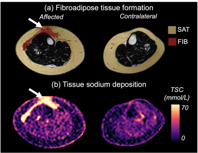 |
21 | Multi-nuclear 1H and 23Na MRI investigation of fibroadipose tissue sodium in patients with secondary lymphedema
Shannon L Taylor1, Michael D Pridmore2, Maria E Garza2,3, Vanessa Crain2, Sheau-Chiann Chen4, Alaina J Brown5, Yu Luo2, Paula MC Donahue6,7, Manus J Donahue3,8, and Rachelle L Crescenzi1,2
1Biomedical Engineering, Vanderbilt University, Nashville, TN, United States, 2Radiology and Radiological Sciences, Vanderbilt University Medical Center, Nashville, TN, United States, 3Neurology, Vanderbilt University Medical Center, Nashville, TN, United States, 4Biostatistics, Vanderbilt University Medical Center, Nashville, TN, United States, 5Obstetrics and Gynecology; Division of Gynecologic Oncology, Vanderbilt University Medical Center, Nashville, TN, United States, 6Physical Medicine and Rehabilitation, Vanderbilt University Medical Center, Nashville, TN, United States, 7Dayani Center for Health and Wellness, Vanderbilt University Medical Center, Nashville, TN, United States, 8Psychiatry and Behavioral Sciences, Vanderbilt University Medical Center, Nashville, TN, United States Keywords: Cancer, Non-Proton, lymphedema, fibrosis, sodium, inflammation, adipose, lymph Advanced-stage lymphedema is a disease of chronic fluid accumulation and tissue inflammation that results in fibrosis and subcutaneous fat expansion in affected limbs. Assessment of fat and fibrosis currently relies on biopsies or non-specific bedside measures. Noninvasive MRI could be important for understanding pathophysiology and evaluating emerging therapies. We cross-sectionally evaluated multi-nuclear proton and sodium MRI in patients with lower-extremity secondary lymphedema. Results reveal subcutaneous adipose tissue expansion throughout the affected calf, locoregional fibrosis, and elevated sodium content in fibroadipose tissues particularly in the anterior calf, thereby highlighting the relevance of these technologies for interrogating tissue health in lymphedema. |
|
1665.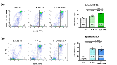 |
22 | Mammary tumor COX-2 expression in immunocompromised mice contributes to splenic MDSC expansion and alters splenic metabolism
James Dion Barnett1, Marie-France Penet1,2, Santosh Kumar Bharti1, Yelena Mironchik1, Balaji Krishnamachary1, and Zaver M Bhujwalla1,2,3
1The Russell H. Morgan Department of Radiology and Radiological Science, Johns Hopkins University School of Medicine, Baltimore, MD, United States, 2Sidney Kimmel Comprehensive Cancer Center, Baltimore, MD, United States, 3Department of Radiation Oncology and Molecular Radiation Sciences, Baltimore, MD, United States Keywords: Cancer, Preclinical, Spleen, MDSC, Cancer, COX-2 Tumor COX-2 expression in breast cancer has been associated with increased aggressiveness. Here we have investigated the effects of tumor COX-2 levels on spleen metabolism and on the expansion of myeloid derived suppressor cells (MDSCs) that potentiate cancer progression. Studies were performed with human triple negative breast cancer cells in immune suppressed mice and mouse breast cancer cells in immune competent mice. COX-2 levels significantly altered splenic metabolism and MDSC expansion in immune suppressed mice but not in immune competent mice that may have implications for breast cancer patients that are immune compromised. |
|
1666. |
23 | Standardized volume and IVIM parameters of spleen in patients with acute leukemia: the value of evaluating tumor burden and prognosis
Wenjin Bian1, Jinliang Niu1, jianting Li1, and Wenqi Wu1
1The Second Hospital of Shanxi Medical University, Taiyuan, China Keywords: Cancer, Diffusion/other diffusion imaging techniques, spleen This study investigated the values of standardized volume and intravoxel incoherent motion (IVIM) parameters of spleen in evaluating tumor burden and prognosis in newly diagnosed acute leukemia (AL). Eighty-five AL patients and seventy-four healthy volunteers were recruited and underwent IVIM in the abdomen on a 3.0T MRI system. The results showed standardized volume and IVIM parameters of spleen were associated with tumor burden and treatment response in AL, and 218.1cm3 was the threshold of standardized spleen volume for predicting treatment response, which indicated that they can be potentially useful in tailoring the individualized treatment plan for each patient. |
|
1667.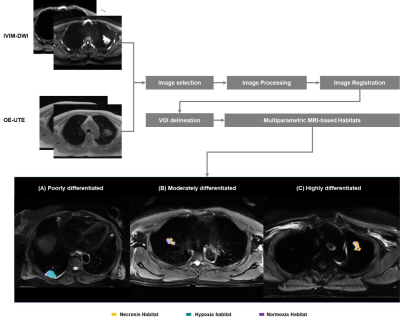 |
24 | Habitat Analysis Based on Multiparametric MRI Predicts Lung Adenocarcinoma Subtypes
Gaofeng Shi1, Qi Wang1, Hui Feng1, Hui Liu1, Mengyu Song1, Xinyue Liang2, and Yongming Dai2
1Department of Radiology, Fourth Hospital of Hebei Medical University, Shijiazhuang, China, 2Central Research Institute, United Imaging Healthcare, Shanghai, China Keywords: Cancer, Lung As a promising approach to analyze tumor heterogeneity through image features, habitat analysis has been applied for a variety of tumors, such as lung adenocarcinoma (LUAD). To our knowledge, the previous applications, mainly based on CT and PET images, would potentially hold a limited impact on the power of habitats analysis. In this study, our goal is to extend the applications of habitat analysis in LUAD with multi-parametric MR images, which would be obtained routinely longitudinally and to guide specific therapies. |
|
1668. |
25 | Prediction of visceral pleural invasion in early-stage non-small cell lung cancer by T2WI
Ning Zhang1, Bei Liu1, Gaofeng Shi1, Qian Xu1, Hui Feng1, Hui Liu1, Chen Zhang2, and Fan Yang1
1Hebei Medical University Fourth Affiliated Hospital, Shijiazhuang, China, 2MR Scientific Marketing, Siemens Healthineers, Beiing, China Keywords: Cancer, Lung This study investigated the clinical value of MRI T2WI for VPI in early-stage NSCLC. Four MRI signs of VPI in lung cancer were summarized by MR T2WI of patients with early-stage NSCLC suspected of VPI. Among them, category 3 signs and category 4 signs had higher positive predictive values. This indicates that T2WI is useful in the prediction of VPI in early-stage NSCLC. |
|
1669.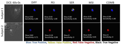 |
26 | Tumor segmentation with nnU-Net on dynamic contrast enhanced MR images of triple negative breast cancer
Zhan Xu1, Sanaz Pashapoor2, Bikash Panthi1, Jong Bum Son1, Ken-Pin Hwang1, Beatriz Elena Adrada2, Rosalind Pitpitan Candelaria2, Mary Saber Guirguis2, Miral Mahesh Patel2, Huong Le-Petross2, Jessica Leung2, Marion Elizabeth Scoggins2, Gary Whitman2, Rania Mohamed2, Deanna Lynn Lane2, Tanya Moseley2, Frances Perez2, Jason White3, Elizabeth Ravenberg3, Alyson Clayborn3,
Huiqin Chen4, Jia Sun4, Peng Wei4, Alastair Thompson5, Anil Korkut6, Lei Huo7, Kelly Hunt8, Stacy Moulder3, Jennifer Litton3, Vicente Valero3, Debu Tripathy9, Wei Yang2, Clinton Yam3, Gaiane Margishvili Rauch2, and Jingfei Ma1
1Department of Imaging Physics, MD Anderson Cancer Center, Houston, TX, United States, 2Departments of Breast Imaging, MD Anderson Cancer Center, Houston, TX, United States, 3Departments of Breast Medical Oncology, MD Anderson Cancer Center, Houston, TX, United States, 4Departments of Biostatistics, MD Anderson Cancer Center, Houston, TX, United States, 5Section of Breast Surgery, Baylor College of Medicine, Houston, TX, United States, 6Departments of Bioinformatics & Computational Biology, MD Anderson Cancer Center, Houston, TX, United States, 7Departments of Pathology, MD Anderson Cancer Center, Houston, TX, United States, 8Departments of Breast Surgical Oncology, MD Anderson Cancer Center, Houston, TX, United States, 9Department of Breast Medical Oncology, MD Anderson Cancer Center, Houston, TX, United States Keywords: Cancer, Breast, TNBC Quantitative image analysis of cancers requires accurate tumor segmentation that is often performed manually. In this study, we developed a deep learning model with the self-configurable nnU-Net for automated tumor segmentation on dynamic contrast enhanced MR images of triple negative breast cancers. Our results on an independent testing dataset demonstrated that this nnU-Net based deep learning model can perform automated tumor segmentation with high sensitivity and Dice coefficient. |
|
1670.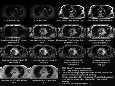 |
27 | Deep Learning Reconstruction: Capability for Image Quality and Staging Accuracy Improvements on Chest MRI in NSCLC Patients
Yoshiharu Ohno1,2, Kaori Yamamoto3, Masato Ikedo3, Masao Yui3, Akiyoshi Iwase4, Yuka Oshima5, Nayu Hamabuchi5, Satomu Hanamatsu5, Hiroyuki Nagata2, Takahiro Ueda1, Hirotaka Ikeda1, Takeshi Yoshikawa1,6, Daisuke Takenaka1,6, Yoshiyuki Ozawa1, and Hiroshi Toyama1
1Radiology, Fujita Health University School of Medicine, Toyoake, Japan, 2Joint Research Laboratory of Advanced Medical Imaging, Fujita Health University School of Medicine, Toyoake, Japan, 3Canon Medical Systems Corporation, Otawara, Japan, 4Fujita Health University Hospital, Toyoake, Japan, 5Fujita Health University School of Medicine, Toyoake, Japan, 6Diagnostic Radiology, Hyogo Cancer Center, Akashi, Japan Keywords: Cancer, Lung, Staging Deep learning reconstruction (DLR) has been applied in routine clinical practice and started to demonstrate its’ potential in different MR examinations. However, no one have evaluated the utility of DLR for chest MRI, yet. We hypothesize that DLR method is useful for chest MRI and improve image quality and diagnostic performance for T and N factor evaluations in non-small cell lung cancer (NSCLC) patients. The purpose of this study was to determine the influence of DLR method on image quality and diagnostic performance for T and N factor evaluations at chest MRI in NSCLC patients. |
|
1671.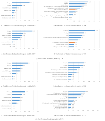 |
28 | Radiomic signature based on enhanced CT and 3T MRI for survival analysis in patients with esophageal squamous carcinoma
Dexuan Li1, Chenglong Wang1, Funing Chu2, Jinrong Qu2, Yang Song3, and Guang Yang1
1Shanghai Key Laboratory of Magnetic Resonance, East China Normal University, Shanghai, China, 2Department of Radiology, The Affiliated Cancer Hospital of Zhengzhou University &Henan Cancer Hospital, Zhengzhou, China, 3MR Scientific Marketing, Siemens Healthcare, Shanghai, China Keywords: Cancer, Tumor We enrolled 478 patients with esophageal squamous carcinoma (ESCC) split them into a training and a test cohort with a ratio of 7 to 3. Radiomic features were extracted from lesions on both MR and CT images and used to build models for predicting disease-free survival (DFS) and overall survival (OS). For both MR and CT images, the radiomic signature combined with clinical variables achieved performance comparable to radiological signature. Over the test cohort, MRI-based models achieved C-index values of 0.707 and 0.663 for DFS and OS predictions, respectively; CT-based models achieved 0.731 and 0.68 for DFS and OS, respectively. |
|
1672.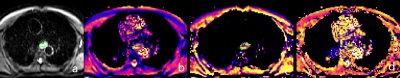 |
29 | Application of fractional order calculus diffusion model in predicting the differentiation in esophageal squamous cell carcinoma
Keke Zhao1, Shaoyu Wang2, Hongkai Zhang1, Jinrong Qu1, and Hailiang Li1
1Henan Tumor Hosptial, Zhengzhou, China, 2MR Scientific Marketing, Siemens Healthineers, Shanghai, China Keywords: Cancer, Cancer, esophageal squamous cell carcinoma A total of 50 patients with locally advanced ESCC were prospectively enrolled in this study. Diffusion Weighted Imaging (DWI) with multiple high b-values was performed before surgery, and a new set of parameters (D, β and m) from a fractional order calculus (FROC) diffusion model were acquired. The grade of differentiation about ESCC was assessed. We found that β and D value exhibited non-significant difference, and m value showed a significant difference between different grades ESCC (P =0.029). |
|
1673.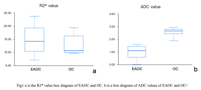 |
30 | R2* and ADC values in differentiating endometriosis-associated ovarian cancer from ovarian cancer
Ye Li1, Ailian Liu1, and Jiazheng Wang2
1The First Affiliated Hospital of Dalian Medical University, Dalian, China, 2Clinical and Technical Support, Philips Healthcare, Beijing, China Keywords: Cancer, fMRI A total of 9 lesions from 7 EAOC patients and 21 lesions from 19 OC patients were collected to compare the differences in R2* and ADC values between the EAOC and OC groups. The results showed that the ADC values were able to discriminate between the two and there was no difference in R2* values. |
|
1674.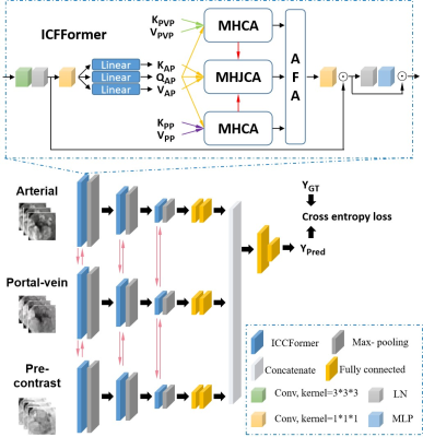 |
31 | Multi-modal fusion with joint conditional transformer for grading hepatocellular carcinoma
Shangxuan Li1, Yanshu Fang2, Guangyi Wang3, Lijuan Zhang4, and Wu Zhou1
1School of Medical Information Engineering, Guangzhou University of Chinese Medicine, Guangzhou, China, 2First Clinical Medical College, Guangzhou University of Chinese Medicine, Guangxhou, China, 3Department of Radiology, Guangdong Provincial People’s Hospital, Guangzhou, China, 4Shenzhen Institutes of Advanced Technology, Chinese Academy of Sciences, Shenzhen, China Keywords: Cancer, Machine Learning/Artificial Intelligence Multimodal medical imaging plays an important role in the diagnosis and characterization of lesions. Transformer pays more attention to global relationship modeling in data, which has obtained promising performance in lesion characterization. We propose a multi-modal fusion network with jointly conditional transformer to realize adaptive fusion of multimodality information and mono-modality feature learning constrained by other modal conditions. The experimental results of the clinical hepatocellular carcinoma (HCC) dataset show that the proposed method is superior to the previously reported multimodal fusion methods for HCC grading. |
|
1675.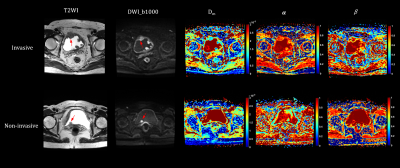 |
32 | Continuous-time random walk diffusion model combined with VI-RADS to predict muscle invasion of bladder cancer
Wei Wang1, Ke Xue2, Yongming Dai2, and Jianxing Qiu1
1Department of Radiology, Peking University First Hospital, Beijing, China, 2MR Collaboration, Central Research Institute, Shanghai United Imaging Healthcare, Shanghai, China Keywords: Cancer, Bladder, diffusion/other diffusion techniques Accurately differentiation of MIBC from non-muscle-invasive bladder cancer (NMIBC) is essential for selecting the optimal treatment plan and obtaining a better prognosis for BCa patients. In this study, the role of the CTRW model, VI-RADS and their combination in characterizing MIBC was evaluated. The CTRW parameters combined with VI-RADS could provide significantly better diagnostic performance for MIBC determination than the VI-RADS score alone. The CTRW model could serve as a compliment to VI-RADS and provide added value for predicting muscle invasion of bladder cancer. |
|
1676.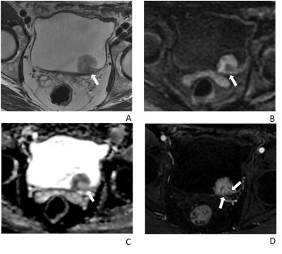 |
33 | Multiparametric MRI Evaluation of Vesical Imaging‑Reporting and Data System (VI‑RADS) for bladder cancer after Neoadjuvant Chemotherapy
Xinxin Zhang1, Yichen Wang1, Sicong Wang2, Jianzhong Shou1, Yan Chen1, and Xinming Zhao1
1National Cancer Center/National Clinical Research Center for Cancer/Cancer Hospital, Chinese Academy of Medical Sciences and Peking Union Medical College, Beijing, 100021, China, Beijing, China, 2GE Healthcare, MR Research China, Beijing, Beijing, China Keywords: Cancer, Bladder Local tumor restaging after neoadjuvant chemotherapy (NAC) treatment is urgently needed. Therefore, the aim of this study was to investigate whether VI-RADS could accurately detect muscle invasion in BC patients after NAC treatment. VI-RADS scores of bladder cancer after NAC were independently assessed by two radiologists. With an optimal cut-off value ≥4, the AUC values for the VI-RADS scores for predicting muscle invasion were 0.91, and 0.94 for reader 1 and reader 2 respectively. VI-RADS could potentially be a restaging tool for patients who underwent NAC. |
|
1677.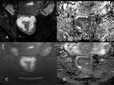 |
34 | To Compare the Application Value of Reduced Field-of-View IRIS-DWI Sequence and TSE-DWI Sequence in Rectal Cancer
Jianwei Zeng1, Haini Zhang1, Yankai Meng1, Lu Han2, Peng Wu2, and Kai Xu1
1Department of Radiology, the Affiliated Hospital of Xuzhou Medical University, Xu zhou, China, 2Philips Healthcare, Shanghai, China Keywords: Cancer, High-Field MRI The rFOV IRIS-DWI and rFOV TSE-DWI sequences are newer techniques in rectal magnetic resonance. In this study, we compared the image quality between reduced field-of-view (rFOV) diffusion weighted imaging (DWI) sequence based on image reconstruction using image-space sampling functions (IRIS) and rFOV turbo spin echo-diffusion weighted imaging (TSE-DWI) sequence. Through subjective and objective image quality analysis, we found different application values of rFOV IRIS-DWI and rFOVTSE-DWI sequences in rectal cancer. |
|
1678.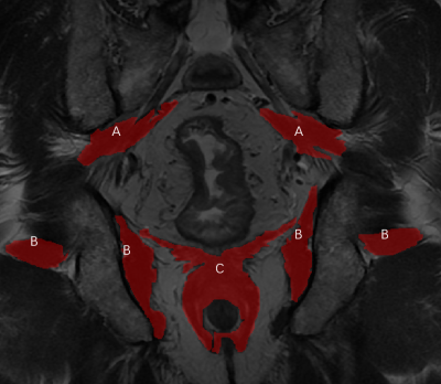 |
35 | The Value of Pelvic Skeletal Muscles to Identify the Low-Risk Rectal Cancers with Poor Prognosis
Yu Fu1, Jiayi Gao1, Mingyang Li1, and Huimao Zhang1
1The First Hospital of Jilin University, Changchun, China Keywords: Cancer, Muscle The low-risk rectal cancer (RC) has no need for postoperative treatment after total mesorectal excision (TME). However, there are some low-risk RC patients without postoperative-treatments had subsequent metastasis and recurrence. Skeletal muscles are gaining more attention which shown to be associated with morbidity and mortality in caners. Therefore, this study established a radiomics model based on pelvic skeletal muscles on MRI to identify the low-risk rectal cancer (RC) with poor prognosis. Our research shows that the novel radiomic signatures could be used to predict disease-free survival (DFS) in low-risk RC to help clinicians improve the treatment decision making followed TME. |
|
1679.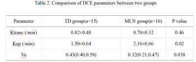 |
36 | The value of dynamic contrast-enhanced MRI for discriminating tumor deposition from metastatic lymph nodes adjacent to rectal cancer
WEN JUN HU1, Anliang Chen1, and AIlian Liu1
1The First Affiliated Hospital of Dalian Medical University, Dalian, China Keywords: Cancer, DSC & DCE Perfusion Tumor deposits (TD) and N stage are powerful prognostic factors for predicting the overall disease-free survival of rectal cancer patients. However, rectal cancer patients with TD are not the same as those with metastatic lymph node(MLN) in terms of treatment measures and prognosis. TD has similar performance to MLN on conventional CT/MRI images, which makes it difficult to distinguish them. Results of this study indicate DCE-MRI can effectively differentiate TD and MLN adjacent to rectal cancer, and the higher Kep and lower Ve values of rectal cancer with MLN may indicate its’ more aggressive. |
|
1680.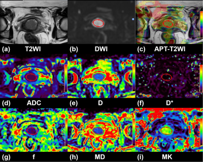 |
37 | Added-value of 3D amide proton transfer MRI in assessing cervical cancer: a comparison with multiple model diffusion-weighted imaging
Shujian Li1, Jieliang Lin2, and Jingliang Cheng1
1the First Affiliated Hospital of Zhengzhou University, Zhengzhou, China, 2Advanced Technical Support, Philips Healthcare, Beijing, China Keywords: Cancer, CEST & MT, Cervical cancer This study compared the diagnostic performances of mean, minimum, and maximum values of APT SI for preoperative identifying the prognostic factors (tumor stage, subtype, grade and LVSI status) of cervical cancer with those of multi-model DWI, and examined the additive value of 3D-APT imaging combined with multi-model DWI for the preoperative characterization of cervical cancer. The results showed that 3D-APT imaging was comparable with multi-model DWI for cervical cancer typing and grading. Adding APT to DWI can significantly improve the diagnostic performance with an AUC of 0.908 to predict tumor subtype, and an AUC of 0.903 to predict histological grade. |
|
| 1681. | 38 | APTw combined with mDixon‑Quant imaging to distinguish the differentiation degree of cervical squamous carcinoma
Xing Meng1, Ailian Liu2, and Shifeng Tian2
1Dalian Women and Children’s Medical Group, Dalian, China, 2the First Affiliated Hospital of Dalian Medical University, Dalian, China Keywords: Cancer, Uterus APTw and mDixon-Quant imaging were evaluated in this study for discrimination of the differention degree of CSC by histopathology. The APTw and R2* values of the poorly differentiated group were significantly higher than those of the well/moderately differentiated group. And the combination of APTw and R2* values showed a high diagnostic efficacy in discrimination of CSC with different differention degrees. |
|
1682.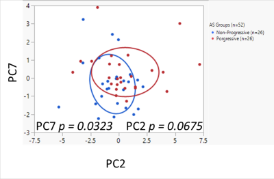 |
39 | Serum Biomarkers for Risk Stratification of Prostate Cancer Patients on Active Surveillance by Untargeted 1H MRS Metabolomics
Leo L. Cheng1,2, Florian Rumpf 3, Matteo Sanchez-Dahl Gonzalez4, and Adam S. Feldman5
1Pathology, Radiology, Athinoula A. Martinos Center for Biomedical Imaging, Massachusetts General Hospital, Charlestown, MA, United States, 2Radiology, Athinoula A. Martinos Center for Biomedical Imaging, Harvard Medical School, Charlestown, MA, United States, 3Athinoula A. Martinos Center for Biomedical Imaging, Massachusetts General Hospital, Charlestown, MA, United States, 4Pathology, Athinoula A. Martinos Center for Biomedical Imaging, Massachusetts General Hospital, Charlestown, MA, United States, 5Urology, Massachusetts General Hospital, Boston, MA, United States Keywords: Cancer, Prostate, Spectroscopy HRMAS 1H MRS was employed for an untargeted investigation of metabolism in prostate cancer patients on AS (n=52). Serum samples from two matched patient groups, progressive (n=26) and non-progressive (n=26) were measured with short and long T2 filter to investigate metabolites in the lipoprotein and LMWM regions respectively. PCA on 46 ROI from data acquired by HRMAS 1H MRS with short T2 resulted in a metabolomic profile, PC7, that separated the progressive and non-progressive groups (p = 0.0323). The findings of such studies could revolutionize healthcare by improving the diagnosis of diseases and contributing to multi-omics integration for precision medicine. |
|
1683.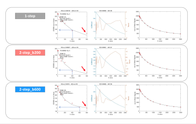 |
40 | Comparison of fitting methods for Non-Gaussian diffusion and IVIM MRI parameter estimation in two mice xenograft models
Yuko Someya1,2, Mami Iima1, Sawako Hayami3, Hirohiko Imai4, Hiroaki Takishima3, Denis Le Bihan5,6, Tomomi Nobashi1, Tsuyoshi Ohno1, and Yuji Nakamoto1
1Department of Diagnostic Imaging and Nuclear Medicine, Kyoto University Graduate School of Medi, Kyoto, Japan, 2Diagnostic Radiology, Kobe City Medical Center General Hospital, Kobe, Japan, 3Kyoto University Faculty of Medicine, Kyoto, Japan, 4Department of Systems science, Graduate School of Informatics, Kyoto University Graduate School of Medi, Kyoto, Japan, 5CEA-Saclay Center, Paris-Saclay, NeuroSpin, Gif-sur-Yvette, France, 6Human Brain Research Center, Kyoto University Graduate School of Medicine, Kyoto, Japan Keywords: Cancer, Diffusion/other diffusion imaging techniques The optimal fitting method (1-step or 2-step) and b-value threshold (200 or 600 s/mm2) in analyzing IVIM/diffusion data was investigated using two different mice xenograft models (breast and colon cancer). The threshold of b=600 rather than 200s/mm2 was found better in these models, as many cases showed non negligible residual IVIM components left with b=200s/mm2. From simulations, it appears that a too low b value threshold leads to an overestimation of ADC0 and underestimation of K and fIVIM .Those results suggest that the b-value threshold must be checked depending on the tissue types. |
|
 Back to Meeting Home
 Back to the Program-at-a-Glance
The International Society for Magnetic Resonance in Medicine is accredited by the Accreditation Council for Continuing Medical Education to provide continuing medical education for physicians.
 Back to Meeting Home
 Back to the Program-at-a-Glance
 View Presentation Video