Digital Poster
Urinary Tract Imaging Updates I
ISMRM & ISMRT Annual Meeting & Exhibition • 03-08 June 2023 • Toronto, ON, Canada

| Computer # | |||
|---|---|---|---|
3625.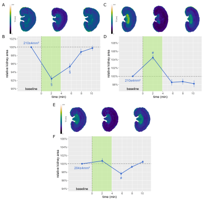 |
41 |
Size Matters: Automated Quantification of Acute Changes in
Kidney Size with Deep Dilated U-Net Segmentation of Dynamic
Parametric MRI
Tobias Klein1,2,
Thomas Gladytz1,
Jason M. Millward1,
Kathleen Cantow3,
Luis Hummel3,
Erdmann Seeliger3,
Sonia Waiczies1,
Christoph Lippert2,4,
and Thoralf Niendorf1,5
1Berlin Ultrahigh Field Facility (B.U.F.F.), Max Delbrück Center for Molecular Medicine, Berlin, Germany, 2Digital Health - Machine Learning Research Group, Hasso Plattner Institute for Digital Engineering, University of Potsdam, Potsdam, Germany, 3Institute of Translational Physiology, Charité - Universitätsmedizin, Berlin, Germany, 4Hasso Plattner Institute for Digital Health, Icahn School of Medicine at Mount Sinai, New York City, NY, United States, 5Experimental and Clinical Research Center, a joint cooperation between the Charité Medical Faculty and the Max Delbrück Center for Molecular Medicine, Berlin, Germany Keywords: Kidney, Preclinical Deep learning algorithms enable fast kidney segmentation, which is crucial to establish renal size as a (pre)clinical biomarker for renal diseases. Tackling this challenge, a novel deep dilated U-Net (DDU-Net) was trained, validated, and tested on preclinical ground truth data, benchmarked on simulated data against an analytical model, and applied to longitudinal in vivo MRI scans acquired in rats, with pathophysiological interventions mimicking clinically realistic scenarios. Our DDU-Net reached a Dice score of 0.98 on the ground truth, outperformed the analytical approach, and facilitated rapid detection of acute changes in kidney size upon acute pathophysiological interventions. |
|
3626.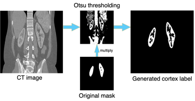 |
42 |
Transfer learning of renal cortex segmentation from CT to MRI:
facilitated with automatic labeling
Chang Ni1,
Xin Mu1,
Yanbin Li2,
Haikun Qi3,4,
and Jeff L. Zhang1
1Vascular and Physiologic Imaging Research (VPIR) Lab, School of Biomedical Engineering, ShanghaiTech University, Shanghai, China, 2Central Research Institute,UIH Group, Shanghai, China, 3School of Biomedical Engineering, ShanghaiTech University, Shanghai, China, 4Shanghai Clinical Research and Trial Center, Shanghai, China Keywords: Kidney, Segmentation Segmentation of renal cortex in MR images is important but challenging. In this study, we proposed to pre-train a ResUNet model with CT images and to use an automatic method for labeling renal cortex for the training data. Such method with transfer learning and automatic labeling performed well in segmenting renal cortex in MR images, with a DICE similarity of 0.85 and volume error of 14%±5%. The proposed method would make labeling of renal cortex for training dataset much more efficiently, and we further confirm the power of transfer learning technique in segmenting renal MR images. |
|
3627.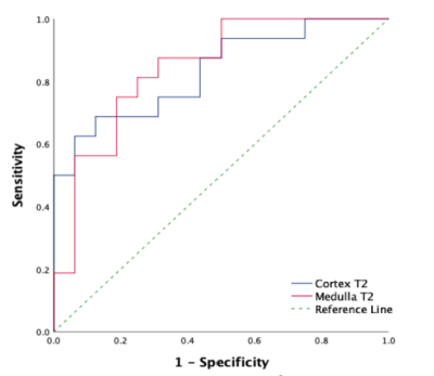 |
43 |
The Value of T2 mapping Sequence in Evaluating Renal Damage in
Patients with Liver Cirrhosis
Ye Ju1,
Yue Wang1,
Nan Wang1,
and Ailian Liu1
1the First Affiliated Hospital of Dalian Medical University, Dalian, China Keywords: Kidney, fMRI Patients with liver cirrhosis are easily complicated with renal injury, and it is often difficult to find the symptoms of early renal injury in time, which eventually leads to poor prognosis. T2 mapping, as a quantitative MRI technique, can provide quantitative information about the content and composition of water and collagen fibers [1]. The results of this study show that T2 mapping can effectively evaluate the renal damage of liver cirrhosis, so T2 mapping is expected to be an effective way to quantitatively and non-invasively evaluate the renal damage of liver cirrhosis. |
|
3628.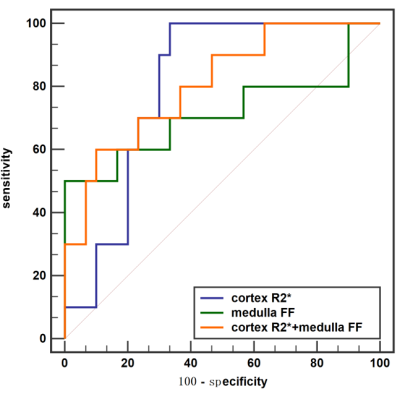 |
44 |
Value of mDIXON-Quant mDixon-Quant sequence Imaging in
Evaluation of Renal Function Damage in Patients with Liver
Cirrhosis
Yue Wang1,
Ye Ju1,
Nan Wang1,
and Ailian Liu1
1the First Affiliated Hospital of Dalian Medical University, Dalian, China Keywords: Kidney, Liver Patients with liver cirrhosis are easy to be complicated with renal function injury, and symptoms of early renal injury are hidden and often difficult to be detected in time, which may ultimately lead to poor prognosis. mDixon-Quant imaging, a novel fat quantitative MRI technique was used to evaluate the degree of renal function injury in patients with cirrhosis. Results showed that mDixon-Quant imaging can be an effective method for noninvasive diagnosis and early assessment of renal function injury in liver cirrhosis. |
|
3629. |
45 |
Perfusion MRI using dynamic contrast-enhanced MRI and arterial
spin labeling for the characterization of small renal masses:
preliminary study.
Haitham Al-Mubarak1,
Octavia Bane1,2,
Paul Kennedy1,
Philip Robson1,
Jordan Cuevas1,2,
Arthi Reddy2,
Kristina Prachanronarong2,
Kirolos Meilika3,
Amir Horowitz4,
Bernd Kuhn5,
Steven Sourbron6,
Ketan Badani3,
Bachir Taouli1,2,
and Sara Lewis1,2
1BioMedical Engineering and Imaging Institute, Icahn School of Medicine at Mount Sinai, New York, NY, United States, 2Department of Diagnostic, Molecular and Interventional Radiology, Icahn School of Medicine at Mount Sinai, New York, NY, United States, 3Department of Urology, Icahn School of Medicine at Mount Sinai, New York, NY, United States, 4Precision Immunology Institute/Tisch Cancer Institute, Icahn School of Medicine at Mount Sinai, New York, NY, United States, 5Siemens Healthcare, Erlangen, Germany, 6University of Sheffield, Sheffield, United Kingdom Keywords: Cancer, DSC & DCE Perfusion Functional imaging using dynamic contrast-enhanced magnetic resonance imaging (DCE-MRI) and arterial spin-labeling (ASL), which is a non-contrast technique, have been proposed to quantitatively assess perfusion and vascular flow in small renal masses (SRM) and may provide complementary information. In our initial series, we found significant differences in certain DCE-MRI quantitative and semi-quantitative parameters for renal cell carcinoma (RCC) versus benign tumors (Vp, wash-in, wash-out TTP and AUC) and between ccRCC versus non-clear cell (non-cc)RCC (Ve), while no differences existed between groups for ASL. Certain pharmacokinetic parameters derived from DCE-MRI appear promising for SRM characterization |
|
3630.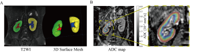 |
46 |
Morphological and Functional Evaluation of Diabetic Kidney
Disease Cohorts by using T2-Weighted and Diffusion Weighted MRI
Kaixuan Zhao1,2,3,
Sheng Li4,
Zhigang Wu5,
Thoralf Niendorf6,
Wenjian Wang4,
and Zaiyi Liu1,3
1Department of Radiology, Guangdong Provincial People’s Hospital, Guangdong Academy of Medical Sciences,Guangzhou 510080, China, Guangzhou, China, 2Guangdong Cardiovascular Institute, Guangdong Provincial People's Hospital, Guangdong Academy of Medical Sciences, Guangzhou 510080, China, Guangzhou, China, 3Guangdong Provincial Key Laboratory of Artificial Intelligence in Medical Image Analysis and Application, Guangdong Provincial People's Hospital, Guangdong Academy of Medical Sciences, Guangzhou 510080, China, Guangzhou, China, 4Division of Nephrology, Guangdong Provincial People’s Hospital, Guangdong Academy of Medical Sciences, Guangzhou, China, Guangzhou, China, 5Philips Healthcare Beijing Ltd., Shenzhen, China, 6Berlin Ultrahigh Field Facility (B.U.F.F.), Max Delbrueck Center for Molecular Medicine in the Helmholtz Association, Berlin, Germany, Berlin, Germany Keywords: Kidney, Kidney Diabetic Kidney Disease (DKD) is the leading cause of end-stage renal disease worldwide. Morphological evaluation of renal compartment volume changes and functional evaluation of renal ADC changes provide comprehensive insight into DKD disease progression. This study compares renal compartment volume changes including: 1) parenchyma volume ,2) pelvis volume, 3) whole kidney volume and 4) parenchyma volume percentage and the functional parameter ADC for DKD groups with different CKD staging. Our results demonstrate that cortical/medullary ADC, parenchyma volume and whole kidney volume are correlated with eGFR and suggest that the cortical ADC is a stronger predictor of eGFR in DKD. |
|
3631.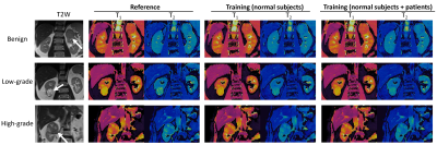 |
47 |
Accelerating 2D Kidney Magnetic Resonance Fingerprinting using
Deep Learning–Based Tissue Quantification
Huay Din1,
Christina J. MacAskill2,
Sree Harsha Tirumani2,3,
Pew-Thian Yap4,
Mark Griswold2,3,
Chris Flask2,3,
and Yong Chen1,3
1Case Western Reserve University, Cleveland, OH, United States, 2Department of Radiology, Case Western Reserve University, Cleveland, OH, United States, 3Department of Radiology, University Hospitals Cleveland Medical Center, Cleveland, OH, United States, 4Department of Radiology, University of North Carolina at Chapel Hill, Chapel Hill, NC, United States Keywords: Kidney, Quantitative Imaging, Cancer In this pilot study, we developed and optimized a spatially-constrained convolutional neural network to accelerate the scan time for 2D kidney Magnetic Resonance Fingerprinting (MRF). Our results suggest that an acceleration factor of 3 can be achieved with the proposed method, which shortens the 2D breath-hold MRF scan from 15 sec to 5 sec. In addition, the deep learning based approach can be applied for T1 and T2 quantification of both normal renal tissues and pathologies including renal cell carcinoma. |
|
3632.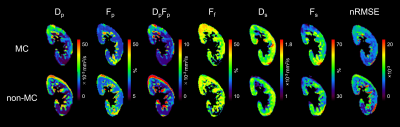 |
48 |
Improved accuracy of triexponential diffusion analysis of the
kidney using motion-corrected reconstruction for free-breathing
DWI
Yuki Makino1,
Naoki Ohno2,
Johannes M Peeters3,
Masami Yoneyama4,
Yu Ueda4,
Tosiaki Miyati2,
Yukihiro Matsuura1,
Toshifumi Gabata5,
and Satoshi Kobayashi1,2,5
1Radiology Division, Kanazawa University Hospital, Ishikawa, Japan, 2Faculty of Health Sciences, Institute of Medical, Pharmaceutical and Health Sciences, Kanazawa University, Ishikawa, Japan, 3Philips Healthcare, Best, Netherlands, 4Philips Japan, Tokyo, Japan, 5Department of Radiology, Kanazawa University Hospital, Ishikawa, Japan Keywords: Kidney, Diffusion/other diffusion imaging techniques The clinical application of triexponential intravoxel incoherent motion analysis in the kidney has been limited because the blurring and misregistration of images caused by respiratory motion during acquisition. To solve this issue, we applied retrospective motion-corrected (MC) reconstruction to free-breathing (FB) DWI of the kidney for accurate triexponential diffusion analysis. Our results showed that triexponential analysis using MC FB-DWI can improve the measurement accuracy and repeatability of triexponential diffusion parameters of the kidney. |
|
3633. |
49 |
3D Multichannel Convolutional Neural Network for Differentiating
Benign and Malignant Solid Renal Masses with/without Bias Field
Correction
Peter Wawrzyn1,
Ruben Ngnitewe Massa1,
Jamal Gardezi1,
and Andrew Wentland1,2,3
1Department of Radiology, University of Wisconsin Madison, Madison, WI, United States, 2Department of Biomedical Engineering, University of Wisconsin Madison, Madison, WI, United States, 3Department of Medical Physics, University of Wisconsin Madison, Madison, WI, United States Keywords: Kidney, Machine Learning/Artificial Intelligence, Bias Field Correction A deep learning model was developed for the differentiation of benign and malignant solid renal masses. A representative and balanced dataset was used, and model performance was evaluated with and without bias field correction (BFC). The model input contained multiple channels for various MRI-based tissue contrast weightings. The model achieved an accuracy and AUC of 74% and 71% with BFC and 80% and 59% without BFC. This work showed that a convolutional neural network can be trained to differentiate benign from malignant renal masses with a high degree of accuracy and that BFC improves AUC. |
|
3634. |
50 |
Preliminary study on the influence of renal motion amplitude on
APTw image quality of normal kidney
Ruirui Ma1,
Min Jia1,
Xia Wang1,
Wei Zhang1,
Na Zhao1,
Gang Tian1,
Chanjuan Yu1,
Xiuzheng Yue2,
and Yuedong Han1
1Xi'an GaoXin Hospital, Xi'an, China, 2Philips Healthcare, Beijing, China Keywords: Kidney, CEST & MT Respiratory movement is the main reason for upper and middle abdominal organ movement, which significantly affects the quality of MR imaging.This study aims to analyze the influence of renal motion amplitude on renal APT imaging through free breathing (FB), and breath holding (BH) based on multi-phase balanced turbo fast field echo (B-TFE). The results showed that renal motion amplitude was negatively correlated with APTw image quality scores. The renal motion amplitude of bilateral kidneys of BH mode was significantly reduced compared with FB, and the APT image quality of BH mode was better. |
|
3635.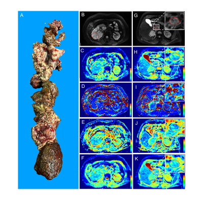 |
51 |
Renal Cell Carcinoma: Predicting Venous Tumor Thrombus
Consistency via IVIM-Diffusion-weighted MR Imaging and Apparent
Diffusion Coefficient
Jian Zhao1,
Hai-Yi Wang1,
Mei-Feng Wang1,
Huan-Huan Kang1,
Hui-Ping Guo1,
Shao-Peng Zhou1,
and Hui-Yi Ye1
1Department of Radiology, Chinese PLA General Hospital, Beijing, China Keywords: Kidney, Diffusion/other diffusion imaging techniques Problem: Evaluation of VTT consistency through preoperative images has not been reported yet.Methods: Intravoxel incoherent motion (IVIM)-derived parameters and apparent diffusion coefficient (ADC) were employed to predict VTT consistency of RCC. Results: Dt of the primary tumor and VTT generated the area under the ROC curve (AUC) values of 0.758 (95%CI: 0.671, 0.832) and 0.712 (95%CI: 0.622, 0.792) for differentiating friable and solid VTT. The AUC value of the model combining Dp and Dt of VTT and Dt of the primary tumor reached 0.886 (95%CI: 0.814, 0.937). Conclusions: IVIM parameters can effectively predict the VTT consistency of RCC. |
|
3636.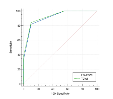 |
52 |
Diagnostic Performance and Interobserver Agreement of Clear Cell
Likelihood Score Using T2-Weighted Imaging with Fat Suppression
Technique
Yu-Wei Hao1,
Hui-Yi Ye1,
and Hai-Yi Wang1
1Department of Radiology, the first Medical Centre of Chinese PLA General Hospital, Beijing, China Keywords: Kidney, Kidney, Clear cell likelihood score; Clear cell renal cell carcinoma Whether T2-weighted imaging with or without fat suppression technique affects the performance of Clear Cell Likelihood Score v2.0 (ccLS v2.0) for diagnosing ccRCC in SRMs is not known. The final results showed that the tumor signal intensity between T2-weighted imaging with and without fat suppression was statistically significant (P < 0.001), but diagnostic performance and interobserver agreement of ccLS v2.0 in the diagnosis of ccRCC based on the two imaging techniques was comparable (P>0.05). Fat-suppressed T2-weighted imaging is also applicable to ccLS v2.0. This finding is helpful to expand the clinical application scope of ccLS and increase its clinical universality. |
|
3637.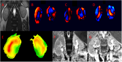 |
53 |
Correlation of Monopolar ADC and Bipolar ADC measurements to MRE
Estimates in Healthy Human Kidney
Gregory McClanahan1,2 and
Arunark Kolipaka1,2
1Biomedical Engineering, Ohio State University, Columbus, OH, United States, 2Radiology, Ohio State University, Columbus, OH, United States Keywords: Kidney, Elastography, MRI, MRE, Diffusion Imaging Kidney disease and injury can be assessed using tissue stiffness measurements determined by Magnetic Resonance Elastography (MRE), a method that uses MRI. Similarly, diffusion measurements such as apparent diffusion coefficient (ADC) can also provide useful information regarding tissue health. Our goal is to assess the health of the cortex, medulla, and whole kidney by comparing ADC measurements from monopolar and bipolar diffusion gradient schemes against MRE derived stiffness values. Preliminary results indicate a strong correlation between MRE derived stiffness values and monopolar derived ADC measurements and a fair correlation between MRE derived stiffness values and bipolar derived ADC measurements. |
|
3638.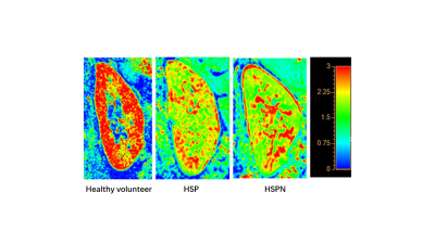 |
54 |
The feasibility of distinguishing HSPN from HSP in children
using intra-voxel incoherent motion diffusion weighted imaging
Hang Su#1,
Gang Zhang#1,
Jingjing Wu1,
Wei Xing1,
Yufu Hu1,
Zhiwei Shen2,
Ke Jiang2,
Yuying Sun1,
Xia Zhang1,
and Xianqing Ren*1
1The First Affiliated Hospital of Henan University of CM, Zhengzhou, China, 2Philips healthcare, Beijing, China Keywords: Kidney, Kidney Currently, the therapeutic significance of IVIM-DWI has been examined in numerous kidney illnesses such as transplanted kidney, renal tumor, and renal failure; however, the application in purpura nephritis is uncommon. As a result, the purpose of this study was to investigate the clinical feasibility of IVIM-DWI in differentiating purpura nephritis (HSPN) from allergic purpura (HSP) in children by using D and K values obtained from a small-field DWI sequence with respiratory trigger |
|
3639.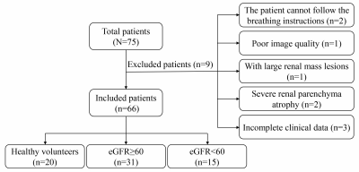 |
55 |
Noninvasive assessment of the renal function, the Oxford
classification and the prognostic risk stratification of IgAN by
DWI and BOLD MRI
Ping Liang1,
Chuou Xu1,
Daoyu Hu1,
and Zhen Li1
1Radiology, Tongji Hospital, Tongji Medical College, Huazhong University of Science and Technology, Wuhan, China Keywords: Kidney, Diffusion/other diffusion imaging techniques Purpose To explore whether IVIM-DWI and BOLD-MRI can assess the renal function, Oxford classification and prognostic risk stratification of IgAN patients. Methods This retrospective study included 46 IgAN patients and 20 healthy volunteers for renal MRI examinations. T2* values on BOLD images and ADC, Dt, Dp, fp values on IVIM-DWI images were measured. Results: Cortical Dt generated the largest area under the curve for differentiating 5-year risk scores ≤ 10% from 5-year risk scores > 10%, and for differentiating T0 from T1/T2. Conclusions IVIM-DWI and BOLD-MRI can assess the renal function, Oxford classification, and prognostic risk stratification of IgAN patients. |
|
3640.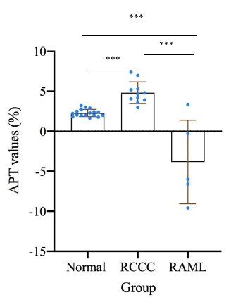 |
56 |
Preliminary exploration of 3D-APTw imaging in renal tumor
applications
Xia Wang1,
Min Jia1,
Xiuzheng Yue2,
Queenie Chan2,
Ruirui Ma1,
Wei Zhang1,
Shanshan Wang1,
Gang Tian1,
Nan He1,
Jiang guang He3,
and Yuedong Han1
1Department of Radiology, Xi'an GaoXin Hospital, Xi'an, China, 2Philips Healthcare, Beijing, China, 3Department of Urology Surgery, Xi'an GaoXin Hospital,, Xi'an, China Keywords: Kidney, CEST & MT APTw imaging focuses on cranial tumor studies with significant clinical value, with fewer studies in mid-upper abdomen, because of the movement of organs and B0 fields inhomogeneous. Previously used showed that intermittent breath-hold (IBH) could to improve the image quality of 3D-APTw imaging in healthy adults. In this study, we used the IBH respiratory compensation mode to initially explore the feasibility of 3D-APTw imaging of renal tumors. The findings suggested that 3D-APTw imaging with IBH can be not only used for imaging renal tumors, but also showed potential values in renal tumors diagnosis and differential diagnosis. |
|
3641.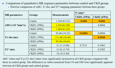 |
57 |
Quantitative MR Images of Renal Parenchymal Disease: An
Experimental In Vivo Study Using Rat Chronic Kidney Disease
Models
Sang Youn Kim1 and
Jeong Yeon Cho1
1Radiology, Seoul National University Hospital, Seoul, Korea, Republic of Keywords: Kidney, Quantitative Imaging Quantitative MRI sequences could be a noninvasive assessment modality in the diagnosis and evaluation of CKD. In particular, T1ρ may be a suitable MR sequence parameter to assess renal parenchymal fibrosis in a quantitative manner. Moreover, monitoring the change in common metabolites using MRS may reflect the alteration of osmolality in the renal medulla in CKD. |
|
3642.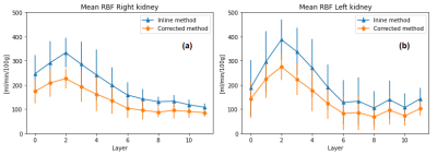 |
58 |
ABSOLUTE QUANTIFICATION OF RENAL ASL IN DOGS
Luis Carlos Sanmiguel Serpa1,
Amber Hillaert2,
and Pim Pullens3
1Radiology, Ghent University, AALTER, Belgium, 2Morphology, Imaging, Orthopedics, Rehabilitation and Nutrition, Ghent University, Ghent, Belgium, 3Ghent Institute of Functional and Metabolic Imaging, Ghent University, Ghent, Belgium Keywords: Kidney, Arterial spin labelling Arterial Spin Labeling (ASL) is a noninvasive fMRI technique that may evaluate canine renal condition. To date, however, the parameters to estimate the renal blood flow (RBF) have not been calculated on dogs. Therefore, λ: blood-tissue water partition coefficient in kidneys and canine at 3T were measured. Measured λ was 0.91[mL/g] and was 1463 ms. With these new values RBF maps were calculated and then compared against the RBF maps given by the scanner. This pilot study demonstrates the relevance of the ASL parameters estimation for the RBF maps calculation in canine kidney. |
|
3643. |
59 |
3D-pASL and DWI for detection of the function of transplanted
kidneys with 1.5T MRI
LING ZHANG 1,
Sheng Xie1,
Xiuzheng Yue2,
Zhenshan Ding1,
Xiaoqi Pu1,
and Yiwei Li1
1China-Japan friendship hospital, Beijing, China, 2Philips Healthcare, Beijing, China Keywords: Kidney, Transplantation Kidney Transplanted is the therapyfor patients with end-stage renal disease. Arterial spin labeling (ASL) uses magnetic labeling of water from inflowing blood as an endogenous tracer to acquire maps of absolute regional perfusion. The purpose of this study was to evaluate the blood flow of transplanted kidney allograft using a 1.5T MRI with ASL technique and determine whether renal allograft cortical perfusion by ASL could be a non-invasive biomarker for identifying kidney allograft function. Our data showed DWI and 3D-pCASL could be quantitative non-invasive tools for the clinical translation of identifying kidney allografts with subclinical pathology in further study. |
|
 Back to Meeting Home
 Back to the Program-at-a-Glance
The International Society for Magnetic Resonance in Medicine is accredited by the Accreditation Council for Continuing Medical Education to provide continuing medical education for physicians.
 Back to Meeting Home
 Back to the Program-at-a-Glance
 View Presentation Video