Digital Poster
Urinary Tract Imaging Updates II
ISMRM & ISMRT Annual Meeting & Exhibition • 03-08 June 2023 • Toronto, ON, Canada

Digital Poster
Urinary Tract Imaging Updates II
| Computer # | |||
|---|---|---|---|
3800.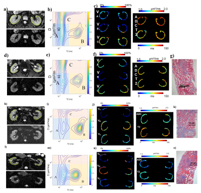 |
41 |
Exploration of interstitial fibrosis in chronic kidney disease
by diffusion-relaxation correlation spectrum MR imaging
Fang Liu1,
Wentao Hu2,
Yongming Dai2,
Minfang Zhang3,
and Yan Zhou1
1Department of Radiology, Renji Hospital, School of Medicine, Shanghai Jiao Tong University, Shanghai, China, 2MR Collaboration, Central Research Institute, United Imaging Healthcare, Shanghai, China, 3Department of Nephrology, Renji Hospital, School of Medicine, Shanghai Jiao Tong University, Shanghai, China Keywords: Kidney, Diffusion/other diffusion imaging techniques Non-invasive evaluation of interstitial renal fibrosis would help monitoring chronic kidney disease (CKD) progression and prognosis prediction. This study was aimed to evaluate renal interstitial fibrosis by diffusion relaxation correlated spectrum imaging (DR-CSI). Correlation was found between DR-CSI volume fractions and interstitial fibrosis level (IFL). VB illustrated best diagnostic performance among MRI indicators. We suggest DR-CSI is of potential value in the evaluation of renal interstitial fibrosis. |
|
3801.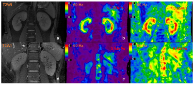 |
42 |
Three-Dimensional Multifrequency MR Elastography: clinical
application for Chronic Kidney Disease
Shan Pi1,
Jonathan M. Scott2,
Yin Li3,
Huiquan Wen1,
Matthew C. Murphy2,
Jingbiao Chen1,
Meng Yin2,
Jun Chen4,
Kevin J Glaser2,
Richard L Ehman2,
Hui Peng3,
and Jin Wang1
1Department of Radiology, the Third Affiliated Hospital, Sun Yat-sen University, Guangzhou, China, 2Department of Radiology, Mayo Clinic, Rochester, MN, United States, 3Department of Nephrology, the Third Affiliated Hospital, Sun Yat-sen University, Guangzhou, China, 4Department of Radiology, The Third Affiliated Hospital, Sun Yat-sen University (SYSU), Guangzhou, China Keywords: Kidney, Elastography Chronic kidney disease (CKD) is increasing in incidence and prevalence worldwide around the world, and early detection of CKD is a major challenge. MR elastography (MRE) is a noninvasive technique capable of quantifying the mechanical properties of tissue that has shown potential for assessing kidney diseases. Our results showedthe stiffness and damping ratio were significantly correlated with eGFR and biopsy score in CKD patients. The stiffness and damping ratio also decreased along with the progression of CKD. MRE using 60-Hz and 90-Hz vibration frequencies can provide potential quantitative biomarkers for evaluating kidney function and pathological injury in CKD patients. |
|
3802. |
43 |
The value of 3D APTw Combined Intravoxel Incoherent Motion in
Evaluation of the Renal Function in Chronic Kidney Disease
Yue Wang1,
Ye Ju1,
Dandan Zheng2,
and Ailian Liu1
1the First Affiliated Hospital of Dalian Medical University, Dalian, China, 2Clinical & Technical Support, Philips Healthcare, China, Shanghai, China Keywords: Kidney, CEST & MT, amide proton transfer Early diagnosis of chronic kidney disease (CKD) is difficult because of its hidden condition, mild symptoms and imperceptible symptoms. Amide proton transfer weighted (APTw) is a new contrast-free MR imaging technique, which can non-invasively detect the changes of amide proton concentration in endogenous mobile proteins and polypeptides in tissues. Intravoxel income motion (IVIM) can simultaneously obtain the microcirculation perfusion and water molecule diffusion of patients. The results showed that APTw and IVIM parameters APT, D* and D had good diagnostic efficiency in evaluating CKD renal function, especially the cortex APT value. |
|
3803. |
44 |
A NEW METHOD TO ANALYSE RENAL PERFUSION: A PROOF OF CONCEPT
Luis Carlos Sanmiguel Serpa1,2,3,
Pieter De Visschere1,2,
Marijn Speeckaert4,5,
and Pim Pullens3,6
1Radiology, Ghent University Hospital, Ghent, Belgium, 2Diagnostic Sciences, Faculty of Medicine and Health Sciences, Ghent University, Ghent, Belgium, 3Ghent Institute of Functional and Metabolic Imaging, Ghent University, Ghent, Belgium, 4Nephrology, Ghent University Hospital, Ghent, Belgium, 5Internal Medicine and Pediatrics, Faculty of Medicine and Health Sciences, Ghent University, Ghent, Belgium, 66IBiTech– Medisip, Ghent University, Ghent, Belgium Keywords: Kidney, Arterial spin labelling To date, renal ASL MRI images are analysed with methods that may underestimate renal perfusion or that may be observer-dependent. In this study, we implemented two analysis methods that take into account the complex morphology of the kidney. One analyses the kidney progressively, from the cortex to the medulla, and the second one, a new algorithm analyses the kidney in radial sections. Our results show that perfusion values change across the kidney and that healthy subjects have higher perfusion values. This study shows the importance of using more robust analysis on kidney ASL MRI images. |
|
3804.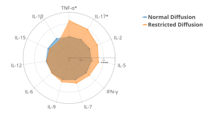 |
45 |
Restricted Water Diffusion in the Renal Cortex is Associated
with Early Developmental Factors and Elevated Urinary Cytokines
in Preadolescents
Navin Michael1,
Liangjian Lu2,
Delicia Shu Qin Ooi3,
Chang-Yien Chan2,3,
Kashthuri Thirumugan1,
Suresh Anand Sadananthan1,
Pottumarthi V. Prasad4,
Marielle Fortier5,
See Ling Loy6,7,
Tan Kok Hian8,9,
Fabian Yap7,10,11,
Yap Seng Chong12,
Keith Godfrey13,
Peter Gluckman14,
Johan G. Eriksson12,15,16,
Yung Seng Lee3,
Karen Moritz17,
Shiao-Yng Chan12,
Mary Wlodek1,18,19,
and S. Sendhil Velan1
1Singapore Institute for Clinical Sciences, Singapore, Singapore, 2Khoo Teck Puat-National University Children's Medical Institute, National University Health System, Singapore, Singapore, 3Department of Paediatrics, Yong Loo Lin School of Medicine, National University of Singapore, Singapore, Singapore, 4Department of Radiology, NorthShore University Health System, Evanston, IL, United States, 5Department of Diagnostic and Interventional Imaging, KK Women’s and Children’s Hospital, Singapore, Singapore, 6Department of Reproductive Medicine, KK Women's and Children's Hospital, Singapore, Singapore, 7Duke-NUS Medical School, Singapore, Singapore, 8Department of Maternal Fetal Medicine, KK Women's and Children's Hospital, Singapore, Singapore, 9Academic Medicine, Duke-National University of Singapore Graduate Medical School, Singapore, Singapore, 10Department of Pediatrics, KK Women's and Children's Hospital, Singapore, Singapore, 11Lee Kong Chian School of Medicine, Nanyang Technological University, Singapore, Singapore, 12Department of Obstetrics and Gynaecology and Human Potential Translational Research Programme, Yong Loo Lin School of Medicine, National University of Singapore, Singapore, Singapore, 13MRC Lifecourse Epidemiology Centre and NIHR Southampton Biomedical Research Centre, University of Southampton and University Hospital Southampton NHS Foundation Trust, Southampton, United Kingdom, 14Liggins Institute, University of Auckland, Auckland, New Zealand, 15Department of General Practice and Primary Health Care, University of Helsinki, Helsinki, Finland, 16Folkhälsan Research Center, Helsinki, Helsinki, Finland, 17School of Biomedical Sciences, Faculty of Medicine, The University of Queensland, Brisbane, Australia, 18School of Molecular Sciences, The University of Western Australia, Crawley, Australia, 19Obstetrics and Gynaecology, University of Melbourne, Parkville, Australia Keywords: Kidney, Diffusion/other diffusion imaging techniques, Intravoxel Incoherent Motion Imaging, Renal Inflammation Developmental factors that impair nephrogenesis can increase risk for chronic kidney disease (CKD). The early stages of CKD are characterized by an increased infiltration of inflammatory cells into the renal interstitium, which can restrict the molecular diffusion of water. We found that maternal gestational diabetes and preterm birth were associated with 2-fold and 4-fold higher risk of having restricted water diffusion (IVIM diffusion coefficient < 10th centile) in the renal cortex of preadolescents, respectively. Preadolescents with restricted diffusion had elevated urinary pro-inflammatory, chemotactic and pro-fibrotic cytokines, without elevations in blood markers of systemic inflammation, which was suggestive of renal inflammation. |
|
3805.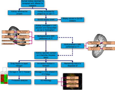 |
46 |
Development of an MR Imaging Protocol for Ex-Vivo Assessment of
Deceased Donor Human Kidneys
Alexander J Daniel1,
Sarah Fawaz2,
Philip Charles3,
Mohamed Elzawahry2,
Jane I Grove4,
David Long5,
James Hunter2,
Maria Kaisar2,
and Susan T Francis1
1Sir Peter Mansfield Imaging Centre, University of Nottingham, Nottingham, United Kingdom, 2Nuffield Department of Surgical Sciences, Oxford University, Oxford, United Kingdom, 3Big Data Institute, Oxford University, Oxford, United Kingdom, 4NIHR Nottingham Biomedical Research Centre, University of Nottingham, Nottingham, United Kingdom, 5Developmental Biology and Cancer Department, Great Ormond Street Institute of Child Health, University College London, London, United Kingdom Keywords: Kidney, Transplantation Kidney transplantation is the preferred treatment for end-stage kidney disease. Donor kidney viability is currently assessed using donor age, medical history, and serum creatinine, but these have limited predictive power. As part of a study to determine if MRI can be used as an alternative, more accurate, measurement of donor kidney viability, we outline a multiparametric MRI protocol (high resolution structural scans, relaxometry (T1, T2 and T2*) mapping, susceptibility-weighted-imaging (SWI), diffusion-weighted imaging (DWI), and magnetisation transfer ratio (MTR)) to study human whole kidneys that have been declined as transplants. MRI measures will be integrated with 3D-histology and tissue proteomics datasets. |
|
3806.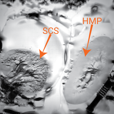 |
47 |
Assessment of Donor Kidney Storage Using a Porcine Model
Alexander J Daniel1,
Mohamed Elzawahry2,
Philip Charles3,
Sarah Fawaz2,
Maria Kaisar2,
James Hunter2,
and Susan T Francis1
1Sir Peter Mansfield Imaging Centre, University of Nottingham, Nottingham, United Kingdom, 2Nuffield Department of Surgical Sciences, Oxford University, Oxford, United Kingdom, 3Big Data Institute, Oxford University, Oxford, United Kingdom Keywords: Kidney, Transplantation Here, we investigate in a porcine model how MR parameters change as donor kidneys are cold stored, by studying the effect of temperature, cold ischaemia time and compare static cold storage (SCS) with hypothermic machine perfusion (HMP).Variation in relaxometry measurements with temperature has been modelled allowing the comparison of in-vivo and ex-vivo datasets, especially important when considering T1 measurements. The effects of cold ischemia time on relaxometry measures have also been quantified allowing this confounding factor to be accounted for in larger studies of human deceased donor organs. Susceptibility Weighted Imaging observed blood products in an SCS porcine kidney. |
|
3807.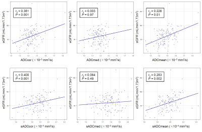 |
48 |
Correlation between Shifted Apparent Diffusion Coefficient of
the Renal Parenchyma and Estimated Glomerular Filtration Rates:
A Pilot Study
Atsushi Nakamoto1,
Hiromitsu Onishi1,
Takahiro Tsuboyama1,
Takashi Ota1,
Hideyuki Fukui1,
Keigo Yano1,
Toru Honda1,
Kengo Kiso1,
Shohei Matsumoto1,
Mitsuaki Tatsumi1,
and Noriyuki Tomiyama1
1Osaka University Graduate School of Medicine, Suita, Japan Keywords: Kidney, Diffusion/other diffusion imaging techniques This retrospective study investigated the correlation between shifted apparent diffusion coefficient (sADC) of the kidney measured using diffusion-weighted imaging obtained with b = 200, 1500 and estimated glomerular filtration rates (eGFR), and the performance in discriminating patients with impaired renal function was compared between sADC and ADC. sADC of the renal cortex showed the highest correlation coefficient in correlation with eGFR. In discriminating cases with eGFR <45, <60, or <90, sADC of renal cortex showed higher area under the curve compared to ADC, although the difference was not significant. sADC might be more useful in predicting renal function than ADC. |
|
3808.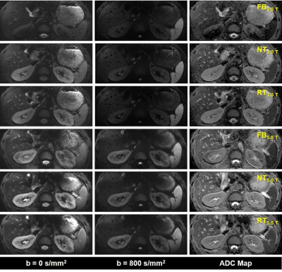 |
49 |
Higher Field Reduced-FOV Diffusion-Weighted-Imaging for
Abdominal Imaging at 5.0 Tesla: Image Quality Evaluation
Compared with 3.0 Tesla
Yunfei Zhang1,
Yongming Dai1,
and Mengsu Zeng2
1MR Collaboration, Central Research Institute, United Imaging Healthcare, Shanghai, China, 2Zhongshan Hospital of Fudan University, Shanghai, China Keywords: New Devices, Diffusion/other diffusion imaging techniques Reduced field-of-view (rFOV) technique is a feasible approach to counter the constraints of conventional DWI. Most recently, the 5.0 T whole-body MRI system was developed, which provides an alternative choice for high field abdominal DWI. This research aims to evaluate the image quality of 5.0 T rFOV-DWI with the 3.0 T rFOV-DWI as the reference. The results indicated that 5.0 T rFOV-DWI had better overall image quality and improved SNR compared to 3.0 T rFOV-DWI, which holds clinical potential for identifying the abdominal abnormalities in routine practice. |
|
3809.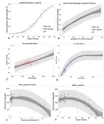 |
50 |
Kidney T2 can be used as a biomarker for disease progression and
functional decline in mouse models of polycystic kidney disease
Florian Schmid1,
Georgios Koukos1,
Matt Sooknah1,
Sanam Assili1,
and Johannes Riegler1
1Calico Life Sciences, South San Francisco, CA, United States Keywords: Kidney, Kidney We investigated quantitative T2 mapping as an MRI biomarker for disease progression in mouse models of autosomal dominant polycystic kidney disease and its correlation with kidney function. Our results show a good correlation between the fraction of the kidney tissue with elevated T2 and cyst load quantified by histology. There was also a good correlation between high T2 values and kidney function decline indicated by reduced glomerular filtration rates. |
|
3810.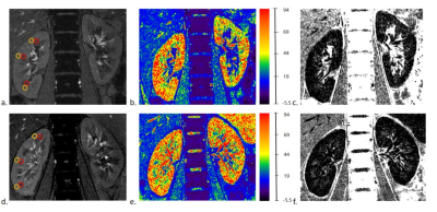 |
51 |
Quantitative assessment of split renal function in early lupus
nephritis using BOLD-MR: an initial study
Ye Wang1,2,
Zexuan Hu3,
Chutong He1,2,
Ruili Wei1,2,
Xinrui Pang1,2,
Fangrong Liang1,2,
Aaron Zhang4,
Yongzhou Xu4,
Shiwen Yuan1,5,
and Ruimeng Yang1,2
1The second Affiliated Hospital Guangzhou, School of Medicine, South China University of Technology, Guangzhou, China, 2Department of Radiology, Guangzhou First People’s Hospital, Guangzhou, China, 3Department of Radiology, TCM-integrated Hospital of Southern Medical University, Guangzhou, China, 4Philips Healthcare, Guangzhou, China, Guangzhou, China, 5Department of Rheumatology, Guangzhou First People’s Hospital, Guangzhou, China Keywords: Kidney, fMRI, split-renal function Lupus nephritis (LN) is one of the most severe manifestations of systemic lupus erythematosus (SLE) that can progress to end-stage kidney disease. This study aimed to detect the split-renal function changes in early-LN by using BOLD-MRI. The results demonstrated the R2*, as a biomarker for tissue oxygenation, was significantly lower in cortex and medulla in the early-LN patients compared to the healthy volunteers. Moreover, The R2* in medulla remained a considerable good power in predicting early-LN, suggesting it has the potential to be employed as a noninvasive imaging biomarker for early-LN patients with SLE. |
|
3811.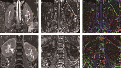 |
52 |
Diffusion tensor imaging-based quantitative assessment of renal
function in early-stage lupus nephritis
Chutong He1,2,
Ye Wang1,2,
Xinrui Pang1,2,
Ruili Wei1,2,
Fangrong Liang1,2,
Yongzhou Xu3,
Aaron Zhang3,
Shiwen Yuan4,5,
and Ruimeng Yang1,2
1The second Affiliated Hospital Guangzhou, School of Medicine, South China University of Technology, Guangzhou, China, 2Department of Radiology, Guangzhou First People’s Hospital, Guangzhou, China, 3Philips Healthcare, Guangzhou, China, 4Department of Rheumatology, The second Affiliated Hospital Guangzhou, School of Medicine, South China University of Technology, Guangzhou, China, 5Department of Rheumatology, Guangzhou First People’s Hospital, Guangzhou, China Keywords: Kidney, Diffusion Tensor Imaging, Lupus nephritis Lupus nephritis (LN) is a substantial cause of death in patients with systemic lupus erythematosus (SLE). The purpose of this study was to explore the value of Diffusion tensor imaging (DTI) in evaluating renal function in patients with early-stage LN. The results showed apparent diffusion coefficient (ADC) values in the cortex and fractional anisotropy (FA) values in the cortex and medulla were significantly lower in early-stage LN patients compared with healthy volunteers. Furthermore, FA values in the cortex and medulla were also independent predictors of early-stage LN, suggesting that FA values can be a potentially sensitive imaging biomarker. |
|
3812.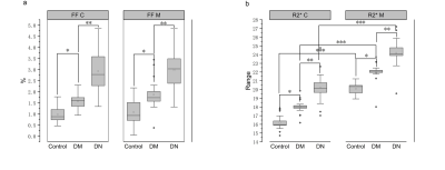 |
53 |
Assessment of renal lipid deposition and abnormal oxygen
metabolism of type 2 diabetes mellitus based on mDixon-Quant
Chun Yang1,2,
Yishi Wang3,
Wei Li1,
and Yuxin Wang1,2
1The First Affiliated Hospital of Shandong First Medical University & Shandong Provincial Qianfoshan Hospital, Jinan, China, 2Shandong First Medical University, Jinan, China, 3Philips Healthcare, Beijing, China Keywords: Kidney, Endocrine, mDixon-Quant, type 2 diabetes mellitus, renal function mDixon-Quant adopts a multi-echo 3D fast GRE sequence that can be used to obtain fat fraction, T2* and R2* mapping in a single breath hold. The variation of deoxyhemoglobin in the kidney can reflect the oxygen metabolism in patients with diabetic nephropathy under pathological and physiological conditions. FF maps can be used for fat quantification. mDixon-Quant provides the assessment both fat metabolism and oxygen metabolism of the kidney at the same time and may be valuable in detecting changes in renal function and pathology. |
|
3813.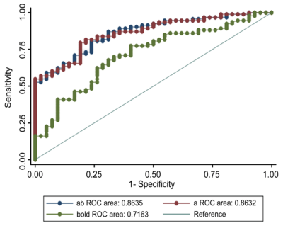 |
54 |
Evaluation of renal allograft function using arterial spin
labeling and blood oxygen level-dependent imaging
Jin Peng1,2,
Yajun Hong3,
Feipeng Zhu4,
Yanjun Li4,
Song Luo5,
Pu-Yeh Wu6,
and Guangming Lu1,5
1The First School of Clinical Medicine, Southern Medical University, Guangzhou, Guangdong, China, 2Department of Interventional Therapy, Chenggong Hospital, Xiamen University, Xiamen, Fujian, China, 3Office of Medical Case Statistics, Department of Medical Affairs, Zhongshan Hospital (Xiamen), Fudan University, Xiamen, Fujian, China, 4Department of Radiology, The First Affiliated Hospital with Nanjing Medical University, Nanjing, Jiangsu, China, 5Department of Radiology, Jinling Hospital, Medical School of Nanjing University, Nanjing, Jiangsu, China, 6GE Healthcare, Beijing, China Keywords: Kidney, Quantitative Imaging, Arterial spin labelling, Blood oxygen level-dependent imaging, Renal allograft In this study, we aimed to assess the renal allografts function after transplantation using ASL and BOLD MRI. We found that RBF and R2* values were significantly lower and higher in the renal transplant injury group, respectively. RBF and R2* were positively and negatively correlated with eGFR, respectively. ROC analysis demonstrated both RBF and R2* reflected injured renal function, and the combination of RBF and R2* showed an AUC of 0.86, comparable to that of RBF alone. In conclusion, compared to BOLD, ASL may be a more promising imaging technique for noninvasive assessment of renal graft function for clinical purposes. |
|
3814. |
55 |
The feasibility of amide proton transfer imaging at 3T for renal
masses: a preliminary study
Yun Xu1,
Qingxuan Wan1,
Xihui Ren1,
Fang Wang1,
Aijun Shen1,
Peng WU2,
and Peijun Wang1
1Tongji Hospital, School of Medicine, Tongji University, Shanghai, China, 2Philips Healthcare, Shanghai, China Keywords: Kidney, CEST & MT, amide proton transfer,RCC This study aims to assess the feasibility of amide proton transfer-weighted (APTw) imaging in depicting renal lesions and its ability to differentiate malignant from benign tumors. The results show that APTw can be used to discriminate renal tumors from normal kidney tissues, as well as to identify benign and malignant renal tumors. However, the technique still needs to be improved to reduce artifacts. |
|
3815. |
56 |
Preliminary exploration of the influence of demographic
characteristics on 3D-APTw imaging of right normal kidney
Gang Tian1,
Xia Wang1,
Ruirui Ma1,
Wei Zhang1,
Na Zhao1,
Min Jia1,
Chanjuan Yu1,
Xiuzheng Yue2,
Queenie Chan2,
and Yuedong Han1
1Department of Radiology, Xi'an GaoXin Hospital, Xi'an Jiao Tong University, Xi'an, Shaanxi, China;, Xi'an, China, 2Philips Healthcare, Beijing, China Keywords: Kidney, CEST & MT APTw imaging is able to detect free protein and peptide molecules in cells non-invasively. The aim of this study was to investigate the influence of age, gender and BMI on the APT values of right normal kidney. The results showed that there was a low correlation of APT values and age or BMI; and the male mean APT values was lower than that in female. The influence of age, gender and BMI factors should be taken into account in scientific research and clinical application of renal APTw imaging. |
|
3816.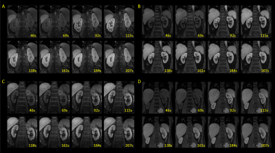 |
57 |
3D free-breathing renal DCE MRI with high spatial and temporal
resolution using Multitasking
Qing Liu1,
Qi Liu2,
Jing Li1,
Zhongqi Zhang2,
Jian Xu2,
Jeff L Zhang3,
and Haoran Sun4
1Radiology, Beichen Hospital, Tianjin, China, 2UIH America, Inc., Houston, TX, United States, 3School of Biomedical Engineering, ShanghaiTech University, Shanghai, China, 4Radiology, Tianjin Medical University General Hospital, Tianjin, China Keywords: Kidney, DSC & DCE Perfusion A 3D, free-breathing renal DCE MRI technique is developed using MRI Multitasking. It produces motion-resolved, high spatial and temporal resolutions images, thus does not require image registration post-processing. The feasibility of this technique in renal DCE MRI is evaluated on healthy subjects. |
|
3817.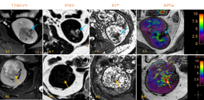 |
58 |
Assessment of the relationship between APTw values and R2* and
PDFF values in renal tumor
Xia Wang1,
Ruirui Ma1,
Wei Zhang1,
Na Zhao1,
Gang Tian1,
Chanjuan Yu1,
Xiuzheng Yue2,
and Yuedong Han1
1Department of Radiology, Xi'an GaoXin Hospital, Xi'an, China, 2Philips Healthcare, Beijing, China Keywords: Kidney, CEST & MT Renal tumors are often containing lipid components, ischemic-hypoxia and hemorrhage. These components can be reflected by the proton density fat fraction (PDFF) and R2* levels of the mDIXON_Quant. Amide proton transfer-weighted (APTw) imaging has made breakthroughs in tumor research, however, its signal intensity is affected by many factors. This study focused on the effect of PDFF and R2* values on APTw values in renal tumors. The preliminary results showed that renal tumor APT values were correlated with PDFF and R2* values. Thus, precision APTw imaging studies of renal tumors need to consider the effects of differential composition. |
|
3818.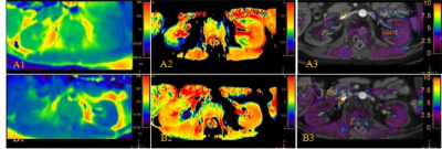 |
59 |
Preliminary exploration of the influence of B0 drift caused by
the change of examination orientations on renal APT images
Wei Zhang1,
Xia Wang1,
Ruirui Ma1,
Na Zhao1,
Gang Tian1,
Chanjuan Yu1,
Min Jia1,
Xiuzheng Yue2,
and Yuedong Han1
1Department of Radiology, Xi'an GaoXin Hospital, Xi'an, China, 2Philips Healthcare, Beijing, China Keywords: Kidney, CEST & MT Amide proton transfer-weighted (APTw) is a promising molecular imaging technique that has been employed in clinics for the detection and grading of tumors. APT value is easily influenced by B0 inhomogeneity causing image artifacts; This variation becomes more complex in abdominal motion organs than that in the brain. This study focused on the effect of variation in B0 field drift with different examination orientations on renal APT image quality. The results showed when head-first and foot-first scanning were used on 3D-APT imaging of normal kidneys, the change of B0 drift on the quality of renal APT image should be noticed. |
|
 Back to Meeting Home
 Back to the Program-at-a-Glance
The International Society for Magnetic Resonance in Medicine is accredited by the Accreditation Council for Continuing Medical Education to provide continuing medical education for physicians.
 Back to Meeting Home
 Back to the Program-at-a-Glance
 View Presentation Video