Digital Poster
Body: Miscellaneous I
ISMRM & ISMRT Annual Meeting & Exhibition • 03-08 June 2023 • Toronto, ON, Canada

| Computer # | |||
|---|---|---|---|
4263.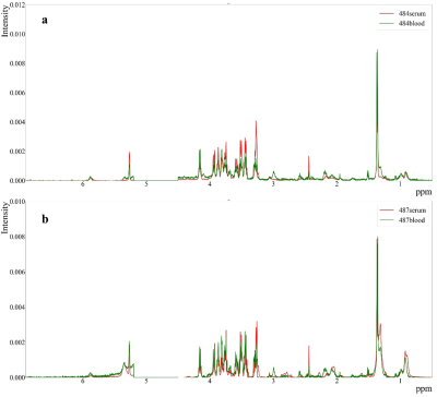 |
1 |
A Strategy of Whole Blood Metabolomics Based on Magnetic
Resonance Spectroscopy by Mixing Blood with Silica Powder
Leo Ling Cheng1,2,
Xiaoyu Wang3,4,
Zehan Liu3,4,
Jessica Duffy4,
Abner Louissaint, Jr.2,4,
and Panteleimon Takis5
1Radiology, Massachusetts General Hospital, Boston, MA, United States, 2Harvard Medical School, Boston, MA, United States, 3Nanjing University, Nanjing, China, 4Molecular Pathology, Massachusetts General Hospital, Boston, MA, United States, 5Imperial College London, London, United Kingdom Keywords: Screening, Spectroscopy, Gelified Biofluids Currently, blood metabolomics analysis has to choose between serum and plasma: the former leaves blood in room temperature and therefore causing metabolic alterations; and the latter has interfering chemicals added during acquisition of samples, which make results complicated. Here, using HRMAS MRS, we studied gel samples of whole mouse blood by mixing them with silica powder. The obtained spectra agreed, in general, with the correlating serum samples, which proved the robustness of this method. Taking advantage of tiny amounts of samples (μl) and simple and rapid pretreatment, our new technique demonstrates a novel approach to metabolomic analysis and clinical applications. |
|
4264.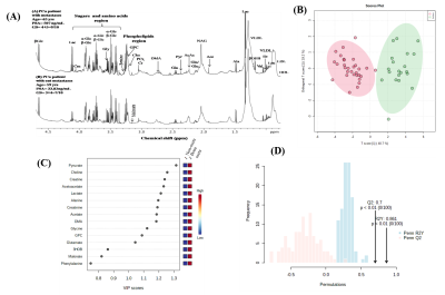 |
2 |
Blood plasma metabolomics study of metastases prostate cancer
patients reflects liver alterations involving carbon and
nitrogen metabolism
Pradeep Kumar1,
Rajeev Kumar2,
Sanjay Thulkar3,
Sanjay Sharma4,
Maroof Ahmad Khan5,
and Virendra Kumar1
1NMR and MRI Facility, AIIMS, New Delhi, India, 2Urology, AIIMS, New Delhi, India, 3Radiology , IRCH, AIIMS, New Delhi, India, 4Radiadiagnosis,RPC, AIIMS, New Delhi, India, 5Biostatistics, AIIMS, New Delhi, India Keywords: Prostate, Blood, Metabolomics Prostate cancer (PCa) with metastases remains incurable diseases requiring early diagnosis and effective treatments. To investigate and integrate the blood plasma metabolomic profiles with clinical parameters to distinguish PCa patients with metastasis from those without bone metastasis using 1H-NMR spectroscopy for establishing potential biomarker/s. Significant changes in liver enzyme ALP, ALT, and AST levels were observed in metastatic PCa. Pathway analysis revealed dysregulation of nitrogen and carbon metabolism. The discovery of metabolites in blood associate with clinical parameters for understanding PCa related pathophysiological mechanisms of bone and liver metastases. |
|
4265.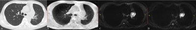 |
3 |
Comparison of multiplexed sensitivity encoding and single-shot
diffusion-weighted echo-planar imaging of the lung: Preliminary
study
Longjiang Fang1,
Wenjing Zhao1,
Yujing Chu1,
Qi Wang1,
and Dmytro Pylypenko2
1Department of Radiology, Weifang People's Hospital, Weifang, China, 2GE Healthcare, Beijing, China Keywords: Lung, Diffusion/other diffusion imaging techniques This study aims to compare multiplexed sensitivity encoding (MUSE) and single-shot diffusion-weighted echo planar imaging (SS DWEPI) techniques in lung MRI. A total of 22 patients with lung lesions were recruited in the study. Qualitative parameters included anatomic details, suscepti-bility artifacts, and diagnostic confidence; quantitative parameters included SNR and ADC of the lesions. The statistical studies demonstrated results that prove MUSE can provide better anatomic detail, lower susceptibility artifacts, higher diagnostic confidence, and improved SNR of the lung. MUSE is a promising technique for both clinical MRI diagnosis and treatment planning for lung lesions. |
|
4266.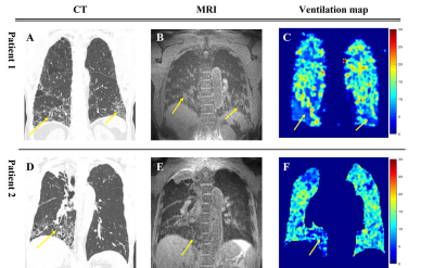 |
4 |
A Preliminary study of Idiopathic Pulmonary Fibrosis using 3D
ventilation map based on 3D Ultrashort Echo time Imaging
Seokwon Lee1,
Ho Yun Lee2,3,
Hye Yun Park4,
Hongseok Yoo4,
Jinil Park5,
Hyonha Kim5,
and Jang-Yeon Park1,5
1Biomedical engineering, Sungkyunkwan University, Suwon, Korea, Republic of, 2Radiology and Center for Imaging Science, Samsung Medical Center, Seoul, Korea, Republic of, 3Health Sciences and Technology, SAIHST, Sungkyunkwan University, Seoul, Korea, Republic of, 4Pulmonary and Critical Care Medicine, Samsung Medical Center, Seoul, Korea, Republic of, 5Intelligent Precision Healthcare Convergence, Sungkyunkwan University, Suwon, Korea, Republic of Keywords: Lung, Lung Idiopathic pulmonary fibrosis (IPF) is a specific form of idiopathic interstitial pneumonia of unknown cause, the most common and fatal of these interstitial lung diseases. A study on the quantitative assessment of IPF severity based on signal intensity in hyperpolarized MRI lung imaging has recently been proposed. In this preliminary study, it was shown that non-contrast-enhanced 3D ultrashort echo-time (UTE) MRI could diagnose ventilation defects in lesions in patients with IPF using UTE-based 3D ventilation maps and histograms in combination with 3D UTE structural images. |
|
4267.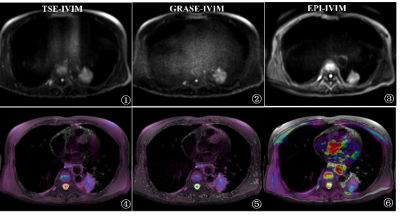 |
5 |
Intravoxel incoherent motion diffusion-weighted imaging of
solitary pulmonary lesions: initial study with gradient-and-spin
echo (GRASE)
Siqiang Lv1,2,
Zhanguo Sun3,
and Xiuzheng Yue4
1Jining Medical University, Jining, China, 2Affiliated Hospital of Jining Medical University, jining, China, 3Affiliated Hospital of Jining Medical University, Jining, China, 4Philips Healthcare, Beijing, China Keywords: Lung, Data Acquisition, SPLs Intravoxel incoherent motion diffusion-weighted imaging (IVIM) could obtain parameters of pure water molecule diffusion and microcirculatory perfusion-related diffusion and more accurately reflects the complexity of the microstructure of the tumor tissues. Gradient-and-spin echo (GRASE), incorporating the gradient echo and spin echo techniques, is a fast-imaging sequence with potential for improved IVIM examinations. The aim of the study was to evaluate the feasibility and image quality of the GRASE-IVIM sequence in evaluating solitary pulmonary lesions (SPLs) by comparing with the EPI-IVIM and TSE-IVIM sequences. Our data showed GRASE-IVIM sequence could be a fast and stable alternative technique to evaluate the SPLs. |
|
4268.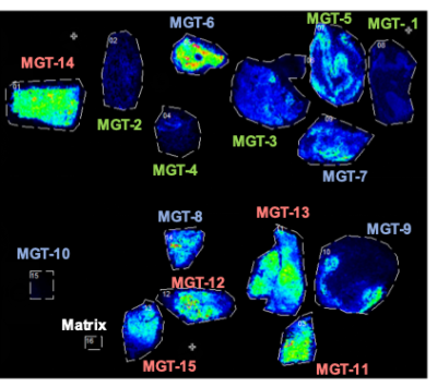 |
6 |
Metabolic Imaging of Pancreatic Ductal Adenocarcinoma
Saleem Yousf1,
Kristine Glunde1,2,
Dalton R. Brown1,
Caitlin Tressler1,
Michael G. Goggins3,4,5,
and Zaver M. Bhujwalla1,2
1Division of Cancer Imaging Research, The Russell H. Morgan Department of Radiology and Radiological Science, Johns Hopkins University School of Medicine, Baltimore, MD, United States, 2Sidney Kimmel Comprehensive Cancer Center, Johns Hopkins University School of Medicine, Baltimore, MD, United States, 3Departments of Pathology, Johns Hopkins University School of Medicine, Baltimore, MD, United States, 4Departments of Oncology, Johns Hopkins University School of Medicine, Baltimore, MD, United States, 5Departments of Medicine, Johns Hopkins University School of Medicine, Baltimore, MD, United States Keywords: Cancer, Metabolism, PDAC Metabolic imaging techniques such as mass spectrometry imaging (MSI) complement magnetic resonance spectroscopy and colocalize metabolite profiles with immunohistological evaluation of tissue sections. Here we have performed matrix-assisted laser desorption/ionization (MALDI)-MSI of normal pancreatic tissue, intraductal papillary mucinous neoplasm (IPMN), and pancreatic ductal adenocarcinoma (PDAC) to characterize the spatial distribution of PDAC metabolites in tissue sections. Our preliminary data with MALDI-MSI identifies significantly increased taurine and significantly decreased creatine, both of which can also be detected with 1H MRS, in PDAC compared to normal tissue. The spatial heterogeneity of metabolite distribution in PDAC tissue can be related to immunohistochemical evaluation. |
|
4269. |
7 |
Early Detection of Pancreatic Cancer by Hyperpolarized Magnetic
Resonance and pO2 Electron Paramagnetic Resonance Imaging
Jose Santiago Enriquez1,2,
Rian M Howell2,3,
Olivereen Le Roux3,
Shivanand Pudakalakatti1,
Prasanta Dutta1,
Florencia McAllister2,3,
and Pratip Bhattacharya1,2
1Cancer System Imaging, UT MD Anderson Cancer Center, Houston, TX, United States, 2UT MD Anderson Cancer Center UT Health Science Center Houston Graduate School of Biomedical Sciences, Houston, TX, United States, 3Clinical Cancer Prevention, UT MD Anderson Cancer Center, Houston, TX, United States Keywords: Cancer, Pancreas, Metabolism, Metabolic Imaging, Hyperpolarized MR (non-gas) There is an unmet need for the early diagnosis of pancreatic cancer. Diagnosis is difficult due to the asymptomatic nature of pancreatic cancer. One way to detect the early stages is to monitor the altered metabolism in premalignant pancreatic lesions in vivo with hyperpolarized metabolic imaging. Here we demonstrate how genetically engineered mouse models were used to detect early stages of pancreatic cancer as we see an increase in the altered metabolism in the pancreatic cancer models compared to control mice. Simultaneously we observe changes in hypoxia levels in these models using electron paramagnetic resonance imaging. |
|
4270.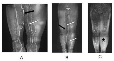 |
8 |
Magnetic Resonance Lymphangiography Manifestations and
Applications in Secondary Lymphoedema
Minge Zhang1,
Jichen Xie1,
Jinbiao Huang1,
Yan Chen1,
Jingjing Wang1,
Liqi Yi2,
Youmao Zheng2,
Shilin Gu2,
Runyu Tang3,
Yunfei Zhang3,
Yongming Dai3,
and Hai Yang1
1Department of Radiology, Taizhou Hospital of Zhejiang Province affiliated to Wenzhou Medical University, Taizhou, China, 2Department of Hand and Foot Surgery, Taizhou Hospital of Zhejiang Province affiliated to Wenzhou Medical University, Taizhou, China, 3Central Research Institute, United Imaging Healthcare, Shanghai, China Keywords: Screening, Visualization, Lymphoedema Imaging Surgical intervention should be considered when extremity lymphedema reached clinical stage II, and the identification of lymphatic vessels from nearby veins is critical for surgery planning. In this study, we reviewed the MRL examinations of patients with secondary lymphedema. The results showed that MRL could be used to characterize the secondary lymphedema pre-operatively, and predefine treatment planning for patients who suffer secondary lymphedema. |
|
4271.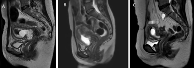 |
9 |
MRI-based scoring model to predict massive hemorrhage during
dilatation and curettage in patients with cesarean scar
pregnancy
Fengleng Yang1,
Huaibo Jing2,
Xiaodan Wang2,
and Zhigang Wang1
1Radiology, Chengdu Women's and Children's Central Hospital, School of Medicine, University of Electronic Science and Technology of China, Chengdu, China, 2Gynecology, Chengdu Women's and Children's Central Hospital, School of Medicine, University of Electronic Science and Technology of China, Chengdu, China Keywords: Urogenital, Urogenital, Cesarean scar pregnancy; Magnetic resonance imaging (MRI); Curettage; Massive hemorrhage; Risk factors Cesarean scar pregnancy is a special type of ectopic pregnancy for which dilatation and curettage (D&C) is one of the main treatment modalities, and uncontrollable hemorrhage is a more dangerous complication during D&C. Accurate preoperative prediction of the risk of intraoperative hemorrhage will help gynecologists draw up precise treatment. The aim of this study is to develop an MRI scoring model for predicting intraoperative hemorrhage during D&C. The study found that cesarean section diverticulum area, uterine scar thickness and gestational sac diameter were independent risk factors for intraoperative hemorrhage. A scoring model was developed, The model possessed decent prediction performance. |
|
4272.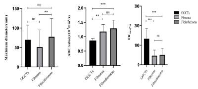 |
10 |
The findings of clinical and imaging features of common ovarian
sex cord stromal tumors with both Ultrasound and MRI
Cheng Meiying1,
Tan Shifang1,
Zhang Lingjie1,
Ren Tian2,
Zhang Chunxiang1,
Wang Kaiyu3,
Li Haiyang4,
Shang Honglei1,
Chang Hui5,
Yang Zhexuan1,
Cao Jing6,
and Zhao Xin1
1Department of Radiology, The Third Affiliated Hospital of Zhengzhou University, Zhengzhou, China, 2Department of Information, The Third Affiliated Hospital of Zhengzhou University, Zhengzhou, China, 3MR Research China, GE Healthcare, Beijing, China, 4Department of Medical Equipment, The Third Affiliated Hospital of Zhengzhou University, Zhengzhou, China, 5Department of Research, The Third Affiliated Hospital of Zhengzhou University, Zhengzhou, China, 6Department of Pathology, The Third Affiliated Hospital of Zhengzhou University, Zhengzhou, China Keywords: Urogenital, MR Value Most of ovarian sex cord stromal tumors (OSCSTs) are benign with good prognosis, but low-grade malignant OSCSTs need special treatment. Correct preoperative diagnoses and accurate classification remain challenging. We retrospectively analyze the clinical and imaging characteristics of OSCSTs and found significant biomarkers such as maximum enhancement ratio (ERmax) and apparent diffusion coefficient (ADC) for the preoperative diagnosis and classification of common OSCSTs. It’s more helpful for preoperative diagnosis by combining of MRI and ultrasound together than by ultrasound alone, and MRI has the similar diagnostic accuracy as the combination. |
|
4273. |
11 |
The value of MR perfusion and diffusion combined with tumor
marker analysis in differentiating benign and malignant ovarian
tumors
Jinsong Bai1,
Haitao Zhang1,
and shaoyu wang2
1Shanxi Hanzhong People's Hospital, Hanzhong, China, 2DI MR SMK, Siemens Healthineers, Shanghai, China Keywords: Pelvis, Cancer, Ovarian cancer The aim of our study was to compare the differential diagnostic efficacy of dynamic contrast enhanced magnetic resonance imaging (DCE-MRI), apparent diffusion coefficient (ADC), serum carbohydrate antigen 125 (CA125), human epididymal secretory protein 4 (HE4) and their combined application in benign and malignant ovarian tumors, and to explore the best method for the diagnosis of ovarian tumors. |
|
4274. |
12 |
APTw Combined With Multiple Models DWI of Endometrial Cancer:
Correlations Between Multimodal Parameters and HIF-1α Expression
Ma Changjun1,
Tian Shifeng1,
Liu Ailian1,
Chen Lihua1,
Lin Liangjie2,
Zhang Xiaoxiao3,
Guo Yinghua4,
and Wang Jiazheng4
1Department of Radiology,, The First Affiliated Hospital of Dalian Medical University, Dalian, China, China, 2Clinical & Technical Support, Philips Healthcare, Beijing, China, China, 3Clinical & Technical Support, Philips Healthcare, Wuhan, China, China, 4Clinical & Technical Support, Philips Healthcare, Shanghai, China, China Keywords: Placenta, Quantitative Imaging HIF-1α is a major transcriptional factor regulating gene expression under hypoxic conditions. The identification of HIF-1α expression is very helpful for the quantitative assessment of tumor hypoxia, on which the therapeutic response is predicted or regimens are adjusted . |
|
4275. |
13 |
Readout-segmented echo-planar imaging in the evaluation of anal
fistula: Comparison with single-shot echo-planar
diffusion-weighted imaging
Yuru Zhong1,
jiao bai1,2,
Jian Shu1,
Fugang Han1,
Jun Li1,
Dongmei Zhao1,
and Yunzhu Wu3
1Radiology Department, The Affiliated Hospital of Southwest Medical University, LuZhou, China, 2Radiology Department, The affiliated Hospital of Southwest Medical University, LuZhou, China, 3SIEMENS Healthineers Ltd.Shanghai, Shanghai, China Keywords: Pelvis, Diffusion/other diffusion imaging techniques To assess the image quality of anal fistula by RESOLVE DWI compared to SS-EPI DWI, and explore the value of RESOLVE DWI for diagnosis and classification of anal fistula. Qualitative and quantitative evaluation of image quality showed that RESOLVE DWI group was superior than SS-EPI DWI group.The accuracy of RESOLVE DWI sequence in the diagnosis of the internal orifice, branch fistula, and classification was higher than in SS-EPI DWI for anal fistula. RESOLVE DWI can provide higher quality images of anal fistula than SS-EPI DWI. |
|
4276.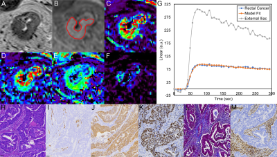 |
14 |
Identification of Imaging Marker for Tumor Tissue Composition in
Rectal Carcinoma Using Dynamic Contrast-Enhance MRI
Jie YUAN1,
Mengxiao Liu2,
Songhua Zhan1,
and Wenli Tan1
1Shuguang Hospital Affiliated to Shanghai University of Traditional Chinese Medicine, Shanghai, China, 2Siemens Healthineers Ltd, Shanghai, China Keywords: Digestive, Cancer Dynamic contrast-enhanced (DCE) MRI incorporates arterial input function and pharmacokinetic models to assess tissue perfusion, vasculature, capillary permeability, and interstitial space volume. It can be used to assess tumor vascularization, which can help determine tumor aggressiveness and the degree of angiogenesis and monitor therapy. However, the relationship between pathological characteristics of rectal cancer and the quantitative analysis of DCE remains unclear. |
|
4277. |
15 |
Effect of Compressed SENSE on 3D T2-weighted sequence for Rectum
Imaging with a deep learning constrained Compressed SENSE
Reconstruction
Ying Qiu1,
Yi Zhu2,
Dandan Guo1,
and Ke Jiang2
1Department of radiology, First Hospital of Jilin University, ChangChun, China, 2Philips Healthcare, BeiJing, China Keywords: Urogenital, Pelvis Rectal MRI examination requires high resolution three-dimensional (3D) sequences to observe the morphology, structure and the location of the lesions. However, 3D sequences may lead to longer scan time ,patient discomfort and image quality problem. In this study, we investigated the use of a deep learning-based reconstruction algorithm (CS-AI) to highly accelerate 3D rectum MRI. The result showed that CS-AI reconstruction can use the same scan time with sufficient image quality compared to SENSE and might be clinically useful in assessment of rectum cancer. |
|
4278.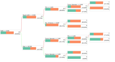 |
16 |
VI-RADS combined with decision tree model preoperatively predict
the pathological grade of bladder cancer with high (≥3) and low
(≤2) scores
Bohong Cao1,
Qing Li1,
Shuai Jiang1,
Yunfei Zhang2,
Yongming Dai2,
and Jianjun Zhou1
1Zhongshan Hospital, Fudan University, Shanghai, China, 2MR Collaboration, Central Research Institute, United Imaging Healthcare, Shanghai, China Keywords: Urogenital, Bladder Bladder cancer pathology grading is currently obtained mainly by invasive cystoscopic biopsy or surgical pathology. This study aimed to investigate the effectiveness of Vesical Imaging Reporting and Data System (VI-RADS) in the diagnosis of high-grade bladder cancer (HG-BC) and explore a new preoperative non-invasive grading prediction system. We demonstrate that most bladder cancers with a VI-RADS score ≥3 are high-grade and that the decision tree model is a good predictor of pathological grading in patients with VI-RADS ≤2. Thus, VI-RADS can be a grouping imaging biomarker for noninvasive prediction of bladder cancer grade. |
|
4279.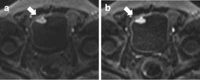 |
17 |
Comparison of integrated slice-specific dynamic shimming and
echo-planar imaging diffusion-weighted imaging in bladder cancer
detection and characterization
yujiao zhao1,
cong you1,
cheng zhang1,
feifei qu2,
jinxia zhu2,
and Thomas Benkert3
1The First Central Clinical College of Tianjin Medical University, tianjin, China, 2MR collaborations, Siemens Healthcare, beijing, China, 3MR Application Predevelopment, Siemens Healthcare GmbH, Erlangen, Germany Keywords: Urogenital, Bladder, bladder cancer This study investigated the image quality and lesion characterization of bladder lesions on integrated slice-specific dynamic shimming (iShim) diffusion-weighted imaging (DWI) compared with single-shot echo-planar imaging (ss-EPI) DWI. The qualitative and quantitative image quality analyses were compared between the 2 DWI techniques. The diagnostic agreements of lesion characterization, including submucosal stalk and myometrial invasion, were also evaluated on DWI images compared with pathologic results. The results indicated that iShim-DWI provided better bladder tumor detection and image quality than ss-EPI DWI, suggesting that iShim-DWI might serve as a competent technique in bladder imaging. |
|
4280.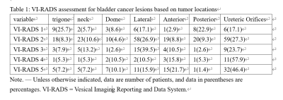 |
18 |
Diagnostic performance of the vesical imaging-reporting and data
system for detecting muscle-invasive bladder cancer based on
tumor locations.
Xiangyu Wang1,
Fan Lin1,
and Kan Deng2
1Shenzhen Second People's Hospital, Shenzhen, China, 2Philips Healthcare, Guangzhou, China Keywords: Pelvis, Tumor The VI-RADS criterion was reported to have limitations, particularly for tumors location. So we evaluated the diagnostic performance of Vesical Imaging-Reporting and Data System (VI-RADS) scoring and diagnostic accuracies based on tumor locations. VI-RADS had superior diagnostic performance for detecting MI, especially in tumors located at the bladder lateral, posterior walls and ureteric orifices. |
|
4281. |
19 |
Amide proton transfer values in testicular spermatogenic
function evaluation: a preliminary study
Guanglei Tang1,
Wenhao Fu1,
Kan Deng2,
and Jian Guan1
1The First Affiliated Hospital, Sun Yat-Sen University, Guangzhou, China, 2Philips Healthcare, Guangzhou, China Keywords: Urogenital, fMRI In this study, we determined APT of normal testes possible variations with age, and to assess the feasibility of APT in testicular spermatogenic function evaluation. Our results showed that APT of normal testicular tissue decrease with advancing age, and could be promising in evaluation of male infertility. |
|
4282. |
20 |
An add-on local external shim array to improve the main magnetic
field homogeneity in the prostate
Carlijn Tenbergen1,
Angeliki Stamatelatou1,
Sahar Nassirpour2,
Paul Chang2,
and Tom Scheenen1,3
1Department of Medical Imaging, Radboud University Medical Center, Nijmegen, Netherlands, 2MR Shim, Reutlingen, Germany, 3Erwin L. Hahn Institute, Essen, Germany Keywords: Prostate, Shims, Diffusion, Spectroscopy B0-field inhomogeneities can influence (spectroscopic) image quality of the prostate, causing geometric distortion in diffusion weighted EPI images as well as line broadening in MRSI voxels. We added a 16-element external local shim array around the pelvis to improve B0-field homogeneity within the prostate and evaluated its effects compared to standard shimming in 9 healthy volunteers. Resulting B0-maps showed a comparable or reduced frequency variation across subjects, with a significant decrease of MRSI citrate line widths. DWI did not show a robust effect over the group of subjects. |
|
 Back to Meeting Home
 Back to the Program-at-a-Glance
The International Society for Magnetic Resonance in Medicine is accredited by the Accreditation Council for Continuing Medical Education to provide continuing medical education for physicians.
 Back to Meeting Home
 Back to the Program-at-a-Glance
 View Presentation Video