Digital Poster
Vessel Wall & Lumen Imaging I
ISMRM & ISMRT Annual Meeting & Exhibition • 03-08 June 2023 • Toronto, ON, Canada

| Computer # | |||
|---|---|---|---|
1506.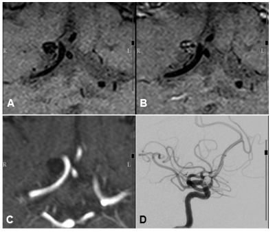 |
41 | Demonstration of Intracranial Aneurysm Healing after Endovascular Coiling Evaluated by 3D T1 SPACE Magnetic Resonance
Sho-Jen Cheng1, Xue-Zhe Lu2, and Yi-Hsin Wang1
1Department of Medical Imaging, Taipei Medical University Hospital, Taipei, Taiwan, 2Siemens Healthcare Limited, Taipei, Taiwan Keywords: Vessel Wall, Vessels Angiography is a gold standard tool in the follow-up of the treated aneurysms to identify if there is aneurysm recanalization need further management. Despite non-invasive MR-based assessments be reported, the pathological changes of aneurysm on MRI was not clear. In this study, we employed three-dimensional variate flip angle MRI to evaluate the endothelial healing in ten cases with intracranial aneurysms after endovascular management. HRVWI routinely demonstrate the enhancement pattern of the aneurysmal neck not only the aneurysmal wall in both cross-sectional and time-serial follow up, increasing our confidence in making the decision whether to retreat or not. |
|
1507.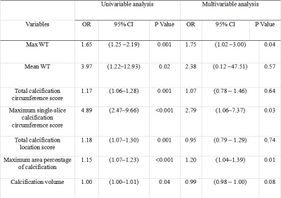 |
42 | Relationship beween Plaque burden, calcification features and low carotid stent expansion rate by high-resolution MR vessel wall imaging
Meng Yu Sun1, Wei Yu1, and Xiu Jian Lian2
1Radiology, Beijing Anzhen Hospital, BeiJing, China, 2Philips Healthcare, BeiJing, China Keywords: Vessel Wall, Atherosclerosis To quantitatively evaluate carotid plaque burden and components and to explore the association between carotid plaque burden and compositions and carotid stent expansion rate (SER) by using carotid high-resolution magnetic resonance vessel wall imaging (HR-VWI). Maximum wall thickness (OR 1.75; 95% CI, 1.02-3.00, P=0.04), maximum single-slice calcification circumference score (OR 2.79, 95% CI 1.06 - 7.37, P=0.03), and maximum area percentage of calcification (OR 1.20, 95% CI 1.04 - 1.39, P=0.01) as predictors of the low SER. Larger culprit plaque, larger calcification area, and greater calcification circumference were independently associated with a low SER after carotid artery stenting. |
|
1508.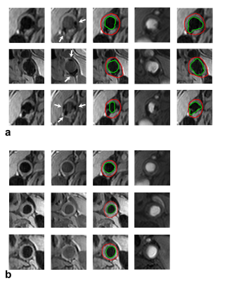 |
43 | Automatic segmentations for carotid vessel lumen and wall with arterial calcifications using spatially registered black- and gray-blood images
Bo Li1,2
1Center Laboratory, The Third Affiliated Hospital of Nanchang University, Nanchang, China, 2Radiology Department, The First Hospital of Nanchang City, Nanchang, China Keywords: Vessel Wall, Segmentation We investigated automatic segmentations for the carotid vessel lumen and wall with the presence of arterial calcifications using spatially registered black- and gray-blood images. CS-siBLAG sequence was used to provide black- and gray-blood images. A K-means algorithm was employed to segment carotid artery lumen on black-blood images and calcifications on gray-blood images. A distance transform and an active contours model was used to segment the vessel wall. This method improves lumen segmentation, since it avoids the over-segmentation of vessel lumen by means of subtracting calcifications obtained on gray-blood images from the vessel lumen segmentation obtained on black-blood images. |
|
1509.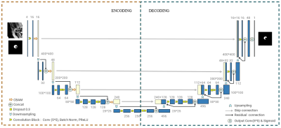 |
44 | Deep Learning-Based Automatic Segmentation of Arterial Plaques in MR Vessel Wall Images
Long Yang1,2, Xiong Yang3, Zhenhuan Gong3, Yufei Mao3, Guanxun Cheng4, Ke Wu3, Cheng Li3, Ye Li1, Dong Liang1, Xin Liu1, Hairong Zheng1, Zhanli Hu1, and Na Zhang1
1Lauterbur Research Center for Biomedical Imaging, Shenzhen Institute of Advanced Technology, Chinese Academy of Sciences, Shenzhen, China, 2School of Computer Sciences, University Sains Malaysia, Penang, Malaysia, 3Department of Image Advanced Analysis of HSW BU, Shanghai United Imaging Healthcare Co., Shanghai, China, 4Department of Radiology, Peking University Shenzhen Hospital, Shenzhen, China Keywords: Vessel Wall, Atherosclerosis, atherosclerotic plaque, morphological quantitative assessment Manual segmentation of atherosclerotic plaque for quantitative assessment is a time-consuming process. In this study, a convolutional neural network based automatic segmentation method named Vessel-Segnet was proposed for quantitative evaluation of lumen, vessel wall and plaque based on MR vessel wall images. The proposed method achieved the best segmentation performance with the highest dice similarity coefficient and the lowest average surface distance among six models. In terms of morphological quantitative evaluation, the proposed method achieved excellent agreement with manual method. Overall, the proposed method can quickly and accurately realize the segmentation of lumen, vessel wall and plaque for quantitative evaluation. |
|
1510.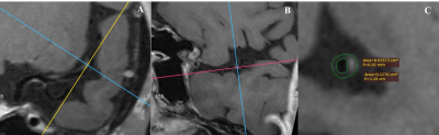 |
45 | The impact of intracranial atherosclerotic on the load and progress of cerebral small vessel disease
Lizhu yuerong1
1radiology, Beijing Chaoyang Hospital of Capital Medical University, Beijing, China Keywords: Vessel Wall, Brain This study hypothesized that the load and the progression of cSVD in the unilateral hemisphere is correlated with the features of ipsilateral intracranial large arterial vessel wall lesions. Using the fluctuations of WHMs at baseline and follow-up to reflect the severity of cSVD. VW-HRMRI, the most sensitive non-invasive ways to evaluate the lesions of intracranial vascular, was used to assessment the morphological and enhancement characteristic of the culprit plaque. The result suggested that mild stenosis and positive remodeling of MCA may associated with WHM progression. |
|
1511.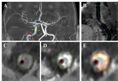 |
46 | The risk prediction of ischemic stroke recurrence: a MR vessel wall imaging study
Fei Zhou1, Maoxue Wang1, Ruijing Xin1, Tianshu Yang2, Yongming Dai3, Na Zhang4, Zhanli Hu4, Xin Zhang1, and Bing Zhang1
1Department of Radiology, The Affiliated Drum Tower Hospital of Nanjing University Medical School, Nanjing, China, 2Shenzhen United Imaging Research Institute of Innovative Medical Equipment, Shenzhen, China, 3MR Collaboration, Central Research Institute, United Imaging Healthcare, Shanghai, China, 4Paul C. Lauterbur Research Center for Biomedical Imaging, Shenzhen Institute of Advanced Technology, Chinese Academy of Sciences, Shenzhen, China Keywords: Vessel Wall, Vessels Symptomatic cerebral vascular stenosis diseases include acute ischemic infarction and transient ischemic syndrome. Patients with these diseases often have a high risk of recurrence in the short term, which may have serious adverse consequences. High-resolution MR vessel wall imaging (HR-VWI) technique has provided new possibilities to assess the threat of stroke recurrence due to stenosis caused by intracranial plaque. The present study confirms that plaque morphological parameters obtained by HR-VWI can provide an effective prediction of stroke recurrence risk in patients with intracranial arterial stenosis. |
|
1512.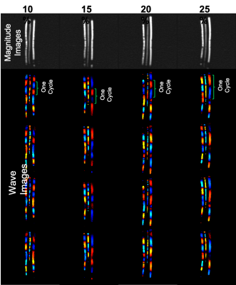 |
47 | Feasibility of Measuring Magnetic Resonance Elastography-derived Stiffness in Aortic Dissection
Adnan A. Hirad1, Faisal fakhouri2, Brian Raterman3, Dakota Gonring1, Arunark Kolipaka3, and Doran Mix1
1University of Rochester, Rochester, NY, United States, 2King Saud University, Riyadh, Saudi Arabia, 3The University of Ohio, Columbus, OH, United States Keywords: Vessel Wall, Blood vessels Type-B aortic dissection represents a serious medical emergency with up to a 50% associated 5-year mortality caused by thoracic aorta, dissection-associated aneurysmal (DAA) degeneration, and rupture. Unfortunately, conventional diagnostic methods cannot distinguish high-risk DAAs that benefit from surgical intervention from stable DAAs. Our goal is to use DAA stiffness measured with magnetic resonance elastography (MRE) as a biomarker to distinguish high-risk DAAs from stable DAAs. This is a feasibility study using MRE to 1) measure stiffness in hydrogel phantoms with human-like dissection geometries and 2) demonstrate the first successful application of MRE to the thoracic aorta of a human volunteer. |
|
1513.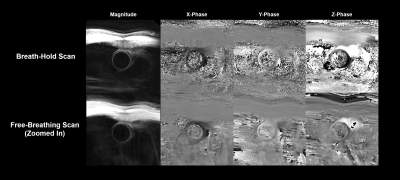 |
48 | Imaging Ascending Aortic Wall Stretch Using Breath-Held Displacement Encoding with Stimulated Echoes MRI: An Intra-scan Reproducibility Study
Huiming Dong1, Joseph Leach1, Ang Zhou1, Megan Ballweber1, Frederick H. Epstein2, Laing Ge3, Elaine Tseng3, David Saloner1, and Dimitrios Mitsouras1
1Radiology and Biomedical Imaging, University of California, San Francisco, San Francisco, CA, United States, 2University of Virginia, Charlottesville, VA, United States, 3Surgery, University of California, San Francisco, San Francisco, CA, United States Keywords: Vessel Wall, Preclinical, Aortic Strain; DENSE MRI In vivo assessment of the mechanical properties of the aorta stands to offer valuable information to evaluate and predict the progression of cardiovascular diseases. Displacement encoding with stimulated echoes (DENSE) is a non-invasive phase-contrast MRI technique that has been adapted to measure ascending aortic wall stretch. Previous studies utilizing long respiratory-gated acquisitions are prone to artifacts from residual cardiac or respiratory motion. In this study, we investigated the reproducibility of a novel breath-held aortic DENSE imaging protocol. The proposed breath-held protocol was highly reproducible (COV=3.27% and LCCC=0.97). Finally, the measured aortic stretch followed the expected relationship with age. |
|
1514.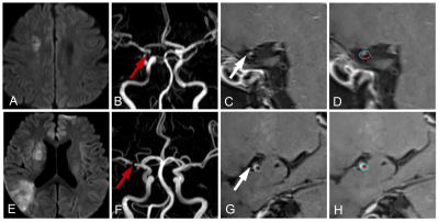 |
49 | High-resolution MR vessel wall imaging combined with Mediterranean diet adherence screener for evaluation of prognosis of ischemic stroke
Xiaochun Wang1 and Wenqiao Zheng1
1First Hospital of Shanxi Medical University, Taiyuan, China Keywords: Atherosclerosis, Stroke To evaluate the prognosis of ischemic stroke (IS) by plaque characteristics based on high-resolution MR vessel wall imaging combined with Mediterranean diet adherence screener (MEDAS). IS patients were divided into two groups according to their prognosis. Clinical data and plaque characteristics were obtained and compared between groups. The correlation between these characteristics and prognosis was investigated by multivariate analysis. The ROC curves were further performed to determine their predictive performance. NIHSS score, MEDAS score, and plaque enhancement ratio were independent predictors of IS prognosis. Noteworthy, the combined model of three parameters showed significant superiority in the prognosis evaluation of IS. |
|
1515.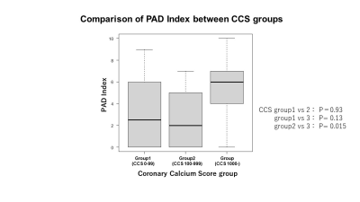 |
50 | Usefulness of Combined Evaluations of Coronary Calcification and Peripheral Nonenhanced MRA for the Risk Assessment of Future Vascular Events
Seigo Yoshida1, Katsumi Nakamura1, Akiyoshi Yamamoto1, Hidetoshi Akashi2, and Tetsuo Imamura3
1Radiology, Tobata Kyoritsu Hospital, Fukuoka, Japan, 2Vascular Surgery, Tobata Kyoritsu Hospital, Fukuoka, Japan, 3Surgery, Tobata Kyoritsu Hospital, Fukuoka, Japan Keywords: Atherosclerosis, Cardiovascular Feasibility of a combined evaluation of coronary artery calcification by plain CT and severity of PAD assessed by nonenhanced MRA was evaluated for predicting the risk of future cerebrovascular and cardiovascular events. The result showed patients who had coronary calcium score of 1000 or higher and more advanced PAD were associated with 67% more subsequent cerebrovascular and cardiovascular events. The possibility of assessing these events in a noninvasive manner has been demonstrated, and early intervention may lead to improved patient outcomes. |
|
1516.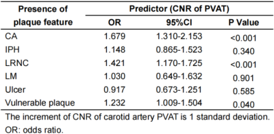 |
51 | Contrast-to-noise ratio of carotid artery perivascular adipose tissue on MRI: A potential indicator for carotid vulnerable plaque
Shuwan Yu1, Ran Huo2, Huiyu Qiao1, Zihan Ning1, Rui Shen1, Ning Xu1, and Xihai Zhao1
1Center for Biomedical Imaging Research, Tsinghua University, Beijing, China, 2Peking University Third Hospital, Beijing, China Keywords: Atherosclerosis, Inflammation, Carotid artery PVAT, Vulnerable plaques Inflammation of carotid artery perivascular adipose tissue (PVAT) is associated with atherosclerotic disease. This study investigated the association between carotid artery PVAT and atherosclerotic plaques using multi-contrast MR vessel wall imaging. The contrast-to-noise ratio (CNR) of PVAT was measured on TOF MRA images. We found that the CNR of carotid artery PVAT measured by TOF MRA is independently associated with carotid vulnerable plaque features, suggesting that the CNR of PVAT might be a potential indicator for vulnerable atherosclerotic plaques. |
|
1517.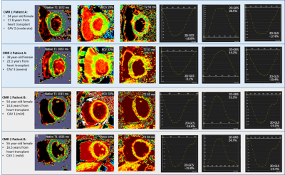 |
52 | Longitudinal study investigation of CMR measures and cardiac allograft vasculopathy over time
Sandra Quinn1, Chantelle Sanchez1, Ozden Kilinc1, Kai Lin1, Kambiz Ghafourian2, Daniel C Lee2, Esther E Vorovich2, Clyde W Yancy2, Vera H Rigolin2, Jon W Lomasney3, Bradley D Allen1, James C Carr1, and Michael Markl1
1Department of Radiology, Feinberg School of Medicine, Northwestern University, Chicago, IL, United States, 2Department of Medicine, Division of Cardiology, Feinberg School of Medicine, Northwestern University, Chicago, IL, United States, 3Department of Pathology, Feinberg School of Medicine, Northwestern University, Chicago, IL, United States Keywords: Myocardium, Transplantation, cardiac allograft vasculopathy CAV is a leading cause of mortality in HTx patients. Diagnosis and grading of CAV is dependent on repeated ICA which carries risk of major complications. CMR has the potential to be useful in CAV surveillance without the risk of ICA. This study has demonstrated that in a group of long-term heart transplant patients with evidence of CAV progression who undergo serial CMR and ICA within a close timeframe, 2D global peak strain parameters and 2D systolic strain rate correlates with CAV severity grade. FTS may therefore prove useful for surveillance of patients for CAV. |
|
1518.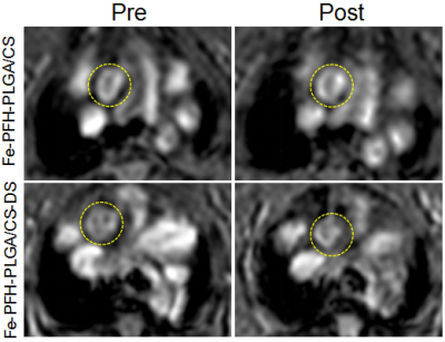 |
53 | Multifunctional Nanoparticles for Detection and Treatment of Atherosclerotic Vulnerable Plaques
Man Ye1
1Renmin Hospital of Wuhan University, Wuhan, China Keywords: Atherosclerosis, Molecular Imaging The instability of atherosclerotic plaque is seriously harmful to human health. The nanoparticle molecular probe we prepared can simultaneously achieve magnetic resonance and ultrasound imaging, which is expected to reflect the pathophysiological characteristics of plaque at the molecular level. The phase change induced by low intensity focused ultrasound (LIFU) can cause the apoptosis of macrophages in plaque,which has the potential for phase change ablation of plaque.1 It provides a simple, effective, safe and non-invasive new method for early diagnosis, treatment and efficacy monitoring of vulnerable plaque. |
|
1519.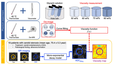 |
54 | Diffusion Weighted-Viscosity Imaging for Atherosclerotic Plaques
Mayuka Seguchi1, Yuki Kanazawa1, Tosiaki Miyati2, Masafumi Harada1, Mitsuharu Miyoshi3, Yuki Matsumoto1, Hiroaki Hayashi2, Yasuhisa Kanematsu1, and Yasushi Takagi1
1Tokushima University, Tokushima, Japan, 2Kanazawa University, Kanazawa, Japan, 3GE Healthcare Japan, Hino, Japan Keywords: Atherosclerosis, Diffusion/other diffusion imaging techniques We developed a Dw-viscosity imaging method to identify atherosclerotic plaques. First, we conducted experiments to determine ADC and viscosity changes of four different concentrations of glycerin solution, and determined the viscosity model. Second, a clinical study was carried out, and Dw-viscosity imaging was performed. As a result, we found that there was a significant difference between with and without hemorrhage findings (P < 0.01). Dw-viscosity imaging enables us to obtain more detailed information about atherosclerotic plaques such as intraplaque hemorrhage. |
|
1520.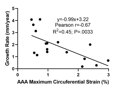 |
55 | MR Feature Tracking-Derived Circumferential Strain of the Aortic Wall Correlates to Abdominal Aortic Aneurysm Growth Independent of Diameter
Huiming Dong1, Joseph Leach1, Ang Zhou1, Teodora Chitiboi2, Megan Ballweber1, Warren Gasper3, David Saloner1, and Dimitrios Mitsouras1
1Radiology and Biomedical Imaging, University of California, San Francisco, San Francisco, CA, United States, 2Siemens Healthineers AG, Erlangen, Germany, 3Surgery, University of California, San Francisco, San Francisco, CA, United States Keywords: Vessels, Vessels, Aortic Strain; MR Feature Tracking Recent studies are increasingly highlighting aortic wall stress, strain, and other mechanical descriptors as relevant factors associated with abdominal aortic aneurysm (AAA) progression. The goal of this prospective patient study was to employ MR feature tracking to measure the maximum circumferential strain in AAAs and study investigate its relationship with AAA growth. The feature tracking-measured AAA circumferential strain was lower than that of the normal aorta, indicating that the AAA wall is less compliant than the non-aneurysmal aorta. Furthermore, AAA strain was associated with the growth rate of the aneurysm, independent of AAA maximum diameter. |
|
1521.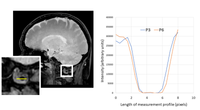 |
56 | 3D Isotropic black-blood cine MRI of intracranial arteries
Niranjan Balu1, Kaiyu Zhang2, Thomas S Hatsukami3, and Chun Yuan1,4
1Radiology, University of Washington, Seattle, WA, United States, 2Bioengineering, University of Washington, Seattle, WA, United States, 3Surgery, University of Washington, Seattle, WA, United States, 4University of Utah, Salt Lake City, UT, United States Keywords: Vessels, Data Acquisition Pulsatile motion of intracranial arteries may provide useful diagnostic information of intracranial vascular pathology. We develop an isotropic 3D black-blood whole brain MRI method for assessment of intracranial vascular pulsation. Comparison of the black-blood with 2D bright-blood PC-MRA and assessment of luminal boundary changes across the cardiac cycle suggest that pulsation of large intracranial arteries can be detected and measured by black-blood cine MRI. |
|
1522.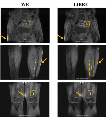 |
57 | Three-dimensional Black-blood Thrombus Imaging (BTI) with LIBRE Fat suppression for the diagnosis of Deep Vein Thrombosis
Zeping Liu1, Zehe He2, Guoxi Xie1, Liping Liao2, Mingxia Tan2, Qizeng Ruan2, Lanbin Huang2, Qingchun Li2, Yuhui Nie1, Anyan Gu1, Zhuoneng Zhang1, and Yi Sun3
1Guangzhou Medical University, Guangzhou, China, 2The First People’s Hospital of Qinzhou, Qinzhou, China, 3Siemens Healthineers, Shanghai, China Keywords: Vessels, Thrombo-Embolic Deep vein thrombosis (DVT) can lead to life-threatening pulmonary embolism. Black-blood thrombus imaging (BTI) technique can accurately identify DVT and provide additional information for thrombus staging. However, non-uniform fat suppression of BTI with conventional fat saturation preparation is obvious due to the field inhomogeneities and large field of view (FOV) imaging, which can affect the accuracy of detecting and staging of thrombus. To address this issue, a field inhomogeneity insensitive BTI technique was developed by incorporating LIBRE pulses for fat free and large FOV thrombus imaging. |
|
1523. |
58 | Pathological changes of the coronary microvascular dysfunction pig models based on diffuse tensor imaging (DTI)
Zilong Ren1, Didi Wen2, Jianxiu Lian3, and Minwen Zheng4
1Department of Radiology, Xijing Hospital, Fourth Military Medical University, Xi'an, China, 2Department of Radiology, Xijing Hospital, Forth Military Medical University, Xi'an, China, 3Philips Healthcare China, Xi'an, China, 4Xijing Hospital, Fourth Military Medical University, Xi'an, China Keywords: Heart, Animals To investigate whether diffuse tensor imaging (DTI) can be used as a predictor for coronary microvascular dysfunction (CMD). Pig models with CMD had higher mean diffusivity (MD), lower FA, and lower E2A compared with normal pigs (mean MDnormal = 1.50 ± 0.02, mean MDCMD = 1.65 ± 0.02, P = 0.001; mean FAnormal = 0.35 ± 0.01, mean FACMD = 0.27 ± 0.01, P < 0.001; mean E2Anormal = 53.33 ± 0.88, mean E2ACMD = 46.57 ± 1, P = 0.004). Cardiac DTI technology could add benefits of offering microstructure change and cardiac remodeling parameters for CMD pig models. |
|
1524.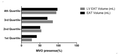 |
59 | Association Between Epicardial Adipose Tissue volume and Microvascular Obstruction with ST-segment elevation myocardial infarction Patients
Dan Mu1, Xiuzheng Yue2, and Xiance Zhao2
1Department of Radiology, Affiliated Nanjing Drum Tower Hospital of Nanjing University Medical School, nanjing, China, 2Philips Healthcare, Shanghai, China Keywords: Atherosclerosis, Cardiovascular Epicardial adipose tissue (EAT) is a novel factor for risk stratification of coronary artery disease. EAT in microvascular obstruction formation in patients with ST-segment elevation myocardial infarction (STEMI) remain unclear. This study aimed to evaluate the correlation between EAT and MVO volume detected by CMR on a clinical 1.5T CMR system with a 28-channel coil array in STEMI patients. |
|
1525.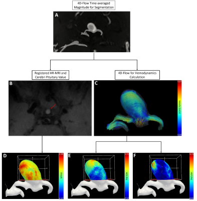 |
60 | Vertical Dynamic Pressure: a promising biomarker for describing inflammation in intracranial aneurysm
Haining Wei1, Mingzhu Fu1, Yudi Tang2, Peng Liu3, and Rui Li1
1Center for Biomedical Imaging Research, Tsinghua University, Beijing, China, 2Beijing Neurosurgical Institute, Capital Medical University, Beijing, China, 3Tiantan Hospital, Beijing, China Keywords: Data Processing, Velocity & Flow, Intracranial Aneurysm Increasing histopathological evidence suggested that the inflammation processes and hemodynamics in vessel wall may mediate the growth and rupture of IA. For hemodynamics, previous studies reported that lower wall shear stress (WSS) is the most highlighted parameter associated with inflammation. Some studies also explored the dynamic pressure distribution at the sites of aneurysms based on computational fluid dynamics. Vertical dynamic pressure(VDP) can be seen as a possible measure for the force of the fluid impinging on the vessel wall. In this study, we propose to investigate the correlation of two hemodynamic parameters(WSS, VDP) with inflammation by advanced MRI techniques. |
|
 Back to Meeting Home
 Back to the Program-at-a-Glance
The International Society for Magnetic Resonance in Medicine is accredited by the Accreditation Council for Continuing Medical Education to provide continuing medical education for physicians.
 Back to Meeting Home
 Back to the Program-at-a-Glance
 View Presentation Video