Digital Poster
New Developments & Clinical Applications of Flow MRI II
ISMRM & ISMRT Annual Meeting & Exhibition • 03-08 June 2023 • Toronto, ON, Canada

Digital Poster
New Developments & Clinical Applications of Flow MRI II
| Computer # | |||
|---|---|---|---|
3819.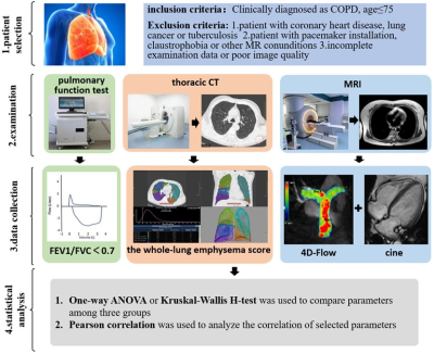 |
61 |
Evaluation of pulmonary artery hemodynamic changes in COPD
patients with different emphysema volume by using 4D-Flow MRI
Jiwei Sun1,
Hong Zhang1,
Anhong Yu1,
Jianxiu Lian2,
Yufan Gao1,
Wei Yuan1,
and Yapeng Yang1
1Radiology, Tianjin Chest Hospital, Tianjin, China, 2Philips Healthcare, Beijing, China Keywords: Flow, Blood, hemodynamic Few studies quantitatively evaluate pulmonary artery hemodynamic changes caused by emphysema. 34 COPD patients were divided into two groups according to emphysema volume and 12 healthy volunteers were matched. Hemodynamic parameters were calculated by 4D-Flow MRI and right ventricular function indicators were evaluated for three groups. The right systolic pressure drop and RVEF were lower in moderate and severe emphysema group but the right EL was higher. The whole-lung emphysema score correlated negatively with RVEF and the right systolic pressure drop. 4D-Flow combined with cine image can comprehensively evaluate the pulmonary artery hemodynamics and right ventricular function of COPD patients. |
|
3820.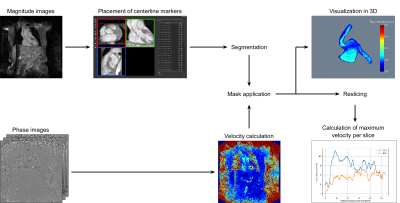 |
62 |
3D phase contrast blood flow measurements in the coronary
arteries
Denise Lichthardt1,
Jens Wetzl2,
Michaela Schmidt2,
Peter Speier2,
Armin M. Nagel1,
and Daniel Giese2
1Friedrich-Alexander-University Erlangen-Nürnberg, Erlangen, Germany, 2Siemens Healthcare GmbH, Erlangen, Germany Keywords: Flow, Cardiovascular Phase contrast MRI enables flow measurement in the coronary arteries. Herewith we propose a 3D Flow sequence and present a novel 3D visualization and quantification tool. Whole heart 3D PC measurements were performed in more than 20 healthy volunteers. An isotropic resolution of 1.2x1.2x1.2mm3 was used, as well as an undersampling factor of 14 in combination with a compressed sensing reconstruction. Our postprocessing tool was used for the 3D visualization of blood flow information as well as quantification of flow velocities per slice perpendicular to the vessel centerline.
|
|
3821.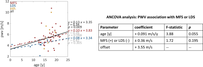 |
63 |
Quantitative characterization of hemodynamic parameters in
pediatric patients with connective tissue disorders
Aparna Sodhi1,
Ethan Johnson2,
Elizabeth Weiss2,
Haben Berhane2,
Dr. Joshua Robinson3,
Dr. Andrada Popescu1,
Dr. Michael Markl2,
and Dr. Cynthia Rigsby1
1Department of Medical Imaging, Ann & Robert H Lurie Children's Hospital, Chicago, IL, United States, 2Department of Radiology, Northwestern University, Chicago, IL, United States, 3Department of Cardiology, Ann & Robert H Lurie Children's Hospital, Chicago, IL, United States Keywords: Flow, Cardiovascular, Marfan Syndrome, Loeys-Dietz Syndrome, Vascular Ehlers-Danlos syndrome Quantitative overview of 4D flow derived aortic hemodynamic parameters such as kinetic energy, peak velocity, and pulse wave velocity in a cohort of pediatric patients with connective tissue disorders such as Marfan Syndrome (MFS), Loeys-Dietz Syndrome (LDS), and Vascular Ehlers-Danlos Syndrome (EDS), using a fully automated pipeline. A statistically significant trend of increased pulse wave velocity - an important measure of vessel stiffness, with increasing age was observed for this cohort. No association was found between PWV and all mean aortic diameters when controlled for age. |
|
3822.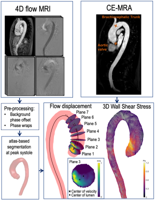 |
64 |
Flow Displacement and Wall Shear Stress in Individuals with
Mild-to-Moderate Aortic Dilation and Tricuspid Aortic Valves
Chiara Trenti1,2,
Filip Hammaréus1,
Tino Ebbers1,2,
Eva Swahn1,3,
Lena Jonasson1,
and Petter Dyverfeldt1,2
1Department of Health, Medicine and Caring Sciences (HMV), Linköping University, Linköping, Sweden, 2Center for Medical Image Science and Visualization (CMIV), Linköping University, Linköping, Sweden, 3Department of Cardiology in Linköping, and Department of Health, Medicine and Caring Sciences, Linköping University, Linköping, Sweden Keywords: Flow, Vessels, Aorta Altered hemodynamics may play a role in the development of ascending aortic aneurysms. In this study, we sought to investigate wall shear stress and flow displacement in patients with tricuspid aortic valve and mild-to-moderate dilatation of the ascending aorta, and age-and sex- matched non-dilated controls. Dilated patients had lower average velocity and wall shear stress in the ascending aorta, and higher flow displacement. This study shows that individuals with dilatation have altered hemodynamics even in an early stage of the disease. |
|
3823.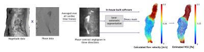 |
65 |
Reproducibility of wall shear stress in abdominal aortic
aneurysms estimated by 4D flow magnetic resonance imaging
Eva Aalbregt1,2,
Aart J. Nederveen2,
Kak Khee Yeung1,
R. Nils Planken2,
Ron Balm 1,
and Pim van Ooij2
1Vascular Surgery, Amsterdam University Medical Centers, Amsterdam Cardiovascular Sciences, Vrije Universiteit Amsterdam, Amsterdam, Netherlands, 2Radiology & Nuclear Medicine, Amsterdam University Medical Centers, Amsterdam Cardiovascular Sciences, University of Amsterdam, Amsterdam, Netherlands Keywords: Flow, Quantitative Imaging, abdominal aortic aneurysm The aim of this study was to assess the reproducibility and inter- and intra-observer agreement for estimating wall shear stress (WSS) in abdominal aortic aneurysms (AAAs) based on 4D flow magnetic resonance imaging (MRI). Asymptomatic AAA patients were included and scanned twice with a one-week interval. Phase-contrast angiographies were acquired by voxel-wise multiplication of magnitude and phase data. Based on statistical analyses the reproducibility was good. An inverse correlation was found between WSS and lumen diameter. Both vorticity and intraluminal thrombus volume were associated with minimum WSS in AAA. 4D flow MRI is robust for estimating WSS in AAAs. |
|
3824.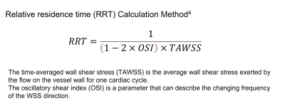 |
66 |
Patients with liver cirrhosis have more stagnant portal venous
blood flow compared to non-cirrhotic subjects: analysis using 4D
flow MRI
Ryota Hyodo1,
Yasuo Takehara1,2,
Takashi Mizuno3,
Kazushige Ichikawa4,
Yoji Ishizu5,
Yasuhiro Ogura6,
and Shinji Naganawa7
1Radiology, Nagoya University Graduate School of Medicine, Nagoya, Japan, 2Fundamental Development for Advanced Low Invasive Diagnostic Imaging, Nagoya University Graduate School of Medicine, Nagoya, Japan, 3Radiological Technology, Nagoya Uviversity Hospital, Nagoya, Japan, 4RadiologicalTechnology, Nagoya Uviversity Hospital, Nagoya, Japan, 5Gastroenterology and Hepatology, Nagoya University Graduate School of Medicine, Nagoya, Japan, 6Transplantation Surgery, Nagoya Uviversity Hospital, Nagoya, Japan, 7Nagoya University Graduate School of Medicine, Nagoya, Japan Keywords: Flow, Velocity & Flow Although liver cirrhosis is an independent risk factor for portal vein thrombosis (PVT), the individual thrombotic risk assessment is complex. Recently, 4D flow MRI has begun to assess portal blood flow; however, there have been no reports of the use of RRT, an indicator of blood flow stagnation, in the portal system. RRT may be used to measure the risk of PVT in liver cirrhosis. In this study, we evaluated the mean RRT value of the main PV calculated with 4D flow MRI and found that the value was significantly higher in cirrhotic patients than in non-cirrhotic subjects. |
|
3825. |
67 |
The Value of 4D Flow MRI Combined with Precontrast T1 mapping
Model in Identifying High-risk Varices of Cirrhosis Patients
Xiaohuan Li1,
Zhou Wei1,
Zhiyuan Chen1,
Dongjing Zhou1,
Shuping Zhang1,
Ruhang Huang1,
Yunzhu Wu2,
and Yupin Liu*1
1Department of radiology, The Second Affiliated Hospital of Guangzhou University of Chinese Medicine, Guangzhou, China, 2MR Scientific Marketing, Siemens Healthineers Ltd, Shanghai, China Keywords: Flow, Velocity & Flow Noninvasive identification of high-risk varices (HRV) in cirrhosis patients is of significantly value because it could help avoid some unnecessary upper gastrointestinal endoscopy and repeat in a short time to evaluate the efficiency of non-selective beta blockers therapy. This study aims to evaluate the value of 4D flow MRI combined with precontrast T1 in identifying HRV. Our results indicated that the model including SMV forward volume, SMV maximum blood flow, precontrast liver T1 mapping, and precontrast spleen T1 mapping could effectively identify HRV of cirrhosis patients. |
|
3826. |
68 |
Evaluation of turbulent Kinetic energy and Viscous energy loss
on 4D flow MRI in patients with hypertrophic cardiomyopathy
Tetusro Sekine1 and
Shinichiro Kumita1
1Nippon Medical School, Tokyo, Japan Keywords: Flow, Cardiovascular The purpose was to validate whether turbulent kinetic energy (TKE) and viscous energy loss (VEL) reflect the condition of hypertrophic cardiomyopathy (HCM). We included consecutive 32 HCM patients. They were classified into HOCM (HCM with left ventricular outflow tract obstruction [LVOTO]) (n=21) and HNCM (HCM without LVOTO) (n=11) based on cardiac ultrasound. Both TKE and VEL were significantly higher in HOCM than HNCM. The strong correlation was observed between normalized LVmass and normalized TKEpeak in asymptomatic HCM (nTKEpeak r=0.879, p<0.001), but not in symptomatic HCM. The TKEpeak had the strongest correlation to each disease condition than the others. |
|
3827.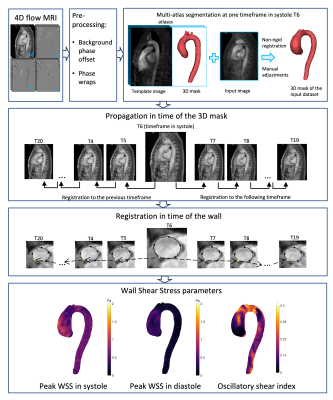 |
69 |
Oscillatory Wall Shear Stress in patients with Bicuspid Aortic
Valve and Aortic Regurgitation
Chiara Trenti1,2,
Petter Dyverfeldt1,2,
Paul W.M. Fedak3,
James A. White3,
and Julio Garcia3,4,5,6
1Department of Health, Medicine and Caring Sciences (HMV), Linköping University, Linköping, Sweden, 2Center for Medical Image Science and Visualization (CMIV), Linköping University, Linköping, Sweden, 3Department of Cardiac Sciences, Cumming School of Medicine, University of Calgary, Calgary, AB, Canada, 4Stephenson Cardiac Imaging Centre, Libin Cardiovascular Institute, Calgary, AB, Canada, 5Department of Radiology, University of Calgary, Calgary, AB, Canada, 6Alberta Children’s Hospital Research Institute, University of Calgary, Calgary, AB, Canada Keywords: Flow, Vessels, Aorta Individuals with bicuspid aortic valve (BAVs) and aortic regurgitation have higher rate of aortic complications compared to individuals with stenotic and functioning BAVs. We sought to investigate wall shear stress (WSS) and oscillatory shear index (OSI) in the ascending aorta of BAVs with functioning valves, stenotic and regurgitant valves, as well as controls with tricuspid aortic valves. BAVs with regurgitation had similar WSS compared to all other groups, but OSI was almost twice as high. This finding may be related to the pendulum volume between the aorta and the left ventricle. Studying OSI may improve the understanding behind BAV aortopathy. |
|
3828.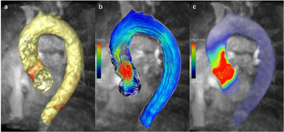 |
70 |
Assessment of Altered Hemodynamics in Patients with Hypertrophic
Obstructive Cardiomyopathy using 4D Flow MRI
Yun Zhao1,
Lu Huang1,
Xiaoyue Zhou2,
and Liming Xia1
1Department of Radiology, Tongji Hospital, Tongji Medical College, Huazhong University of Science and Technology, Wuhan, China, 2MR Collaboration, Siemens Healthineers Ltd, Shanghai, China Keywords: Flow, Blood, 4D flow MRI; Hemodynamics; Energy loss; Hypertrophic Obstructive Cardiomyopathy; Hypertrophic Obstructive Cardiomyopathy (HOCM) is characterized by dynamic obstruction of blood flow in the left ventricular outflow tract. 4D flow MRI can be used for comprehensive evaluation of cardiac and aortic hemodynamics by visualization of the complex spiral LVOT 3D blood flow patterns. This study used 4D flow MRI to evaluate the degree of LVOT obstruction and the flow energy loss in patients with HOCM. The results showed that spiral flow and viscous energy loss were associated with the LVOT pressure gradient, and played a role in structural remodeling of the left ventricle. |
|
3829. |
71 |
Association between angioarchitecture and flow parameters in
ruptured and unruptured arteriovenous malformation: a pilot
study by 4D flow MR
Yuting Wang1,
Haining Wei2,
Meixiong Cheng3,
Yishuang Wang1,
Yunzhu Wu4,
Mingzhu Fu2,
and Rui Li2
1Radiology, Sichuan Provincial People's Hospital, Chengdu, China, 2Center for Biomedical Imaging Research Beijing, Qinghua University, Beijing, China, 3Neurosurgery, Sichuan Provincial People's Hospital, Chengdu, China, 4Siemens Healthineers Ltd., Shanghai, China Keywords: Flow, Velocity & Flow, arteriovenous malformation The hemodynamics of cerebral AVM likely vary with lesion angioarchitecture and the rupture status, which couldn’t be reflected by morphology-based imaging. In this pilot study, 9 patients with AVM had their complicated feeding/draining patterns visualized by 4D-flow MR. AVM tend to have heterogeneous hemodynamics even with the same Spetzler-Martin grade or similar angioarchitecture. It tends to have smaller flow of the feeding artery after rupture. AVM with deep vein drainage tends to have diffused nidus and higher wall shear stress adjacent to the nidus. Higher flow of the feeding artery was associated with higher dynamic pressure and larger nidus volume. |
|
3830. |
72 |
Hemodynamic analysis of intracranial aneurysms before and after
flow diverter stents implantation based on 4D Flow MRI
Yuxin Liu1,
Haining Wei1,
Tian Bing2,
Hou Yuxi2,
Mingzhu Fu1,
and Rui Li1
1Center for Biomedical Imaging Research, Tsinghua University, Beijing, China, 2Changhai Hospital, Shanghai, China Keywords: Flow, Velocity & Flow, Hemodynamics Intracranial aneurysm has become an important issue because of the high morbidity and the high mortality rate due to aneurysm rupture. Recently, the flow diverter stent has been considered as an effective device for the treatment of aneurysms by modifying the hemodynamics of the aneurysm. 4D Flow MRI can can offer three-dimensional visualization of blood flow pattern and a comprehensive analysis of hemodynamics. In this study, we calculated and analyzed the hemodynamics in aneurysm hemodynamics before and after the implantation of flow diverter stents based on 4D Flow MRI. |
|
3831.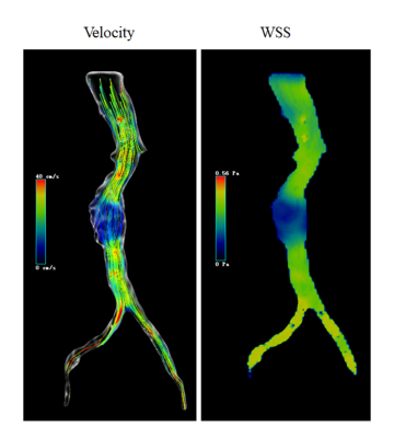 |
73 |
Hemodynamics of common iliac arteries is associated with the
risk of abdominal aortic aneurysm: assessment using 4D flow MRI
Wen Zeng1,
Chunchao Xia1,
Xiaoyong Zhang2,
and Zhenlin Li1
1West China Hospital, Sichuan University, Chengdu, China, 2Clinical Science, Philips Healthcare, Chengdu, China, Chengdu, China Keywords: Flow, Cardiovascular, Abdominal aortic aneurysm Studies have confirmed the interaction between iliac arteries and abdominal aortic aneurysms. Four-dimensional (4D) flow MRI can reveal the hemodynamic changes of common iliac arteries in abdominal aortic aneurysm and health controls. In this study, we tried to explain the relationship between the changes of iliac artery hemodynamics and the asymmetry of the left and right iliac artery blood flow and the occurrence and development of abdominal aortic aneurysms in patients. |
|
3832. |
74 |
Evaluation of diastolic ventricular interdependence using
simultaneous left and right heart transvalvular real time phase
contrast CMR
Simon Thalén1,
Peder Sörensson2,
Daniel Giese3,
Andreas Sigfridsson1,
and Martin Ugander1,4
1Department of Clinical Physiology, Karolinska Institutet and Karolinska University Hospital, Stockholm, Sweden, 2Department of Cardiology, Karolinska Institutet and Karolinska University Hospital, Stockholm, Sweden, 3Siemens Healthineers, Erlangen, Germany, 4Kolling Institute, Royal North Shore Hospital, and Charles Perkins Centre, Sydney, Australia Keywords: Flow, Velocity & Flow, Ventricular interdependence Diastolic ventricular interdependence occurs in several clinical settings, most notably pericardial effusion and constrictive pericarditis. It is measured using Doppler echocardiography or invasive cardiac catheterization where the respiratory variation in velocity or pressure is measured. In pericardial effusion it is an early sign of hemodynamic significance which is not always correlated to the volume of effusion. In constrictive pericarditis, it is used to separate constrictive and restrictive physiologies. In this study a method to measure diastolic ventricular interdependence by simultaneously quantifying the respiratory variation in mitral and tricuspid early inflow velocities using real time phase contrast CMR is presented. |
|
3833.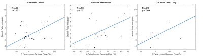 |
75 |
Interval Changes in 4D Flow-Derived Hemodynamic Parameters
Stratify Type B Aortic Dissection Patients and Correlate with 2D
Through-Plane Flow
Josh Engel1,
Ozden Kilinc1,
Justin Baraboo2,
Elizabeth Weiss1,2,
Christopher Mehta3,
Andrew Hoel4,
S. Chris Malaisrie3,
Michael Markl2,
and Bradley Allen1
1Radiology, Northwestern University, Chicago, IL, United States, 2Biomedical Engineering, Northwestern University, Chicago, IL, United States, 3Cardiac Surgery, Northwestern Medicine, Chicago, IL, United States, 4Vascular Surgery, Northwestern Medicine, Chicago, IL, United States Keywords: Vessels, Velocity & Flow, Aortic Dissection, 4D Flow We investigated whether changes in 4D flow-derived hemodynamic parameters stratified type B aortic dissection patients and sought to validate a 4D flow-derived 2D planar false lumen flow method. Patient scans with baseline and follow-up 4D flow at least 120 days apart were manually segmented into true and false lumens. Voxel-wise parametric maps and 2D forward and reverse flow in the descending aorta were calculated. Changes in voxel-wise hemodynamic parameters significantly correlated with aortic growth rate, with changes in false lumen reverse flow as the most robust parameter. 2D planar flow correlated with voxel-wise flow, but not aortic growth. |
|
3834.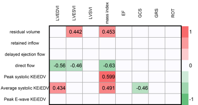 |
76 |
Ventricular flow analysis and its Association with cadiac
function and structure of hypertrophic cardiomyopathy: 4D flow
CMR study
Yuying Chen1,
Yang Peng1,
Yu Feng1,
Jun Yuan1,
Lin Peng1,
Mengzhu Wang2,
and Ning Jin3
1The First Affiliated Hospital of Sun Yat-sen University, Guangzhou, China, 2MR Scientific Marketing, Siemens Healthineers Ltd., Guangzhou, China, 3Siemens Medical Solutions USA, Inc. Customer Solutions Group,Siemens Healthineers, MR Scientific Marketing, Siemens Healthineers Ltd., Chicago, IL, United States Keywords: Cardiomyopathy, Cardiomyopathy, 4D flow This study aimed to investigate the potential of ventricular flow parameters derived from four‑dimensional (4D) flow cardiovascular magnetic resonance (CMR) to accurately support assessment of hypertrophic cardiomyopathy (HCM). Our results show that reduced left ventricular (LV) residual volume was associated with LV dysfunction. Higher LV retained inflow and lower delayed ejection inflow were observed in obstructive HCM patients. Flow components and kinetic energy correlated significantly with LV functional and remodelling CMR parameters. These findings suggest that 4D flow CMR may be potential method to explain the relationship between hemodynamic and pathophysiological changes in HCM. |
|
3835. |
77 |
Quantitative Mitral Valvular Regurgitation Hemodynamics Analysis
Using 4D flow MRI and Echocardiography: Characteristics of Flow
Momentum
Jeesoo Lee1,
Adarsh Aratikatla1,
Erik Wu2,
Taimur Safder3,
Gloria Ayuba3,
James Thomas3,
and Michael Markl4
1Radiology, Northwestern University, Chicago, IL, United States, 2Northwestern University, Chicago, IL, United States, 3Cardiology, Northwestern Memorial Hospital, Chicago, IL, United States, 4Radiology, Northwestern University, chicago, IL, United States Keywords: Valves, Velocity & Flow, Mitral valvular regurgitation, 4D flow MRI, Jet momentum Quantitative hemodynamic assessment of mitral valvular regurgitation (MVR) is critical for evaluating severity of the disease, but has been challenging with conventional echocardiographic and cardiac MRI techniques. 4D flow MRI is a promising tool that enables direct retrospective quantification of MVR hemodynamics. This study proposes a regurgitant flow momentum as a novel and reliable fluid dynamic parameter for MVR assessment using 4D flow MRI. The conservation characteristics of MVR jet flow momentum and its relationship with LA enlargement were investigated in 44 patients using 4D flow MRI and echocardiography. |
|
3836.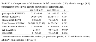 |
78 |
Effect of age on the evaluation of left ventricular kinetic
energy from 4d-flow CMR
Jingyu Zhang1,
Di Tian1,
Ziqi Xiong1,
Yifan He1,
and Zhiyong Li1
1The First Affiliated Hospital of Dalian Medical University, Dalian, China Keywords: Heart, Heart Four-dimensional flow cardiovascular magnetic resonance imaging (4d-flow CMR) left ventricular kinetic energy (LVKE) parameter Peak A- wave KEiEDV increased, KEiEDV E/A ratio decreased with age. This suggests that 4d-flow CMR LVKE parameters can respond to age-induced changes in LV diastolic function. |
|
3837.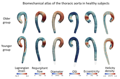 |
79 |
Exploration of age-related hemodynamic and mechanical changes in
the human thoracic aorta using an atlas based approach
Elodie Piot1,
Nicolas Duchateau1,
Marine Menut2,
Benyebka Bou-Said2,
Patrick Clarysse1,
Philippe Douek1,3,
Karl Kunze4,
Rene Botnar5,
Sara Boccalini6,
Claudia Prieto5,
and Monica Sigovan1
1CREATIS, Lyon, France, 2INSA de Lyon, Lyon, France, 3Departement of Radiology, HCL, Lyon, France, 4Siemens Helthineers, London, United Kingdom, 5School of Biomedical Engineering and Imaging Sciences, King’s College London, London, United Kingdom, 6Department of Radiology, HCL, Lyon, France Keywords: Data Processing, Blood vessels Understanding the hemodynamic involvement in a vascular pathology requires inter-subject comparisons that are not straightforward due to variability in terms of aorta morphology. Hemodynamic atlases can facilitate detection of intra-group characteristics. We propose here a workflow to create a hemodynamic atlas using 4D Flow MRI. In addition, we propose to investigate the aorta wall stiffness using non-rigid image registration and inverse mechanical modeling. This type of analysis is expected to improve pathophysiological understanding of vascular disease, by enabling the investigation of potential correlations between hemodynamic and wall mechanical properties at each point of the aorta. |
|
 Back to Meeting Home
 Back to the Program-at-a-Glance
The International Society for Magnetic Resonance in Medicine is accredited by the Accreditation Council for Continuing Medical Education to provide continuing medical education for physicians.
 Back to Meeting Home
 Back to the Program-at-a-Glance
 View Presentation Video