Digital Poster
Technical Solutions in Cardiovascular Imaging & Image Processing II
ISMRM & ISMRT Annual Meeting & Exhibition • 03-08 June 2023 • Toronto, ON, Canada

Digital Poster
Technical Solutions in Cardiovascular Imaging & Image Processing II
| Computer # | |||
|---|---|---|---|
5123.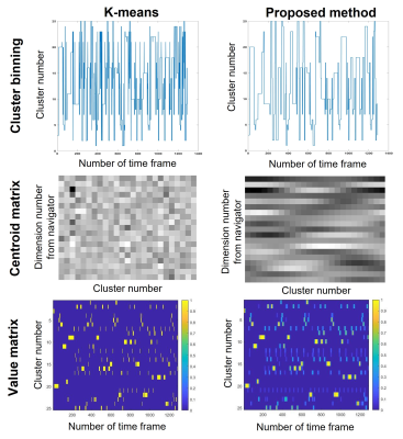 |
21 |
Data-driven clustering for ECG-free cine MRI with robustness
against irregular cardiac motion
Zhengyang Ming1,2,
Arutyun Pogosyan3,
Anthony G. Christodoulou4,5,
J. Paul Finn1,2,
Dan Ruan1,4,6,
and Kim-Lien Nguyen1,2,3,4
1Physics and Biology in Medicine Graduate Program, University of California,Los Angeles, Los Angeles, CA, United States, 2Department of Radiological Sciences, David Geffen School of Medicine at UCLA, Los Angeles, CA, United States, 3Division of Cardiology, David Geffen School of Medicine at UCLA and VA Greater Los Angeles Healthcare System, Los Angeles, CA, United States, 4Department of Bioengineering, University of California, Los Angeles, Los Angeles, CA, United States, 5Biomedical Imaging Research Institute, Cedars-Sinai Medical Center, Los Angeles, CA, United States, 6Department of Radiation Oncology, David Geffen School of Medicine at UCLA, Los Angeles, CA, United States Keywords: Arrhythmia, Machine Learning/Artificial Intelligence Both ECG-gating and self-gated approaches are used for cardiac motion binning in segmented cardiac cine MRI. Typical self-gated methods reduce ECG-dependency by assuming periodic cardiac motion, but may be less reliable in the presence of irregular cardiac motion. We propose a novel clustering algorithm that incorporates regularization in both temporal and cluster dimension to provide robustness against irregular cardiac motion in complex arrhythmias such as atrial fibrillation and premature ventricular contraction. Compared with images from k-means clustering, initial validation using the modified algorithm shows higher image quality scores with comparable single-to-noise ratio (SNR) and image sharpness. |
|
5124. |
22 |
A Data Augmentation Framework to Improve R-peak Detection in ECG
Recorded in MRI Scanners
Maroua Mehri1,2,
Pierre Aublin3,
Guillaume Calmon1,
Freddy Odille1,3,4,
and Julien Oster3,4
1Epsidy, Nancy, France, 2University of Sousse, National School of Engineers, LATIS-Laboratory of Advanced Technology and Intelligent Systems, Sousse, Tunisia, 3IADI, INSERM U1254 and Université de Lorraine, Nancy, France, 4CIC-IT 1433, INSERM, Université de Lorraine and CHRU Nancy, Nancy, France Keywords: Heart, Data Analysis, Machine learning/Artificial intelligence Detecting R-peaks in ECG using deep learning (DL) requires large, annotated datasets. Such datasets with the MRI-specific magneto-hydrodynamic (MHD) effect, do not exist currently. We propose a robust data augmentation framework using records available online, adding realistic MHD artifacts to augment the training dataset. MHD artifacts were modelled from eight 3T MRI-ECG records, and added to 75 non-MRI-ECG records. The R-peak detection was evaluated on six 3T MRI-ECG records. Compared to a DL model trained without data augmentation, the number of false positives and missed detections were reduced by 57.6% and 16.4%, the overall error was decreased by 25%. |
|
5125.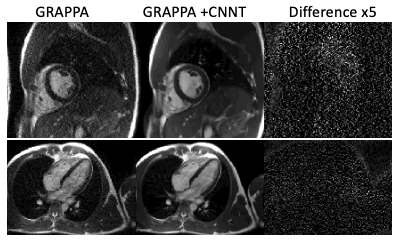 |
23 |
CNNT denoising for cine imaging at 0.55T with higher
acceleration rates
Hui Xue1,
Ahsan Javed1,
Rajiv Ramasawmy1,
Azaan Rehman1,
Peter Kellman1,
and Adrienne E Campbell-Washburn1
1National Heart, Lung, and Blood Institute, National Institutes of Health, Bethesda, MD, United States Keywords: Heart, Machine Learning/Artificial Intelligence Low field MRI systems are promising to increase the accessibility of cardiac MRI, at the expense of lower SNR. Here, we present the application of a g-factor-savvy denoising for cine imaging at 0.55T to increase useable acceleration rate. We used a model that combines convolutional neural networks and transformer model. The denoising network uses complex 2D+time images in SNR-units and g-factor maps as inputs and was trained with 3T data. The percent mean myocardial SNR gain at 0.55T across 9 healthy volunteers was 97±31% (R=2) and 122±22% (R=3), with no indication of overt temporal or spatial smoothing. |
|
5126.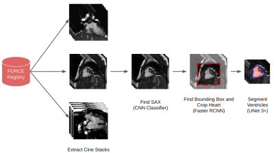 |
24 |
Deep Learning Pipeline for Preprocessing and Segmenting Cardiac
Magnetic Resonance of Single Ventricle Patients from an Image
Registry
Tina Yao1,2,
Nicole St. Clair3,
John Gold3,
Gabriel Miller3,
David Schidlow3,
Sunil Ghelani3,
Rahul Rathod3,
Jennifer Steeden1,
and Vivek Muthurangu1
1Institute of Cardiovascular Science, University College London, London, United Kingdom, 2Institute of Health Informatics, University College London, London, United Kingdom, 3Department of Cardiology, Boston Children's Hospital, Boston, MA, United States Keywords: Heart, Machine Learning/Artificial Intelligence We have created an end-to-end machine learning pipeline that takes cardiac magnetic resonance scans straight from a registry of single ventricle patients, performs image classification, calculates bounding boxes, and segments the ventricles. The clinical utility of the pipeline is that there is very little human preprocessing required from the clinicians. The pipeline has great robustness as it is trained on multicenter data from different countries, with different scanners, image sizes and aspect ratios, patient ages (relating to heart sizes), and the inherent variability of single ventricle patients. Heart metrics calculated from our pipeline can guide treatment for single ventricle patients. |
|
5127.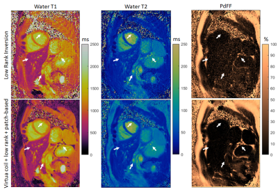 |
25 |
Improved T1, T2 and PDFF mapping with rosette MRF using
virtual-coil + low-rank + patch-based regularization
Gastao Cruz1,
Yuchi Liu1,
Evan Cummings1,
Jesse Hamilton1,
Vikas Gulani1,
and Nicole Seiberlich1
1Department of Radiology, University of Michigan, Ann Arbor, MI, United States Keywords: Heart, MR Fingerprinting In this work, T1/T2/PDFF mapping with rosette cardiac MRF is improved by exploiting the idea of virtual coils along with subspace constrained reconstruction. The Hermitian symmetry of k-space is explicitly included in the forward model via virtual coils and combined with global low-rank models. This model is further combined with a regularizer leveraging prior information from dictionaries, patch-similarity, and locally low-rank. This virtual-coil + low-rank + patch-based approach is used to jointly reconstruct multi-echo rosette MRF data for improved quality of the final T1, T2, and PDFF maps. |
|
5128.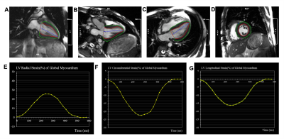 |
26 |
Cardiac Magnetic Resonance Feature Tracking for Detection of
Acute Cardiac Allograft Rejection after Heart Transplantation
Yunling Li1,
Kai Yang2,
Xiuyu Chen2,
Jianxiu Lian3,
Yong Sun1,
and Shihua Zhao2
1Department of Cardiology, The Second Affiliated Hospital of Harbin Medical University, Harbin, China, HarBin, China, 2Department of Magnetic Resonance Imaging, Fuwai Hospital, State Key Laboratory of Cardiovascular Disease, National Center for Cardiovascular Diseases, Beijing, China, 3Beihang University, Beijing, China Keywords: Heart, Cardiovascular, Acute Cardiac Allograft Rejection Acute cardiac allograft rejection is a major cause of morbidity and mortality after heart transplantation. Reliable non-invasive diagnostic techniques for acute cardiac allograft rejection (ACAR) are unavailable. Cardiac magnetic resonance feature tracking can detect global functional changes in the longitudinal, radial, and circumferential directions during the early stages of graft rejection. We found that cardiac magnetic resonance feature tracking (CMR-FT)-derived global longitudinal strain (GLS), global circumferential strain (GCS), and global radial strain (GRS) are safe, noninvasive, and gadolinium contrast-free parameters for ACAR diagnosis. CMR-FT may provide a method that can reduce the dependency on invasive EMBs after heart transplantation. |
|
5129.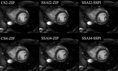 |
27 |
Deep-learning-based super-resolution technique for cine cardiac
magnetic resonance
Satonori Tsuneta1,
Satoru Aono2,
Rina Kimura1,
Jihun Kwon3,
Takuya Aoike2,
Masami Yoneyama3,
Kinya Ishizaka2,
Noriyuki Fujima1,
and Kohsuke Kudo4
1Department of Diagnostic and Interventional Radiology, Hokkaido University Hospital, Sapporo, Japan, 2Department of Radiological Technology, Hokkaido University Hospital, Sapporo, Japan, 3Philips Japan, Ltd., Tokyo, Japan, 4Department of Diagnostic Imaging, Hokkaido University Graduate School of Medicine; Global Center for Biomedical Science and Engineering, Faculty of Medicine, Hokkaido University, Sapporo, Japan Keywords: Myocardium, Data Processing Cine cardiac magnetic resonance (CMR) imaging is an optimal cardiac volumetric analysis method because of its high contrast resolution. However, its spatial resolution is limited owing to prolonged scanning and breath-holding. Although compressed sensing–sensitivity encoding (Compressed SENSE; CS) and its deep-learning-based advancement (SmartSpeed AI; SSAI) can reduce the scan time, the spatial resolutions remain unchanged. Herein, we investigated the effect of a deep-learning-based super-resolution technique (SmartSpeed Precise Image; SSPI) on the cine CMR visual image quality in comparison with CS and SSAI with conventional zero-filling interpolation; resultantly, the SSPI significantly improved the visual image quality scores. |
|
5130. |
28 |
Detection of Congenital Heart Disease in MR images using Machine
Learning
Dominik Daniel Gabbert1,
Lennart Petersen1,2,
Abigail Burleigh1,
Simona Boroni Grazioli1,
Sylvia Krupickova3,
Reinhard Koch2,
Anselm Sebastian Uebing1,
Monty Santarossa2,
and Inga Voges1
1Department of Congenital Heart Disease and Pediatric Cardiology, University Hospital Schleswig-Holstein, DZHK (German Center for Cardiovascular Research), partner site Hamburg/Kiel/Lübeck, Kiel, Germany, 2Multimedia Information Processing Group, Kiel University, Kiel, Germany, 3Departments of CMR and Paediatric Cardiology, Royal Brompton Hospital, London, United Kingdom Keywords: Heart, Machine Learning/Artificial Intelligence, Congenital Heart Disease We present a new method for detection of hypoplastic left heart syndrome (HLHS) based on the spatial arrangement of 7 distinctive anatomical landmarks in CMR images. The method was applied to the axial SSFP CMR scans of 46 patients with HLHS and 33 healthy controls. A tailor-made U-net-like deep convolutional network (CNN) with a shared 3D-convolutional encoder backbone and 7 segmentation heads was used for prediction of landmarks. Classification based exclusively on the coordinates of the detected landmarks had an accuracy of 98.7%. In future studies, the method may be applied to HLHS subgroups or other cardiac diseases. |
|
5131.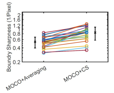 |
29 |
Novel Reconstruction Method to Improve the Sharpness of Free
Breathing Cardiac MR Late Gadolinium Enhancement Images on a
Commercial 0.55T System
Yu Ding1,
Yingmin Liu1,
Chong Chen1,
Ning Jin2,
Rizwan Ahmad1,
and Orlando Simonetti1
1The Ohio State University, Columbus, OH, United States, 2Siemens Healthineers, Columbus, OH, United States Keywords: Myocardium, Low-Field MRI, Late Gadolinium Enhancement Low-field cardiac MR imaging has intrinsically low SNR that degrades the image quality, especially for LGE imaging. Using multiple acquisitions and motion-corrected averaging is a well-established method to improve SNR. However, the ineffectiveness of MOCO at low field leads to blurring. We proposed a novel method that integrates motion correction into compressed sensing and reconstructs a single motion-corrected image. Quantitative sharpness measurements demonstrate that the proposed method improves boundary sharpness, independent of differences in SNR. |
|
5132.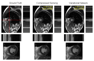 |
30 |
Deep Learning-Based Reconstruction of Accelerated Cardiac Cine
MRI at 0.55T
Marc Vornehm1,2,
Jens Wetzl2,
Daniel Giese2,3,
Jianing Pang4,
Rizwan Ahmad5,
and Florian Knoll1
1Computational Imaging Lab, Friedrich-Alexander-Universität Erlangen-Nürnberg, Erlangen, Germany, 2Magnetic Resonance, Siemens Healthcare GmbH, Erlangen, Germany, 3Institute of Radiology, University Hospital Erlangen, Friedrich-Alexander-Universität Erlangen-Nürnberg, Erlangen, Germany, 4Siemens Medical Solutions USA Inc., Chicago, IL, United States, 5Biomedical Engineering, The Ohio State University, Columbus, OH, United States Keywords: Heart, Low-Field MRI Acceleration of cardiac cine MRI is highly desirable in order to decrease the required breath-hold duration. On low-field MRI systems in particular, this could help make cardiac MRI more widely available. We present a method based on the Variational Network for reconstruction of cardiac cine MRI, trained on data from 1.5 and 3T systems. Reconstructions of retrospectively and prospectively undersampled acquisitions at 0.55T with an acceleration rate of eight are shown and compared to Compressed Sensing reconstructions. Despite the domain shift to low-field data, the neural network achieved an SSIM of 94.6%, which is comparable to the Compressed Sensing results. |
|
5133. |
31 |
“ShortCardiac” - an open-source tool for assessing cardiac
function quickly
Karl Ludger Radke1,
Janina Hußmann1,2,
Lena Röwer1,2,
Dirk Voit3,
Jens Frahm3,
Gerald Antoch1,
Dirk Klee1,
Frank Pillekamp1,2,
and Hans-Jörg Wittsack1
1University Düsseldorf, Department of Diagnostic and Interventional Radiology, Düsseldorf, Germany, 2Department of General Pediatrics, Neonatology and Pediatric Cardiology, University Children's Hospital Düsseldorf, D-40225 Düsseldorf, Germany, Düsseldorf, Germany, 3Biomedical NMR, Max Planck Institute for Multidisciplinary Sciences, D-37070 Göttingen, Germany, Göttingen, Germany Keywords: Heart, Software Tools In this work, we propose a new open-source framework for cardiac functional analysis. Our proposed framework can be used in a pipeline with the commercial software Circle cvi42 as well as the free software ITKSnap and enables a step towards a fully automated, reproducible, and quantitative assessment of MR images in clinical routine. |
|
5134.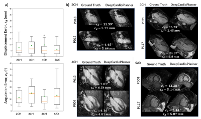 |
32 |
DeepCardioPlanner: Deep Learning-based tool for automatic CMR
planning
Pedro Louro Costa Osório1,
Markus Henningsson2,3,4,
Alberto Gomez Herrero5,6,
Rita G. Nunes1,
and Teresa M. Correia6,7
1Institute for Systems and Robotics - Lisboa and Department of Bioengineering, Instituto Superior Técnico - Universidade de Lisboa, Lisbon, Portugal, 2Division of Cardiovascular Medicine, Department of Medical and Health Sciences, Linköping University, Linköping, Sweden, 3Centre for Medical Image Science and Visualization (CMIV), Linköping University, Linköping, Sweden, 4MR Physics, Perspectum Ltd, Oxford, United Kingdom, 5Ultromics Ltd, Oxford, United Kingdom, 6School of Biomedical Engineering Imaging Sciences, King’s College London, London, United Kingdom, 7Centre for Marine Sciences - CCMAR, Faro, Portugal Keywords: Heart, Machine Learning/Artificial Intelligence, View Planning Cardiac Magnetic Resonance (CMR) is a powerful technique which can be used to perform a comprehensive cardiac examination. However, its adoption is often limited to specialised centres, in part due to the need for highly trained operators to perform the complex procedures of determining the 4 standard cardiac planes: 2-, 3-, 4-chamber and short axis views. To automate view planning, a deep learning-based tool (DeepCardioPlanner) has been proposed to regress the view defining vectors from a rapidly acquired 3D image. It successfully takes advantage of multi-objective learning to allow accurate, fast and reproducible view prescriptions without any operator input. |
|
5135.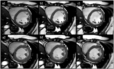 |
33 |
Assessment of cardiac function using free-breathing, motion
corrected artificial intelligence cine: comparison with
conventional cine
Lingping Ran1,
Lu Huang1,
Xianghu Yan1,
Yun Zhao1,
and Liming Xia1
1Department of Radiology, Tongji Hospital, Tongji Medical College, Huazhong University of Science and Technology, Wuhan, China Keywords: Heart, Heart Routine cardiac functional assessment with conventional balanced steady state free precession (bSSFP) cine sequence generally requires multiple breath-holding (BH) and long examination time. The purpose of this study was to evaluate the image quality and bi-ventricular functional analysis using a novel free-breathing (FB) artificial intelligence (AI) cine method with motion corrected (MOCO) in a clinical context. Compared to conventional multiple BHs cine, the proposed FB-MOCO AI cine method achieved comparable image quality with shortened scan times. No significant difference in ventricular volumetric and functional parameters were found, suggesting its potential applicability for clinical applications. |
|
5136.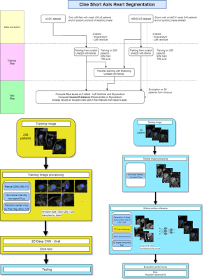 |
34 |
2D U-Net for left heart segmentation for CINE sequences, deal
with new target data: comparison of 3 approaches
Habib Rebbah1,
Guillaume Gautier1,
and Timothé Boutelier1
1Research & Innovation, Olea Medical, La Ciotat, France Keywords: Heart, Segmentation In the case of U-Net based segmentation algorithms, we propose to explore a common question of the medical image research teams: using already existing algorithm or train a new one specific to the target data. We compared for the cardiac CINE case three approaches: using an algorithm trained with external data, train a new one with the taget data from scratch, and fin-tune the first one with the target data. |
|
5137. |
35 |
Unsupervised deep learning with variational autoencoder for
image quality improvement in ultrafast cardiac cine MRI: Initial
results
Yajing Zhang1,
Guohui Ruan1,
Tianyu Han2,
Christiane Kuhl3,
Masami Yoneyama4,
Daniel Truhn3,
and Shuo Zhang3,5
1MR Clinical Science, Philips Health Technology, Suzhou, China, 2RWTH University Aachen, Aachen, Germany, 3Diagnostic and Interventional Radiology, University Hospital RWTH Aachen, Aachen, Germany, 4Philips Japan, Tokyo, Japan, 5Philips Market DACH, Hamburg, Germany Keywords: Myocardium, Cardiovascular Ultrafast imaging with high acceleration in cardiac MRI is of great clinical interest, but so far often results in inferior image quality that prevents its use in routine diagnosis. In this work, we aim to establish an unsupervised deep learning neural network based on vector quantized variational autoencoder for noise reduction and image quality improvement. Initial results on both public and clinical data promise a new approach to the existing methods. Further investigations with focus on its effectiveness of performance in real world applications are warranted. |
|
5138.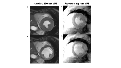 |
36 |
Cine MRI reconstructed from 3T contrast-enhanced free-running 5D
coronary MRA: comparison to standard 2D cine MRI
Haruno Ito1,
Masaki Ishida1,
Masafumi Takafuji1,
Shinichi Takase1,
Yoshiaki Komori2,
Davide Piccini3,4,
Jessica A.M. Bastiaansen4,
Jérôme Yerly4,5,
Matthias Stuber4,5,
and Hajime Sakuma1
1Radiology, Mie University Hospital, Tsu, Mie, Japan, 2Siemens Healthcare K.K., Tokyo, Japan, 3Advanced clinical imaging technology, Siemens Healthineers International AG, Lausanne, Switzerland, 4Department of Diagnostic and Interventional Radiology, Lausanne University Hospital and University of Lausanne, Lausanne, Switzerland, 5CIBM Center for Biomedical Imaging, Lausanne, Switzerland Keywords: Heart, Heart, Cine MRI Cine MRI derived from 3T contrast-enhanced free-running 5D whole-heart coronary MRA (free-running cine MRI) was compared with standard 2D cine MRI in 30 patients with suspected CAD. In a selected patient, temporal width was optimized to 50 ms. Then, in the remaining 29 patients, free-running cine MRI provided good agreement in LV volume and function quantification with standard 2D cine MRI with a good inter-observer reproducibility. The results suggest that free-running cine MRI allows for a shorter scan protocol by skipping the standard 2D cine MRI. |
|
5139.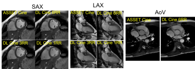 |
37 |
Evaluation of image quality and global cardiac function for deep
learning accelerated cardiac Cine
Xucheng Zhu1,
Suryanarayanan Kaushik2,
Frandics Chan3,
Melany Atkins4,
Prashant Nagpal 5,
Reed Busse2,
and Martin Janich6
1GE Healthcare, Menlo Park, CA, United States, 2GE Healthcare, Waukesha, WI, United States, 3Radiology, Stanford University, Palo Alto, CA, United States, 4Radiological Consultants, Inova Fairfax Hospital, Fairfax, VA, United States, 5Radiology, University of Wisconsin–Madison, Madison, WI, United States, 6GE Healthcare, Munich, Germany Keywords: Heart, Image Reconstruction, Deep learning, reconstruction Cardiac bSSFP Cine is widely used clinically; however, it is time consuming and requires multiple breath-holds. Deep learning-based accelerated Cine (DLCine) is a novel technique combining accelerated variable density sampling and deep learning regularized reconstruction that allows much higher acceleration compared to conventional Cine with parallel imaging. The purpose of this work was to compare image quality and global cardiac function utilizing DLCine versus conventional Cine by three expert readers. The results demonstrate that DLCine can be used to reduce the scan time while maintaining image quality and providing accurate global cardiac function measurement. |
|
5140.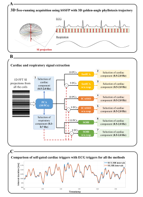 |
38 |
Blind Source Separation Improves the Precision and Robustness of
Self-Gated Motion Extraction in Free-Running 4D Whole-Heart MRI
Isabel Montón Quesada1,
Augustin C. Ogier1,
Jérôme Yerly1,2,
Jonas Richiardi1,
Christopher W. Roy1,
Juerg Schwitter3,
Matthias Stuber1,2,
and Ruud B. van Heeswijk1
1Department of Radiology, Lausanne University Hospital (CHUV) and University of Lausanne (UNIL), Lausanne, Switzerland, 2CIBM Center for BioMedical Imaging, Lausanne, Switzerland, 3Cardiology Service, Cardiovascular Department, Lausanne University Hospital (CHUV) and University of Lausanne (UNIL), Lausanne, Switzerland Keywords: Heart, Image Reconstruction Several cardiac and respiratory motion-resolved free-running acquisition techniques have recently been developed, but automated self-gated motion extraction with principal component analysis (PCA) alone remains challenging and sometimes fails to correctly identify the components related to cardiac motion. We combined three blind source separation algorithms (fastICA, ICASSO and SOBI) with PCA and compared them with gold-standard ECG R-wave triggers in 10 healthy volunteers to determine the most precise and robust self-gated cardiac trigger extraction. Of these tested techniques, PCA combined with SOBI resulted in the lowest variability of the cardiac intervals relative to ECG intervals (p=0.03) and the sharpest images (p=0.03). |
|
5141.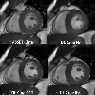 |
39 |
A feasibility study of deep learning cardiac cine comparing
image quality and volumetry with the conventional ASSET cine
Shigeo Okuda1,
Ryo Tsukada2,
Manabu Arai1,
Sari Motomatsu2,
Atsushi Nozaki3,
Xucheng Zhu4,
and Masahiro Jinzaki1
1Radiology, Keio University School of Medicine, Tokyo, Japan, 2Keio University Hospital, Tokyo, Japan, 3GE Healthcare Japan, Tokyo, Japan, 4GE Healthcare, Menlo Park, CA, United States Keywords: Heart, Cardiovascular, Accelerated cine Accelerated cardiac cine was obtained with deep learning reconstructed technique on ten patients (DL Cine). Three series of DL Cine with different parameters were acquired, including reduction factor (RF) of 12 under free breathing (FB), RF of 12 during one breathhold (R12) and RF = 9 dividing the left ventricular short axis into two slabs during each breathhold (R9). The two readers evaluate image quality (IQ) score and measure the cardiac functional parameters and compared them with the conventional ASSET cine. Although the IQ score was smaller than ones of the conventional cine, they are clinically acceptable. We found good correlations between volumetry on the conventional and DL cine. |
|
5142. |
40 |
Reconstruction of Dynamic MRI Tensor Data Using Frequency
Low-rank and Sparsity Prior
Runyu Yang1,
Haozhong Sun1,
and Huijun Chen1
1Center for Biomedical Imaging Research, Tsinghua University, Beijing, China Keywords: Heart, Image Reconstruction Obtaining high spatial and temporal resolution image in dynamic magnetic resonance imaging is a huge challenge. It is effective to use low-rank and sparse prior jointly for dMRI reconstruction. However, the models represented the tensor data into a matrix, which can not completely explore the spatiotemporal correlation information. Besides, using nuclear norm as convex surrogate of the rank function to enforce the low-rank, which may lead to the sub-optimal result. Hence, we proposed the piecewise frequency tensor nuclear norm in the low-rank and joint sparse prior to reconstructed data. The proposed method was tested in cardiac cine and perfusion data. |
|
 Back to Meeting Home
 Back to the Program-at-a-Glance
The International Society for Magnetic Resonance in Medicine is accredited by the Accreditation Council for Continuing Medical Education to provide continuing medical education for physicians.
 Back to Meeting Home
 Back to the Program-at-a-Glance
 View Presentation Video