Digital Poster
New Developments & Clinical Applications of Flow MRI I
ISMRM & ISMRT Annual Meeting & Exhibition • 03-08 June 2023 • Toronto, ON, Canada

Digital Poster
New Developments & Clinical Applications of Flow MRI I
| Computer # | |||
|---|---|---|---|
3644.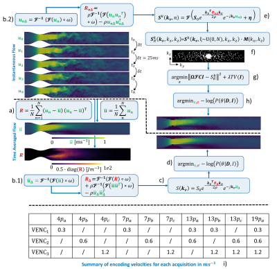 |
61 |
Optimal encoding strategies for turbulence quantification of
undersampled 4D flow MRI
Pietro Dirix1,
Stefano Buoso1,
and Sebastian Kozerke1
1University and ETH Zurich, Zürich, Switzerland Keywords: Flow, Image Reconstruction We expand a previously developed pipeline for generation of synthetic 4D flow MRI of turbulent flows to account for realistic k-space filling and add an MR temporal averaging component to the simulations. We show that temporal averaging in typical undersampled 4D flow MRI is imperfect, leading to inherent scan-to-scan variations of measured turbulence fields. We demonstrate how encoding schemes, when designed appropriately, allow to significantly reduce scan-to-scan variability, increasing the confidence on MRI measurements. |
|
3645.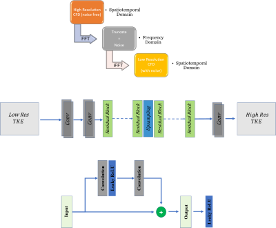 |
62 |
TKE-Net: Deep Learning for Estimation of Super-Resolved
Turbulent Kinetic Energy Maps from 4D-Flow MRI Data
Amirkhosro Kazemi1,
Marcus Stoddard1,
and Amir A. Amini1
1Electrical and Computer Engineering, University of Louisville, Louisville, KY, United States Keywords: Phantoms, Heart, Hemodynamics, Flow, Neural network, Deep learning Variations in velocity derivative fluctuations have been correlated with changes in pressure gradient. Image denoising and super-resolution techniques are required to accurately quantify velocity fluctuations. We propose a novel network, which we call TKE-Net to estimate Turbulent Kinetic Energy (TKE) which utilizes a ResNet convolutional neural network backbone. The network is trained and tested with low-resolution simulated CFD data, as could be derived from low resolution 4D flow in a phantom model of arterial stenosis. The results indicate good accuracy in estimating TKE. The method was also applied to in-vitro 4D flow MRI data in identical geometry. |
|
3646.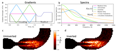 |
63 |
On the interplay of encoding and readout in turbulent flow
imaging
Charles McGrath1,
Pietro Dirix1,
Jonathan Weine1,
and Sebastian Kozerke1
1University and ETH Zurich, Zurich, Switzerland Keywords: Flow, Velocity & Flow, Turbulence The accuracy of turbulent flow quantification using phase-contrast MRI depends on both the turbulence energy spectrum to be imaged and the sampling spectrum of the velocity encoding gradients. In the present work their interplay is studied in detail, revealing erroneous quantification of intravoxel standard deviation and turbulent kinetic energy in particular in cases exhibiting low turbulence frequencies. It is concluded that experimental design for turbulent flow quantification needs to consider the joint effect of velocity encoding and imaging gradients. Keywords: Turbulence, TKE, Phase Constrast, Simulation, Flow, Cardiovascular |
|
3647.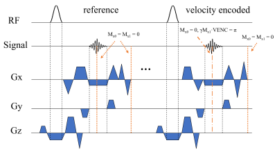 |
64 |
In-Plane Balanced Phase-Contrast Steady-State Free Precession
(PC-SSFP) for All-in-One Diastolic Function Evaluation
Jie Xiang1,
Jerome Lamy2,
Maolin Qiu2,
and Dana C. Peters1,2
1Department of Biomedical Engineering, Yale University, New Haven, CT, United States, 2Department of Radiology and Biomedical Imaging, Yale University, New Haven, CT, United States Keywords: Flow, Quantitative Imaging Diastolic function evaluation requires estimates of early and late diastolic mitral valve flow velocities (E and A), and mitral annulus tissue velocity (e’). Our goal was to develop a bSSFP phase-contrast (PC) sequence (PC-SSFP) for in-plane flow-encoding, for simultaneous recording of E and A, and estimation of e’ based on tracking mitral valve motion on cine magnitude images, in a single breath-hold. Phantom and in vivo experiments showed agreement of PC-SSFP with velocities on GRE-based PC providing similar velocity curves, while achieving the high SNR and contrast of bSSFP cine images. |
|
3648.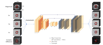 |
65 |
Denoising Vastly Undersampled Radial Portal Venous 4D Flow Data
via Deep Learning
Tarun Naren1,
Oliver Wieben1,2,
Thekla H Oechtering2,3,
Scott B Reeder1,2,
and Kevin M Johnson1,2
1Department of Medical Physics, University of Wisconsin - Madison, Madison, WI, United States, 2Department of Radiology, University of Wisconsin - Madison, Madison, WI, United States, 3Department of Radiology, Universität zu Lübeck, Lübeck, Germany Keywords: Flow, Liver Radially undersampled 4D flow MRI is a promising method for non-invasive mapping of blood flow in the portal venous system. However, collecting sufficient projections to produce clinically viable images can lead to long scan times (10+ minutes) as fewer projections cause undersampling artifacts that appear as structured noise. In this study, we propose a data-driven, deep learning method to denoise vastly undersampled (<10% of full Nyquist sampling) radial 4D flow MRI data in the portal vein. We train a network on a heterogeneous, time-averaged dataset with two levels of undersampling and perform a quantitative hemodynamic analysis to compare results. |
|
3649.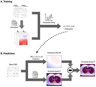 |
66 |
Prediction of Eddy Current-Induced Background Phase Offsets in
2D Phase Contrast-MRI
Julio A. Oscanoa1,2,
Matthew J. Middione2,
Michael Loecher2,
Ana Beatriz Solana3,
Shreyas S. Vasanawala2,
and Daniel B. Ennis2
1Department of Bioengineering, Stanford University, Stanford, CA, United States, 2Department of Radiology, Stanford University, Stanford, CA, United States, 3GE Healthcare, Munich, Germany Keywords: Flow, System Imperfections: Measurement & Correction, Phase-Contrast, Eddy currents We propose a physics-driven approach for prediction of eddy current-induced background phase offsets in 2D PC-MRI. The method leverages the applied gradient waveforms and a circuit model to estimate the system-dependent parameters that govern the background phase. The aim is to use these parameters and the model to predict the background phase of clinical acquisitions from pulse sequence information only. Our method achieved an RMSE of 1.05 cm/s and adequate visual quality. |
|
3650.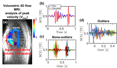 |
67 |
Deep learning-based prediction of aortic hemodynamics obtained
by 4D flow MRI using seismocardiography of chest vibrations
Mahmoud Ebrahimkhani1,
Ethan Johnson1,
Aparna Sodhi2,
Joshua Robinson2,3,
Cynthia Rigsby2,
Bradly Allen3,
and Michael Markl3
1Radiology, Northwestern University, Chicago, IL, United States, 2Ann & Robert H. Lurie Children’s Hospital, Chicago, IL, United States, 3Northwestern University, Chicago, IL, United States Keywords: Flow, Heart We pursued a deep learning approach to investigate the utilization of a wearable seismocardiography (SCG) device to predict measures of flow similar to those obtained using 4D flow MRI. SCG can measure the chest vibrations caused by cardiac mechanical activities such as valve closures and changes of pulsatile flow. We hypothesized that deep learning can be used to infer the pathological changes in blood flow, such as a higher systolic peak velocity (Vmax) in patients with aortic valve diseases, from the SCG signals. |
|
3651.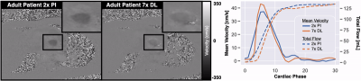 |
68 |
Evaluation of PC-MRI Flow in Adults Using a Deep Learning-Based
Reconstruction Trained on Pediatric Data
Matthew J. Middione1,
Julio A. Oscanoa1,2,
Xianglun Mao3,
Christina R. Ruiz4,
Michael Salerno1,4,5,
Shreyas S. Vasanawala1,
and Daniel B. Ennis1,5
1Department of Radiology, Stanford University, Stanford, CA, United States, 2Department of Bioengineering, Stanford University, Stanford, CA, United States, 3GE HealthCare, Menlo Park, CA, United States, 4Department of Medicine, Stanford University, Stanford, CA, United States, 5Cardiovascular Institute, Stanford University, Stanford, CA, United States Keywords: Flow, Machine Learning/Artificial Intelligence, Phase Contrast We have previously trained a deep learning-based (DL) reconstruction for 2D PC-MRI using fully-sampled (n=194) raw k-space pediatric datasets. This DL-based reconstruction provided up to 9x undersampling with ≤5% error in accuracy and precision of peak velocity and total flow. Herein, we analyze this pediatric trained DL-based reconstruction in adult volunteers (n=3) and adult patients (n=8) and show that our DL-based reconstruction provides ~5% error in accuracy and precision of peak velocity and total flow for up to 7x undersampling. |
|
3652.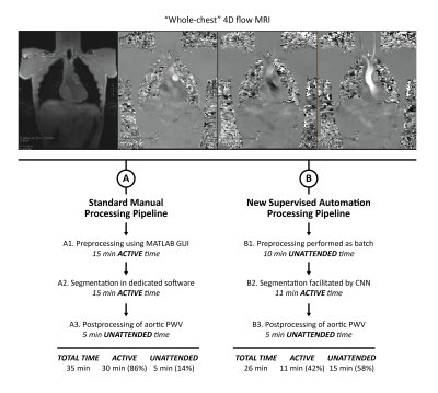 |
69 |
Supervised automation of “whole-chest” 4D flow MRI analysis from
original DICOM files to quantification of aortic pulse wave
velocity
Kelly Jarvis1,
Ethan Johnson1,
Haben Berhane1,
Adam Richter1,
Elizabeth Weiss1,
and Michael Markl1
1Department of Radiology, Northwestern University, Chicago, IL, United States Keywords: Flow, Data Processing Analysis of pulse wave velocity by 4D flow MRI usually involves a manual processing pipeline with multiple steps of active use. We set out to automate the entire pipeline from original DICOMs to parameter quantification for evaluation of “whole-chest” 4D flow MRI. A deep learning algorithm trained on aortic acquisitions was used to facilitate segmentation. The supervised automation pipeline allowed for shorter analysis times and less user interaction, enabling the user to focus on refining aortic segmentations generated by deep learning. Results were comparable, but additional work including adjustment of preprocessing settings and retraining of CNN is warranted for optimization. |
|
3653.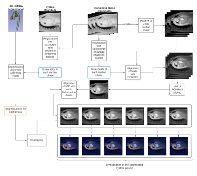 |
70 |
Improving the segmentation over time of 4D flow MRI images,
using a medical image registration neural network VoxelMorph
Aaron Christhoper Ponce1,2,
Sergio Uribe2,3,4,5,
and Julio Sotelo2,5,6
1School of Informatics Engineering, Universidad de Valparaíso, Valparaíso, Chile, 2Millennium Institute for Intelligent Healthcare Engineering, iHEALTH, Santiago, Chile, 3Department of Radiology, School of Medicine, Pontificia Universidad Católica de Chile, Santiago, Chile, 4Institute for Biological and Medical Engineering, Schools of Engineering, Medicine and Biological Sciences, Pontificia Universidad Católica de Chile, Santiago, Chile, 5Biomedical Imaging Center, Pontificia Universidad Católica de Chile, Santiago, Chile, 6School of Biomedical Engineering, Universidad de Valparaíso, Valparaíso, Chile Keywords: Flow, Segmentation The segmentation of large vessels in 4D Flow MRI remains a challenge, due to different problems such as random acquisition noise, low spatial and temporal resolution, velocity aliasing, respiratory motion, phase offsets. For that reason the use of a single segmentation has been standardized to represent the geometry throughout all the cardiac phases. However, recent studies have proposed the use of image registration to be able to solve this problem. In this work, an algorithm based on a medical image registration neural network is proposed to improve the segmentation over time for the cardiac phases of the systole period. |
|
3654.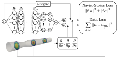 |
71 |
Reconstructing 3D Velocity and Pressure Fields From 2D Flow
Measurements Using Physics Informed Neural Networks
Stefano Buoso1,
Pietro Dirix1,
and Sebastian Kozerke1
1Institute of Biomedical Engineering, ETH Zurich, Zurich, Switzerland Keywords: Flow, Velocity & Flow We propose physics-informed neural networks to augment 2D phase-contrast flow measurements in order to reconstruct full 3D velocity and pressure fields. In this conceptual work, 2D flow measurements are assimilated using the incompressible Navier-Stokes equations defined on the vessel anatomy reconstructed from anatomical scans. The network leverages a low-rank representation of velocity and pressure fields as physical prior and it is demonstrated to reconstruct 3D velocity and pressure gradient fields with good accuracy. |
|
3655.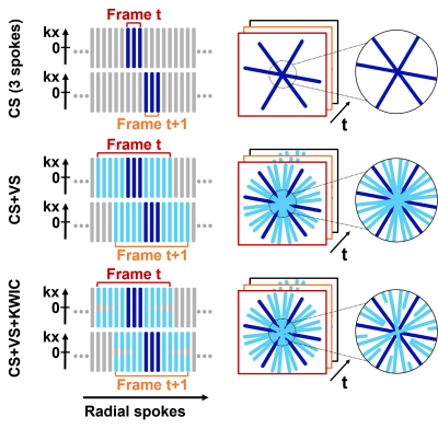 |
72 |
View-Sharing and KWIC Filtering Deblur 64-fold Accelerated
Real-Time Phase-Contrast MRI Obtained with Radial k-space
Sampling
Huili Yang1,2,
Andrine Larsen3,
Joshua D Robinson4,
KyungPyo Hong1,
Cynthia K Rigsby5,
and Daniel Kim1,2
1Radiology, Northwestern University, Chicago, IL, United States, 2Biomedical Engineering, Northwestern University, Evanston, IL, United States, 3Biomedical Engineering, Lehigh University, Bethlehem, PA, United States, 4Pediatrics, Ann & Robert H. Lurie Children’s Hospital, Chicago, IL, United States, 5Medical Imaging, Ann & Robert H. Lurie Children’s Hospital, Chicago, IL, United States Keywords: Flow, Cardiovascular, compressed sensing, real-time imaging, phase-contrast MRI Highly-accelerated real-time phase-contrast (rt-PC) using compressed sensing may underestimate (~17%) velocity measurements due to over regularization. We sought to address this underestimation by incorporating view-sharing (VS) and k-space weighted image contrast (KWIC) filtering. This is particularly important for pediatric patients who have faster heart rates and smaller hearts than adults, it is particularly important to achieve high temporal resolution. The proposed reconstruction achieves 25 ms temporal resolution without significant temporal blurring artifacts. |
|
3656.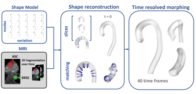 |
73 |
Digital Twinning of Aortae from 2D+time MRI
Gloria Wolkerstorfer1,
Stefano Buoso1,
and Sebastian Kozerke1
1Institute for Biomedical Engineering, University and ETH Zurich, Zurich, Switzerland Keywords: Flow, Data Analysis, Aorta; Shape modelling; Shape reconstruction; Image-based shape reconstruction; Aortic shape reconstruction We propose an automatic pipeline to generate digital twin aortae from 2D+time MRI slices. First, a statistical shape model is fitted to the segmentation of 2D slices over time and then local refinements are applied to closely match dynamics over time. The approach is exemplified on a dataset of 10 aortic stenosis patients on which we quantify the impact of the number of available slices on the reconstructed anatomy. |
|
3657.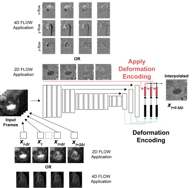 |
74 |
Deformation-ENcoding Transformer (DENT) for High Frame Rate for
Phase Contrast MRI
Manuel A Morales1,
Amine Amyar1,
Siyeop Yoon1,
Jennifer Rodriguez1,
Martin S Maron2,
Ethan J Rowin2,
Shiro Nakamori1,
Jiwon Kim3,
Robert M Judd4,
Jonathan W Weinsaft3,
Warren J Manning1,
and Reza Nezafat1
1BIDMC, Boston, MA, United States, 2Tufts Medical Center, Boston, MA, United States, 3Weill Cornell Medicine, New York, NY, United States, 4Duke University, Durham, NC, United States Keywords: Flow, Cardiovascular Quantification of blood flow using 2D or 4D phase-contrast (PC) MRI is routinely being used to evaluate blood flow in cardiovascular disease. However, PC has only a modest temporal resolution compared to echocardiography. We sought to develop a deformation-encoding transformer (DENT) model for cardiac frame interpolation and evaluate its potential in increasing temporal resolution. DENT was trained using a large multi-center (centers = 3), multi-vendor (vendors = 3) and multi-field strength (1.5T, 3T) cine MRI dataset (patients = 3178). The model was successfully applied to 2D/4D-PC MRI without modifications, enabling a 2-fold gain in temporal resolution. |
|
3658.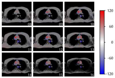 |
75 |
Radial phase-contrast FLASH acquisiton with SSA-FARY utilizing a
Hadarmad-encoding sheme
Vanessa Thiesen1,
Ansgar Adler1,
and Martin Uecker1,2,3
1Institute of Biomedical Imaging, Graz, Austria, 2Institute for Diagnostic and Interventional Radiology of the University Center Göttingen, Göttingen, Germany, 3DZHK (German Center for Cardiovascular Research), Partner Site Göttingen, Göttingen, Germany Keywords: Flow, Velocity & Flow The assessment of flow within a heart cycle places high demands on the temporal resolution of the measurement protocol. With self-gating we can exploit the quasi-periodicity of cardiac and respiratory motion to meet this challenge. We combined a Hadamard flow-encoding scheme together with SSA-FARY to further elaborate on the idea of a flow measurement with a high temporal and spatial resolution. The approach delivered promising results in 2D and can be seen as a basis for further developement towards 3D flow MRI. |
|
3659.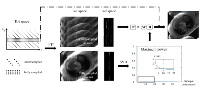 |
76 |
Accelerated phase-contrast for diastolic function evaluation
using k-t PCA methods
Jie Xiang1,
Jerome Lamy2,
Gigi Galiana1,2,
and Dana C. Peters1,2
1Department of Biomedical Engineering, Yale University, New Haven, CT, United States, 2Department of Radiology and Biomedical Imaging, Yale University, New Haven, CT, United States Keywords: Flow, Quantitative Imaging Diastolic function evaluation requires estimates of early and late diastolic mitral valve flow velocities (E and A), normally evaluated by echocardiography, using phase contrast (PC). Our goal was to investigate k-t principal component analysis (PCA), constrained by either global and local principal components, for E and A measurements. Importantly, we found that retaining the central k-space used in estimating the principal components improved the final velocity maps. We compared local PCA with global PCA. Retrospectively undersampled images showed strong agreement with standard PC acquisitions. Higher E and A were observed using higher temporal resolution in prospective k-t PCA PC acquisitions. |
|
3660.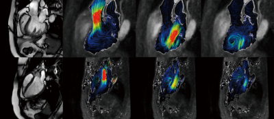 |
77 |
Investigation of 4D Flow CMR technique for left ventricular
hemodynamics in heart failure patients
Guo Jiaxuan1,
Yue Xiuzheng2,
Huang shan2,
and Zhu Li3
1Ningxia medical university, Yinchuan, China, 2Philips Healthcare, Beijing, China, 3General Hospital of Ningxia Medical University, Yinchuan, China Keywords: Heart, Cardiovascular, Heart Failure The number of patients with heart failure is increasing. Our study is based on the 4D flow technique of cardiac magnetic resonance and attempts to find cardiac functional parameters that precede structural changes in the heart at the level of the ventricular inflow and outflow tracts. We have found easier methods for visualizing blood flow and have the potential to further advance clinical diagnosis. |
|
3661.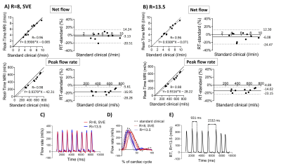 |
78 |
Validation of real-time phase contrast MRI with online
compressed sensing reconstruction in phantom and patients
Tania Lala1,
Lea Christierson2,3,
Petter Frieberg1,
Daniel Giese4,5,
Nina Hakacova 2,
Pia Sjöberg1,
Ellen Ostenfeld1,
and Johannes Töger1
1Clinical Physiology, Department of Clinical Sciences Lund, Skåne University Hospital, Lund University, Lund, Sweden, 2Department of Clinical Sciences Lund, Pediatric Heart Center, Skåne University Hospital, Lund University, Lund, Sweden, 3Department of Biomedical Engineering, Lund University, Lund, Sweden, 4Magnetic Resonance, Siemens Healthcare GmbH, Erlangen, Germany, 5Institute of Radiology, University Hospital Erlangen, Friedrich-Alexander-Universität Erlangen-Nürnberg, Erlangen, Germany Keywords: Flow, Velocity & Flow Real-time Phase Contrast MRI with Compressed Sensing reconstruction is a promising method to image cardiac flow in patients with arrhythmia, where cardiac-gated sequences fail. In this study, we compared it against the gated standard clinical flow MRI using a phantom model and in patients. The phantom underwent pulsatile periodic and non-periodic flow. For in vivo validation, non-arrhythmic patient data (N=10) and data from patient with atrial fibrillation (N=1) were collected from the ascending aorta. Non-periodic flow was captured both in the phantom and in vivo. Real-time MRI showed good accuracy in net flow quantification and underestimated peak flow. |
|
3662.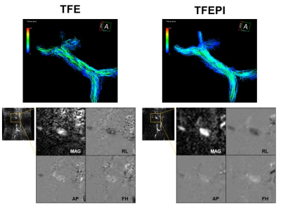 |
79 |
Utility of Noncontrast-enhanced Turbo-field Echo-planar
Imaging-based 4D Flow MRI for Portal Venous System
Naoya Kinota1,2,
Daisuke Abo3,
Daisuke Kato1,
Satonori Tsuneta3,
Noriko Nishioka1,
Kinya Ishizaka4,
Noriyuki Fujima3,
Kazuyuki Minowa5,
and Kohsuke Kudo1,6
1Department of Diagnostic Imaging, Faculty of Medicine and Graduate School of Medicine, Hokkaido University, Sapporo, Japan, 2Department of Dental Radiology, Hokkaido University Hospital, Sapporo, Japan, Japan, 3Department of Diagnostic and Interventional Radiology, Hokkaido University Hospital, Sapporo, Japan, 4Department of Radiological Technology, Hokkaido University Hospital, Sapporo, Japan, 5Department of Radiology, Faculty of Dental Medicine, Hokkaido University, Sapporo, Japan, 6Global Center for Biomedical Science and Engineering, Faculty of Medicine, Hokkaido University, Sapporo, Japan Keywords: Vessels, Velocity & Flow Echo-planer imaging-based three-dimensional cine phase contrast (4D flow) MRI has been reported to achieve good image quality with a short acquisition time in cardiovascular imaging, but its application to the portal venous (PV) system has not been reported. In this study, the turbo-field echo-planar imaging (TFEPI) 4D flow MRI with a turbo-field echo (TFE)-based one in the PV system was compared. The TFEPI sequence showed a better image quality and flow consistency at the splenic and superior mesenteric vein confluence with a shorter acquisition time compared to the conventional TFE-based one. |
|
 Back to Meeting Home
 Back to the Program-at-a-Glance
The International Society for Magnetic Resonance in Medicine is accredited by the Accreditation Council for Continuing Medical Education to provide continuing medical education for physicians.
 Back to Meeting Home
 Back to the Program-at-a-Glance
 View Presentation Video