Digital Poster
Parametric Mapping of the Heart I
ISMRM & ISMRT Annual Meeting & Exhibition • 03-08 June 2023 • Toronto, ON, Canada

Digital Poster
Parametric Mapping of the Heart I
| Computer # | |||
|---|---|---|---|
3994.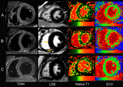 |
61 |
Multiparametric Cardiac Magnetic Resonance Reveals Persistent
Myocardial injury in Patients with Exertional Heat illness
Song Luo1,
Jun Zhang1,
and Wei Qiang Dou2
1Jinling Hospital, Medical School of Nanjing University, Nanjing, China, 2GE Healthcare, Bei Jing, China Keywords: Inflammation, Cardiovascular To explore the clinical potential of multiparametric cardiac magnetic resonance (CMR) in evaluating myocardial injury in patients with exertional heat illness (EHI).This prospective study enrolled 28 male with EHI and 18 age-matched male healthy controls. All subjects underwent CMR, and 9 patients had follow-up CMR measurements 3 months after recovery from EHI. As shown in this study, EHI patients have left ventricular dysfunction, myocardial edema and fibrosis, and persistent myocardial injury at 3-month follow-up after the EHI episode. This knowledge has an important role in guiding patients recovering from EHI back to a return to work, play or duty, respectively. |
|
3995.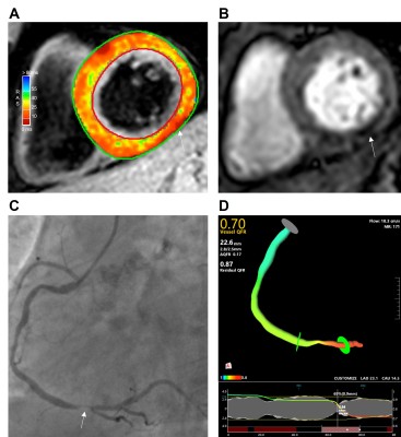 |
62 |
T2*BOLD for evaluating coronary arteries with hemodynamic
changes in stable multivessel coronary artery disease
Lei Zhao1,
Weibo Chen2,
Yongyi Wang1,
Song Xue1,
and Lianming Wu1
1Renji Hospital, Shanghai, China, 2Philips Healthcare, Shanghai, China Keywords: Vessels, Cardiovascular, T2*-mapping This study used T2*BOLD and QFR to evaluate coronary arteries with hemodynamic changes in coronary artery disease. Fifty patients with at least 1 significant coronary artery stenosis (diameter stenosis >50%) and 21 healthy control subjects underwent coronary angiography combined with QFR measurements and CMR. The CMR protocol consisted of T2* mapping and resting perfusion, contrasted with QFR. QFR≤0.80 was considered to indicate the presence of hemodynamic obstruction. Our study revealed that T2* BOLD and QFR have good agreement in detecting hemodynamic changes of stenotic coronary arteries. T2* BOLD is superior to semiquantitative perfusion imaging in analyzing myocardial perfusion without stress. |
|
3996.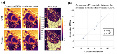 |
63 |
Rapid Myocardial T1 Stress/Rest Reactivity Mapping in
Ferumoxytol-enhanced Cardiac MRI: Initial Results
Hazar Benan Unal1,
Shahriar Zeynali1,
Fei Han2,
Gregory J. Anthony3,
Subha Raman4,
Balaji Tamarappoo5,
Rohan Dharmakumar1,
and Behzad Sharif1
1Krannert Cardiovascular Research Center, Indiana University School of Medicine, Indianapolis, IN, United States, 2Siemens Medical Solutions, Inc., Los Angeles, CA, United States, 3Department of Radiology and Imaging Sciences, Indiana University, Indianapolis, IN, United States, 4Cardiovascular Medicine, IU Health/IU School of Medicine, Indianapolis, IN, United States, 5Indiana University Health, Indianapolis, IN, United States Keywords: Myocardium, Ischemia, dobutamine Myocardial T1 reactivity, defined as the relative T1 change from rest to stress, has been proposed as a marker for detection of ischemic heart disease. SASHA-based T1 mapping provides higher accuracy than MOLLI, but the scan time can be prohibitive under stress. In this work, we investigated the feasibility of accelerated SASHA T1 mapping for ferumoxytol-enhanced dobutamine-stress T1 reactivity studies at 3T in a preclinical setting. We showed that a 2-fold accelerated SASHA T1 mapping can provide sufficiently accurate results compared to conventional SASHA. |
|
3997.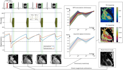 |
64 |
Multiparametric 3D cardiac MR for simultaneous bright- and
black-blood imaging and joint T1/T2 myocardial tissue mapping
Ivan Kokhanovskyi1,2,3,
Michael G Crabb2,
Karl P Kunze2,4,
Radhouene Neji2,4,
Carl Ganter1,
Donovan P Tripp2,
Carlos Castillo-Passi2,5,
Dimitrios C Karampinos1,
Claudia Prieto2,5,6,7,
and Rene M Botnar2,3,5,6,7
1Department of Diagnostic and Interventional Radiology, Klinikum rechts der Isar, Technische Universität München, Munich, Germany, 2School of Biomedical Engineering and Imaging Sciences, King's College London, London, United Kingdom, 3Institute for Advanced Study, Technische Universität München, Garching, Germany, 4MR Research Collaborations, Siemens Healthcare Limited, Frimley, United Kingdom, 5Millennium Institute for Intelligent Healthcare Engineering, Santiago, Chile, 6Escuela de Ingeniería, Pontificia Universidad Católica de Chile, Santiago, Chile, 7Instituto de Ingeniería Biológica y Médica, Pontificia Universidad Católica de Chile, Santiago, Chile Keywords: Heart, Data Acquisition Accurate diagnosis of cardiac disease requires imaging with high spatial resolution and whole-heart coverage. Current clinical protocols acquire contrasts sequentially, in different 2D orientations during several breath-holds leading to long examination times and misregistration artifacts. To address these limitations, we present a novel free-breathing four-heartbeat sequence for simultaneous 3D whole-heart Assessment of Cardiovascular anaTomy via bright- and black-blood Imaging and T1/T2 myocardial tissue quantificatiON (ACTION). Sequence parameters are optimized with Bloch simulations to enhance the mapping sensitivity and to improve the quality of anatomical images. Primarily results in a standardized phantom and in-vivo show potential for comprehensive heart disease detection. |
|
3998.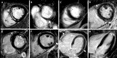 |
65 |
Cardiac magnetic resonance in survivors of critical illness:
myocardial fibrosis and dysfunction are associated with acute
kidney injury
Alexander Isaak1,
Isabel Pomareda1,
Narine Mesropyan1,
Dmitrij Kravchenko1,
Leon Bischoff1,
Daniel Kuetting1,
Ulrike Attenberger1,
Sebastian Zimmer2,
Jens-Christian Schewe3,
Stefan Kreyer3,
and Julian Luetkens1
1Diagnostic and Interventional Radiology, University Hospital Bonn, Bonn, Germany, 2Clinic for Internal Medicine II, Heart Center, University Hospital Bonn, Bonn, Germany, 3Anesthesiology and Intensive Care Medicine, University Hospital Bonn, Bonn, Germany Keywords: Cardiomyopathy, Quantitative Imaging, T1 and T2 Mapping In this prospective study, cardiac magnetic resonance (CMR) was performed in 48 intensive care unit (ICU) survivors of acute critical illness without prior known cardiac disease. CMR revealed previously unrecognized functional abnormalities, including reduced left ventricular ejection fraction and impaired strain values, as well as evidence of focal and diffuse myocardial fibrosis represented by positive late gadolinium enhancement lesions and elevated myocardial T1 mapping and ECV. Findings were more pronounced in ICU survivors who had experienced acute kidney injury. Unrecognized fibrotic and functional myocardial alterations may be a correlate of physical impairment in post-intensive care syndrome. |
|
3999.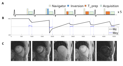 |
66 |
Navigator acceptance window width does not affect accuracy and
precision in free-breathing 2D cardiac MR fingerprinting of the
myocardium at 3T.
Pauline Calarnou1,
Augustin C. Ogier1,
Roger Hullin2,
Philippe Meyer3,
Jérôme Yerly1,4,
and Ruud B. Van Heeswijk1
1Department of Radiology, Lausanne University Hospital and University of Lausanne, Lausanne, Switzerland, 2Cardiology Service, Cardiovascular Department, Lausanne University Hospital (CHUV) and University of Lausanne (UNIL), Lausanne, Switzerland, Lausanne, Switzerland, 3Cardiology Service, Department of Medicine, Geneva University Hospital and University of Geneva, Switzerland, Geneva, Switzerland, 4CIBM, Lausanne, Switzerland Keywords: Myocardium, Relaxometry We implemented and characterized a free-breathing 2D cardiac joint T1-T2 MR fingerprinting technique at 3T named PARMA that includes a lung-liver navigator to minimize through-plane motion. We assessed the effect of rejected navigators on the relaxation times. Joint T1-T2 maps with four different navigator acceptance window widths (NAWWs from ±4mm to ±32mm) were acquired in 6 healthy volunteers and compared to clinical routine techniques. The accuracy and precision of the maps resulting from the different NAWWs did not significantly differ, suggesting that the NAWW can be chosen as a balance between navigator inefficiency and through-plane motion. |
|
4000.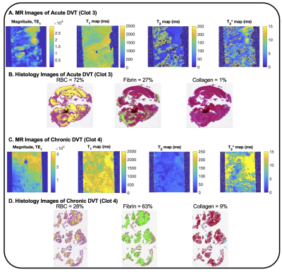 |
67 |
Using Quantitative MR and Histology to Characterize Acute and
Chronic DVT at 9.4T
Caroline D. Jordan1,2,
Kavya Sinha2,
Deborah C. Vela3,
L. Maximillian Buja3,4,
Christof Karmonik2,5,
and Trisha L. Roy2,5
1School of Engineering Medicine, Texas A&M University, Houston, TX, United States, 2Houston Methodist Research Institute, Houston Methodist Hopsital, Houston, TX, United States, 3Department of Cardiovascular Pathology, Texas Heart Institute, Houston, TX, United States, 4Department of Pathology and Laboratory Medicine, University of Texas, Houston, Houston, TX, United States, 5Weill Cornell Medical College, Houston, TX, United States Keywords: Vessels, Tissue Characterization, DVT, clot Deep venous thrombosis (DVT) is common. Mechanical thrombectomy may offer advantages compared to medical therapy alone, but it can be challenging to determine the optimal treatment. Six patients underwent thrombectomy, and the ex vivo clot was scanned at 9.4T. We acquired T1, T2, and T2* maps, and histologic content of red blood cells, fibrin, and collagen. R2 between RBC content and each relaxation time was R2 = 0.58 for T1, R2 = 0.82 for T2, and R2 = 0.69 for T2*. T1, T2, and T2* mapping of ex vivo DVT clots at 9.4T may be feasible for determining clot components. |
|
4001.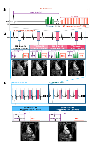 |
68 |
Whole-heart simultaneous CINE T2mapping and coronary MR
angiography
Kazuo Kodaira1,
Masami Yoneyama2,
Michinobu Nagao3,
Mana Kato1,
Takumi Ogawa1,
Yutaka Hamatani1,
Isao Shiina1,
Yasuhiro Goto1,
and Shuji Sakai3
1Department of Radiological Services, Tokyo Women's Medical University, Tokyo, Japan, 2Philips Japan, Tokyo, Japan, 3Department of Diagnostic imaging & Nuclear Medicine, Tokyo Women's Medical University, Tokyo, Japan Keywords: Myocardium, Quantitative Susceptibility mapping T2mapping is generally obtained only in diastole. However, because myocardial edema can affect both diastole and systole, acquisition of T2mapping of different cardiac phases is desirable. In addition, CMRA is also necessary in post-MI. However, scanning all these sequences prolongs exam time and increases patient stress. To overcome this limitation, we report a new T2mapping technique that enables quantitative systolic and diastolic T2mapping and CMRA acquisition in one scan by using dynamic trigger delay. In this study, we demonstrate the feasibility of this approach in healthy volunteer examination. |
|
4002.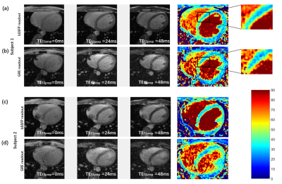 |
69 |
Accelerated 3D free-breathing cardiac T2 mapping with tiny
golden-angle hybrid trajectory and tyGRASP
Jiaojiao Hu1,
Jiantai Zhou1,
Wei Cui1,
Yang Ji2,
Benedictor Alexander Nguchu1,
Yanming Wang1,
Yong Zhang3,
and Bensheng Qiu1
1Center for Biomedical Imaging, University of Science and Technology of China, Hefei, China, 2Wellcome Centre for Integrative Neuroimaging, FMRIB Division, Nuffield Department of Clinical Neurosciences, University of Oxford, Oxford, United Kingdom, 3GE Healthcare, Shanghai, China Keywords: Myocardium, Heart Cardiac diseases are characterized by complex 3D pathological structures, 3D cardiac T2 mapping can be used as a promising tool to describe different pathologies. However, 3D T2 mapping requires a long acquisition time, which makes scanning more sensitive to motion, especially for high spatial resolution imaging. In our study, we aim to develop an accelerated and robust 3D cardiac T2 mapping technique by combining bSSFP-based hybrid radial-cartesian sampling with tiny Golden angle RAdial Sparse Parallel (tyGRASP) MRI at 3 T. The results showed that our method can generate precise T2 values with an average scan time of 6.1±1.0 min. |
|
4003.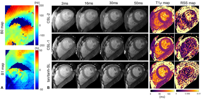 |
70 |
Optimization of spin-lock preparation pulses for B1 and B0
insensitive cardiac T1ρ mapping
Haikun Qi1,2,
Zhenfeng Lv1,2,
Jian Xu3,
and Peng Hu1,2
1School of Biomedical Engineering, ShanghaiTech University, Shanghai, China, 2Shanghai Clinical Research and Trial Center, Shanghai, China, 3UIH America, Inc., Houston, TX, United States Keywords: Myocardium, Tissue Characterization Cardiac T1ρ mapping is a promising technique for assessment of myocardial fibrosis without exogenous contrast agent. However, its wide application is hindered by the sensitivity of T1ρ preparation to B1 and B0 inhomogeneities. In this study, the state-of-the-art constant spin-lock methods including the composite and adiabatic excitation continuous-wave spin-lock methods were investigated using numerical simulations to assess their robustness to field inhomogeneities, and validated in phantoms and a preliminary subject. Two T1ρ preparation modules were found to generate superior T1ρ mapping quality in the presences of B0 and B1 inhomogeneities, indicating the potential of clinical application of cardiac T1ρ MRI. |
|
4004. |
71 |
Ungated and free-breathing radial simultaneous multi-slice
cardiac T1 mapping
Johnathan Le1,2,
Jason Mendes2,
Konstantinos Sideris3,
Josef Stehlik3,
Brent Wilson3,
Edward DiBella1,2,
and Ganesh Adluru1,2
1Biomedical Engineering, University of Utah, Salt Lake City, UT, United States, 2Utah Center for Advanced Imaging Research, University of Utah, Salt Lake City, UT, United States, 3Cardiology, University of Utah, Salt Lake City, UT, United States Keywords: Myocardium, Relaxometry Cardiac T1 mapping has been shown to be a promising method for assessing different cardiomyopathies. Most cardiac T1 mapping methods require ECG gating with long breath holds to capture specific desired cardiac phases and to minimize breathing motion. However, certain cardiomyopathies can make it difficult for patients to maintain a breath hold for the duration of a cardiac T1 mapping sequence. Furthermore, some of these diseases, for example atrial fibrillation with changing R-R intervals can make capturing a specific cardiac phase difficult. Here we propose a radial simultaneous multi-slice (SMS) cardiac T1 mapping sequence without ECG gating or breath holding. |
|
4005. |
72 |
Rapid multiple-breath-holds myocardial CINE T2-mapping with
dynamic multiple trigger delay acquisition
Mana Kato1,
Masami Yoneyama2,
Michinobu Nagao3,
Kazuo Kodaira1,
Takumi Ogawa1,
Yutaka Hamatani1,
Isao Shiina1,
Yasuhiro Goto1,
and Shuji Sakai3
1Department of Radiological Services, Tokyo Women's Medical University, Tokyo, Japan, 2Philips Japan, Tokyo, Japan, 3Department of Diagnostic imaging & Nuclear Medicine, Tokyo Women's Medical University, Tokyo, Japan Keywords: Heart, Heart T2-mapping is generally obtained only in diastole. However, because myocardial edema can affect both diastole and systole, acquisition of T2-mapping of different cardiac phases is desirable, but it prolongs examination time and increases patient stress. To overcome this limitation, we report a new T2-mapping technique using dynamic trigger delay which enables quantitative mapping at multiple cardiac phases in one scan. In this study, we demonstrate the feasibility of this approach in healthy volunteer examination. |
|
4006.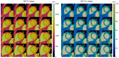 |
73 |
Fast 3D free-breathing simultaneous T1 and T1ρ mapping for
noncontrast myocardial tissue characterization at 3T
Haikun Qi1,2,
Junpu Hu3,
Jian Xu4,
Jiayu Zhu3,
René Rene Botnar5,
Claudia Prieto5,
and Peng Hu1,2
1ShanghaiTech University, Shanghai, China, 2Shanghai Clinical Research and Trial Center, Shanghai, China, 3United Imaging Healthcare, Shanghai, China, 4UIH America, Inc., Houston, TX, United States, 5School of Biomedical Engineering and Imaging Sciences, King's College London, London, United Kingdom Keywords: Myocardium, Tissue Characterization Relaxation parameters T1 and T1ρ have shown promising results for endogenous assessment of myocardial tissue. However, mapping of the whole moving heart is challenging. This study proposes a novel free-breathing joint T1 and T1ρ mapping technique with near-isotropic spatial resolution, whole left ventricle coverage and short acquisition time of 5-6min. A diaphragmatic navigator was leveraged for prospective motion correction and retrospective motion mitigation. The T1ρ preparation module was optimized to be robust to field inhomogeneities and applicable to 3T. Phantom results indicated good accuracy of the proposed technique, and in vivo feasibility was demonstrated in healthy subjects. |
|
4007.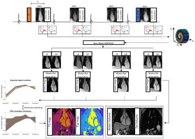 |
74 |
3D whole-heart joint T1/T1ρ mapping and water/fat imaging at
low-field 0.55T scanner
Michael G Crabb1,
Karl P Kunze1,2,
Carlos Castillo-Passi1,3,
Camila Munoz1,
Carlos Velasco1,
Radhouene Neji1,2,
Claudia Prieto1,3,
and Rene M Botnar1,3,4
1School of Biomedical Engineering and Imaging Sciences, King's College London, London, United Kingdom, 2MR Research Collaborations, Siemens Healthcare Limited, Camberley, United Kingdom, 3Institute for Biological and Medical Engineering, Pontificia Universidad Catolica de Chile, Santiago, Chile, 4School of Engineering, Pontificia Universidad Catolica de Chile, Santiago, Chile Keywords: Myocardium, Low-Field MRI Native T1 and T1ρ mapping has shown promising results for the detection of focal and diffuse myocardial fibrosis without requiring contrast agents. However, conventional myocardial maps are acquired sequentially in 2D at 1.5T/3T fields. 3D joint T1/T1ρ mapping has recently been proposed at 1.5T, but not demonstrated at lower field, which could potentially make cardiac MRI more accessible and affordable. Furthermore, lower T1 relaxation times, reduced SAR and fewer B0/B1 inhomogeneities make low-field MRI an attractive alternative for joint T1/T1ρ mapping. Here, we investigate the feasibility of novel 3D whole-heart joint T1/T1ρ mapping on the latest 0.55T MR scanner generation. |
|
4008.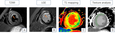 |
75 |
Texture analysis of area-at-risk based on T1-mapping associated
with infarct characteristics following acute STEMI
Jiali Wang1,
Kai Xu1,
Chunfeng Hu1,
Yankai Meng1,
Shuguang Han1,
Peng Wu2,
Lu Han2,
and Yongzhou Xu3
1The Affiliated Hospital of Xuzhou Medical University, Xuzhou, China, 2Philips Healthcare, Shanghai, China, 3Philips Healthcare, Guangzhou, China Keywords: Myocardium, Ischemia, T1 mapping To assess the potential of texture analysis (TA) of area-at-risk (AAR) based on native T1 mapping in predicting the severity of injury in ST-segment elevation myocardial infarction (STEMI) patients. Cine, T1 mapping, and late gadolinium enhancement (LGE) images were analyzed to evaluate cardiac function, and scar characteristics. The predictive value of TA for adverse LV remodeling (ALVR) and large final infarct size was evaluated. Entropy and uniformity (parameters of TA) in AAR of T1 mapping were highly correlated with convalescent infarct size, uniformity independently predicted large final infarct size, however, TA of AAR was not associated with convalescent ALVR. |
|
4009.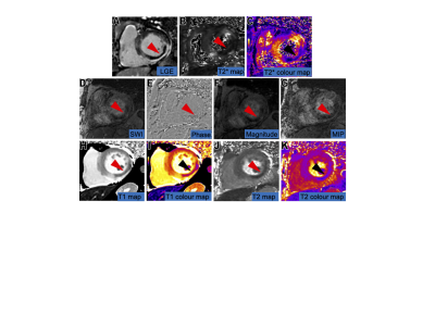 |
76 |
Cardiac MR Susceptibility-weighted Imaging Detects
Intramyocardial Hemorrhage of Acute Myocardial Infarction:
comparison with T1 / T2 mapping
Jinyang Wen1,
Lu Huang1,
Xiaoyue Zhou2,
and Liming Xia1
1Tongji Hospital, Tongji Medical College, Huazhong University of Science and Technology., Wuhan, Hubei, China, China, 2MR Collaboration, Siemens Healthineers Ltd., Shanghai, China, China Keywords: Myocardium, Myocardium, Susceptibility-weighted Imaging Diagnosis of intramyocardial hemorrhage (IMH) in ST-segment elevation myocardial infarction (STEMI) patients following reperfusion treatment is critical for determining prognosis. This study investigated the feasibility of susceptibility-weighted imaging (SWI) to detect IMH in STEMI patients that underwent reperfusion therapy by comparing the diagnostic performance and image quality with T1/T2 mapping. SWI showed excellent diagnostic performance in detecting IMH among STEMI patients that underwent reperfusion treatment and was comparable with T1/T2 mapping. SWI is a sensitive technique to accurately diagnose IMH in STEMI patients following reperfusion treatment. |
|
4010.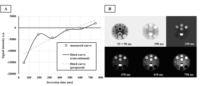 |
77 |
Novel myocardial T1 analysis robust to transient longitudinal
relaxation due to irregular heart rate variability in PCTIP
Yuta Endo1,
Sanae Takahashi1,
Haruna Shibo1,
Kuninori Kobayashi1,
Makoto Amanuma1,
and Shigehide Kuhara1
1Department of Medical Radiological Technology, Faculty of Health Sciences, Kyorin University, Tokyo, Japan Keywords: Myocardium, Relaxometry, Myocardial T1 mapping, PCTIP Myocardial T1 mapping by polarity-corrected inversion time preparation (PCTIP) is expected to reduce measurement underestimation compared to the MOLLI method. However, measurement precision is reportedly reduced, especially for heart-rate variability. We devised an analysis to overcome this problem in PCTIP and showed that it improved measurement accuracy at high heart rates. Conventional analysis has difficulty fitting to this complex T1 relaxation, resulting in less accurate and precise measurement of longer T1. T1 analysis of PCTIP using the proposed analysis showed the potential to achieve accurate and precise T1 measurements, even for irregular heart rate variability. |
|
4011. |
78 |
T1 mapping and extracellular volume for evaluating myocardial
fibrosis in animal models with ischemia with non-obstructive
coronary artery
Zilong Ren1,
Didi Wen2,
Jianxiu Lian3,
Jiantao Liu3,
Zhe Huang3,
and Minwen Zheng4
1Department of Radiology, Xijing Hospital, Fourth Military Medical University, Xi'an, China, 2Department of Radiology, Xijing Hospital, Forth Military Medical University, Xi'an, China, 3Philips Healthcare China, Xi'an, China, 4Xijing Hospital, Fourth Military Medical University, Xi'an, China Keywords: Cardiomyopathy, Animals To investigate whether T1 mapping and extracellular volume (ECV) can be used as predictors of myocardial fibrosis for ischemia with non-obstructive coronary artery disease (INOCA). Results showed pigs models with INOCA had higher native T1 mapping and ECV (mean native T1 mappingnormal = 1068.3 ± 111.5 ms, mean native T1 mappingINOCA = 1291.7 ± 109.2 ms, P = 0.018; mean ECVnormal = 23.0 ± 1.7 %, mean ECVINOCA = 46.6 ± 10.4 5, P = 0.006). T1 mapping technology could be a noninvasive way for assessing the myocardial fibrosis for INOCA models. |
|
4012.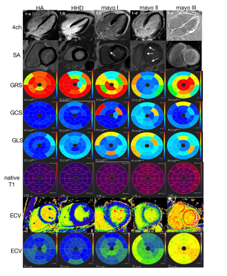 |
79 |
The diagnostic value of multi-parameter CMR for early detection
of light-chain amyloidosis from hypertension cardiomyopathy
patients
Fang Wang1,
Lili Yang1,
Yanbin Yang1,
Xiaowei Ruan1,
Rui Wang1,
and Xiuzheng Yue2
1Peoples hospital of ningxia hui autonomous region, Yinchuan, China, 2Philips Healthcare, Beijing, China, Beijing, China Keywords: Cardiomyopathy, Cardiomyopathy, CMR, light chain amyloidosis, hypertension cardiomyopathy The value of cardiac magnetic resonance imaging in diagnosing light chain amyloidosis has been confirmed, and its characteristic LGE manifestations are an important means to identify myocardial amyloidosis. However, early-stage light-chain amyloidosis patients always have the same symptoms as hypertensive cardiomyopathy in clinical practices. This study aimed to investigate the combined diagnostic efficacy of T1mapping, ECV, and myocardial strain techniques in early myocardial amyloidosis by comparing with healthy controls and patients with hypertensive cardiomyopathy that also caused uniform myocardial thickening. |
|
4013.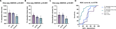 |
80 |
Cardiac MRI including myocardial T2 mapping for the
differentiation of AL and ATTR amyloidosis
Dmitrij Kravchenko1,2,
Alexander Isaak1,2,
Narine Mesropyan1,2,
Claus Christian Pieper1,
Daniel Kuetting1,2,
Leon M. Bischoff1,
Ulrike Attenberger1,
and Julian Luetkens1,2
1Diagnostic and interventional radiology, University Hospital Bonn, Bonn, Germany, 2Quantitative Imaging Laboratory Bonn, Bonn, Germany Keywords: Myocardium, Cardiomyopathy Cardiac amyloidosis can be visualized on CMR, but to date no reliable imaging parameters exist to distinguish the two most common forms of cardiac amyloidosis, AL and ATTR. Retrospective analysis of 37 cardiac amyloidosis patients shows that T2 times are markedly elevated in AL compared to ATTR, and in both compared to a control group of patients with other reasons for cardiac hypertrophy. Myocardial T2 mapping may be a useful CMR parameter for the differentiation of AL and ATTR cardiac amyloidosis.Keywords: amyloidosis, cardiac, MRI, CMR |
|
 Back to Meeting Home
 Back to the Program-at-a-Glance
The International Society for Magnetic Resonance in Medicine is accredited by the Accreditation Council for Continuing Medical Education to provide continuing medical education for physicians.
 Back to Meeting Home
 Back to the Program-at-a-Glance
 View Presentation Video