Digital Poster
Technical Solutions in Cardiovascular Imaging & Image Processing I
ISMRM & ISMRT Annual Meeting & Exhibition • 03-08 June 2023 • Toronto, ON, Canada

Digital Poster
Technical Solutions in Cardiovascular Imaging & Image Processing I
| Computer # | |||
|---|---|---|---|
4967.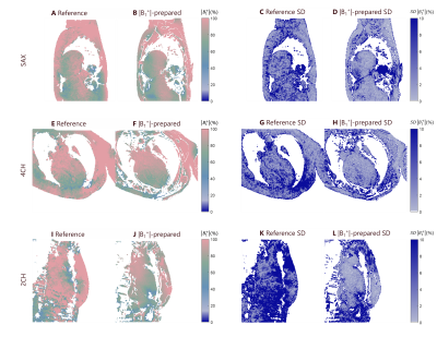 |
21 |
Mapping cardiac |B1+| field with B1+ prepared imaging
Paulina Siuryte1,
Markus Henningsson2,
Christal van de Steeg-Henzen3,
and Sebastian Weingärtner1
1TU Delft, Delft, Netherlands, 2Department of Health, Medicine and Caring Sciences, Linköping University, Linköping, Sweden, 3Holland PTC, Delft, Netherlands Keywords: Heart, Artifacts, cardiac B1+ mapping, transmit field imaging Cardiac MRI at 3T offers gains in SNR, but is strongly affected by field inhomogeneities, including the transmit field B1+. Mapping |B1+| field around the heart is particularly challenging due to the presence of motion, and few techniques exist for robust imaging. Recently, Bloch-Siegert shift mapping was proposed for motion robust cardiac |B1+| mapping, however, it requires long echo times making it more susceptible to artifacts. Here, we introduce a new preparation module for efficient |B1+| mapping. The method shows 0.06% / 0.35% reduced noise variability in phantom and 0.61±0.35% / 0.89±0.63% in-vivo imaging, and visually improved map quality. |
|
4968.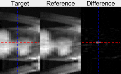 |
22 |
Fast continuous 3D catheter balloon tracking for MRI-guided
cardiac catheterization using 1D slice projection imaging with
GRE stack of slices.
Grzegorz Tomasz Kowalik1,
Radhouene Neji1,2,
Tracy Moon3,
Nina Mellor3,
Reza Razavi1,
Kuberan Pushparajah1,
and Sébastien Roujol1
1School of Biomedical Engineering and Imaging Sciences, King's College London, London, United Kingdom, 2MR Research Collaborations, Siemens Healthcare Limited, Camberley, United Kingdom, 3Guy's and St Thomas' NHS Foundation Trust, London, United Kingdom Keywords: Heart, MR-Guided Interventions An automated catheter tracking approach was developed for real-time continuous catheter visualization during MR-guided cardiac catheterization. 1D projection images are acquired for each slice of a stack providing fast 3D volume screening. These images are first collected in the absence of catheter to create an atlas on background signal across the breathing cycle. During catheter navigation, incoming images are matched to the best images of the atlas, which are then subtracted to suppress background signal. Finally, automated peak detection is applied to extract the catheter balloon location. The technique is successfully demonstrated during in-vitro and in one patient. |
|
4969.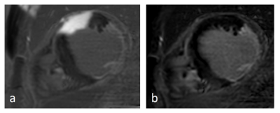 |
23 |
Cardiac MRI at 3.0T in the presence of implantable cardioverter
defibrillators (ICDs)
Theresa Reiter1,
Oliver M Weber2,
Ingo Weiss3,
and Wolfgang Rudolf Bauer1
1Medicine I, University Hospital Wuerzburg, Wuerzburg, Germany, 2Philips GmbH, Hamburg, Germany, 3Biotronik SE Co.KG, Berlin, Germany Keywords: Myocardium, Visualization Performing a cardiac MRI in the presence of active cardiac implants remains a challenge due to the extensive artifact burden caused by the implant. Sequence solutions established at 1.5 T have yet to be transferred to a clinical use at 3T. In this work, we use phantom and volunteer measurements in order to establish an adapted 3T protocol for CINE with TFE and wideband LGE-PSIR sequences that sufficiently suppress image artifacts caused by active implants. The first clinical data show the feasibility of this protocol and its ability to detect scar tissue in patients with implanted ICD. |
|
4970. |
24 |
Real-time Automatic Passive Catheter Tracking During MR-guided
Cardiac Catheterization
Rohini Vidya Shankar1,
Li Huang1,
Radhouene Neji1,2,
Grzegorz Kowalik1,
Alexander Paul Neofytou1,
Ronald Mooiweer1,2,
Tracy Moon3,
Nina Mellor3,
Reza Razavi1,
Kuberan Pushparajah1,
and Sébastien Roujol1
1Biomedical Engineering, King's College London, London, United Kingdom, 2MR Research Collaborations, Siemens Healthcare Limited, Camberley, United Kingdom, 3Guy's and St Thomas' NHS Foundation Trust, London, United Kingdom Keywords: Heart, MR-Guided Interventions, Tracking, Real-time MR-guided cardiac catheterization procedures currently employ passive tracking approaches to follow the gadolinium-filled catheter balloon during catheter navigation. This requires frequent manual tracking and repositioning of the imaging slice, especially when the catheter moves out-of-plane during the procedure. In this study, we developed a novel MRI guidance approach that enables automatic real-time tracking of the catheter balloon and repositioning of the imaging slice for continuous visualization of the balloon during catheter navigation. We first demonstrate the proposed approach in a phantom and subsequently present an initial evaluation in patients. |
|
4971.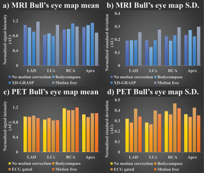 |
25 |
Quantification and evaluation of cardiac motion correction
methods for hybrid PET/MRI
Heeseung Lim1,
Benjamin Wilk1,2,
Gerald Moran3,
Heather Biernaski1,
Jonathan D. Thiessen1,2,4,
and Frank S. Prato1,2,5
1Lawson Imaging, Lawson Health Research Institute, London, ON, Canada, 2Medical Biophysics, Western University, London, ON, Canada, 3Siemens Healthcare Limited, Oakville, ON, Canada, 4Medical Imaging, Western University, London, ON, Canada, 5Physics and Astronomy, Western University, London, ON, Canada Keywords: Heart, Cardiovascular Different motion correction methods for cardiac imaging were compared with no motion correction and free-motion images on a hybrid PET/MRI platform. These data were acquired in eight large animals five days after induction of myocardial infarction. PET/MRI images were acquired under different conditions of cardiac and respiratory motion: BodyCompass™, XD-GRASP, ECG gated. Upon completion of in vivo imaging, the animal was immediately euthanized within the scanner, and motion-free images were acquired. Initial quantitative evaluation of the cardiac images showed variation in mean and standard deviation values between the region of interest and different motion correction methods. |
|
4972.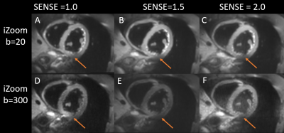 |
26 |
SENSE factor optimization for 2D RF based Reduced Field of View
DWI on Cardiac at 3.0T
Yang Wu1,
Peng Sun2,
Zhigang Wu2,
Xiaoxiao Zhang2,
Jing Zhang2,
and Jiazheng Wang2
1Department of Medical Imaging, Wuhan Asia General Hosipital, Wuhan, China, 2Philips Healthcare, Beijing, China Keywords: Myocardium, Cardiovascular Diffusion MRI delivers unique information about the heart without the use of external contrast agents, Because of B0 inhomogeneity within the thorax and short transverse relaxation durations, which cause substantial distortion and signal loss, it has proven technically problematic. DWI with Zoom imaging with 2D RF pulse (iZoom) could significantly minimize distortion, but it is still vulnerable to field inhomogeneity when it is big. iZoom with SENSE could reduce distortion and signal loss even further. An in vivo research on volunteers with various SENSE factors revealed that combining iZoom with SENSE considerably improves visual quality. |
|
4973.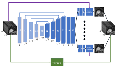 |
27 |
Deep-learning-based Group-wise Motion Correction for Myocardial
T1 Mapping
Eyal Hanania1,
Lilach Barkat1,
Israel Cohen1,
Haim Azhari1,
and Moti Freiman1
1The Technion – Israel Institute of Technology, Haifa, Israel Keywords: Myocardium, Quantitative Imaging, Cardiac, Relaxation Diffuse myocardial diseases can be diagnosed using T1 mapping technique. The T1 relaxation parameter is computed through the pixel-wise model fitting. Hence, pixel misalignment resulted by cardiac motion leads to an inaccurate T1 mapping. Therefore, registration is needed. However, standard registration methods are computationally expensive. To overcome this challenge, we propose a new deep-learning-based group-wise registration approach that register all the different time points simultaneously. Our approach achieved the best median model-fitting R2 compared to baseline methods (0.9846, vs. 0.9651/0.9744/0.9756), and achieve reasonably close T1 value to the expected myocardial T1 value |
|
4974.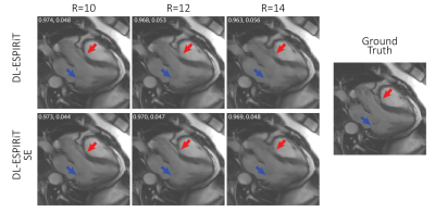 |
28 |
Improving cardiac cine MRI using a deep learning-based ESPIRiT
reconstruction with self attention
Terrence Jao1,
Christopher Sandino2,
and Shreyas Vasanawala1
1Department of Radiology, Stanford University, Stanford, CA, United States, 2Department of Electrical Engineering, Stanford University, Stanford, CA, United States Keywords: Heart, Machine Learning/Artificial Intelligence, Deep Learning, Unrolled, Self Attention, CINE A deep learning based ESPIRiT (DL-ESPIRiT) was recently proposed to reconstruct dynamic MRI data with higher reconstruction accuracy. However, the method still has difficulty resolving fine anatomic structures. We propose incorporating self-attention to the network using a computationally lightweight squeeze-excitation block (DL-ESPIRiT SE), which uses global information to select more important features while suppressing less important ones. We demonstrate improved reconstruction with DL-ESPIRiT SE, which is most pronounced during faster cardiac motion such as in ventricular ejection. |
|
4975.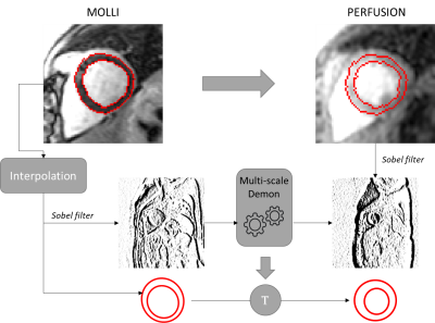 |
29 |
2D U-Net based myocardium segmentation for quantitative
sequences with limited data: demon co-registration approach
Habib Rebbah1 and
Timothé Boutelier1
1Research & Innovation, Olea Medical, La Ciotat, France Keywords: Myocardium, Segmentation Obtaining databases with expert’ manual segmentations to train U-Net is an essential but also a time-consuming operation. We explore here the effect of expanding such a database to other cases with a demon co-registration algorithm. The purpose is to speed up the manual steps but also to allow the development of segmentation algorithms for new cases. We illustrate our approach with MOLLI’ masks expanded to T2 and perfusion data. |
|
4976.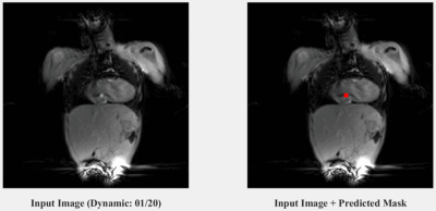 |
30 |
Automatic catheter tracking during cardiac MRI guided cardiac
catheterization using deep learning
Alexander Paul Neofytou1,
Grzegorz Tomasz Kowalik1,
Radhouene Neji1,2,
Reza Razavi1,
Kuberan Pushparajah1,
and Sébastien Roujol1
1School of Biomedical Engineering and Imaging Sciences, Faculty of Life Sciences and Medicine, King's College London, London, United Kingdom, 2MR Research Collaborations, Siemens Healthcare Limited, Camberley, United Kingdom Keywords: Heart, MR-Guided Interventions A deep neural network trained on images with artificially generated catheter signal was developed and enables accurate detection of catheter signal under real-time conditions. Further investigations, with in-line integration, are now warranted to determine the benefit of this technique for improved catheter navigation. |
|
4977.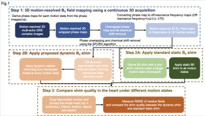 |
31 |
Correcting motion-induced B0 shim failure at 3T CMR using a
deep-learning-enabled 3D motion-resolved B0 shimming
Xinqi Li1,
Yuheng Huang2,
Xingmin Guan2,
Xinheng Zhang2,
Ghazal Yoosefian2,
Xiaoming Bi3,
Fei Han3,
HsuLei Lee1,
Anthony Christodoulou1,
Debiao Li1,
Rohan Dharmakumar2,
Hui Han1,
and Hsin-Jung Yang1
1Biomedical Image Research Institute, Cedars-Sinai Medical Center, Los Angeles, CA, United States, 2Krannert Cardiovascular Research Center, Indiana University School of Medicine, Indianapolis, IN, United States, 3Siemens Healthineers, Malvern, PA, United States Keywords: Myocardium, Shims B0 inhomogeneity is a long-lasting issue for CMR in high-field (3T and above) scanners. B0 shimming is a standard way to improve the B0 field. However, motion-induced field inhomogeneity is an unknown factor in routine practice and compromises cardiac B0 shimming quality. Here, we proposed a motion-resolved cardiac B0 shimming strategy by acquiring motion-resolved cardiac B0 field maps and adopting a modified U-net model for precise and automatic shim field derivation. We showed that dynamic shimming could improve field homogeneity through the respiratory cycle and provide a reliable B0 field for free-breathing CMR at 3T. |
|
4978.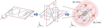 |
32 |
Detection and tracking enhancement using 4-channels local
standalone resonators for catheterized interventional MRI at 3T
Komlan Payne1,
Yunkun Zhao1,
Leslie L. Ying1,
and Xiaoliang Zhang1
1Department of Biomedical Engineering, University at Buffalo, Buffalo, NY, United States Keywords: Vessel Wall, Interventional Devices, Blood vessels Catheter-based RF coils are preferred over conventional external coils due to the weak detection signal from intermediate field region. One of the major issues using catheter RF coils is the design payload (feeding network, preamplifier, balun) associated with the limited space of the catheter. The technique also lacks efficient way to track the catheter during the procedure. To overcome these challenges, we propose 4-channel standalone and wireless resonators to amplify the local B1 field while a body coil is used as the transceiver. The receive B1 fields with and without the local resonators are obtained for field enhancement analysis. |
|
4979.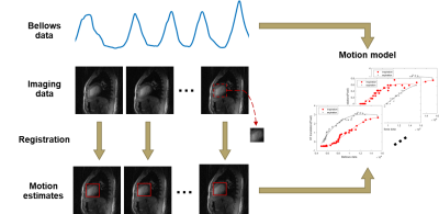 |
33 |
Patient specific respiratory motion correction for cardiac MRI
Jiameng Diao1,
Yang Liu1,
Wenjian Liu1,
Yiming Tao1,
Jianing Geng1,
Shang Cao1,
Jiayu Zhu2,
Junpu Hu2,
Jian Xu3,
Zijian Zhou1,
Haikun Qi1,4,
and Peng Hu1,4
1School of Biomedical Engineering, ShanghaiTech University, Shanghai, China, 2United Imaging Healthcare, Shanghai, China, 3UIH America, Inc., Houston, TX, United States, 4Shanghai Clinical Research and Trial Center, Shanghai, China Keywords: Heart, Motion Correction Magnetic resonance imaging (MRI) is susceptible to respiratory motion-induced artifacts due to the relatively slow data acquisition. These artifacts remain an impediment to clinical application and are further amplified in cardiovascular MRI. Respiratory motion model is an effectively method to reduce motion artifacts. In this study, we proposed a novel patient-specific respiratory motion correction method with more reliable training data and capability to perform spoke-wise correction. Combined with compressed sensing, parallel imaging, image registration and model fitting, this method greatly reduces respiratory motion artifacts. |
|
4980.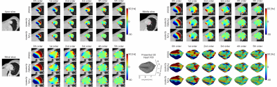 |
34 |
Spherical harmonic shim analysis of B0 distributions in cardiac
imaging planes from 921 subjects
Yun Shang1,
Sebastian Theilenberg1,
Boyu Peng2,
Michael Hock3,
Laura M. Schreiber3,4,
Sachin Jambawalikar1,2,
and Christoph Juchem1,2
1Department of Biomedical Engineering, Columbia University in the City of New York, New York, NY, United States, 2Department of Radiology, Columbia University in the City of New York, New York, NY, United States, 3Chair of Molecular and Cellular Imaging, Comprehensive Heart Failure Center (CHFC), Würzburg, Germany, 4Section of Medical Physics, Department of Radiology, Mainz University Hospital, Mainz, Germany Keywords: Heart, Shims Cardiac functional scans adopting bSSFP sequences suffer from dark band artifacts due to B0 inhomogeneity. The best remedy to mitigate this issue is through cardiac B0 shimming. A limited understanding of the B0 conditions in clinical diagnostic orientations impedes the development of an optimal B0 shim strategy in the human heart. Here we perform a theoretical analysis of spherical harmonic B0 shim at 3 T using a static global approach and slice-specific dynamic shim updating in the short-axis views of human hearts from 921 subjects as a starting point for the development of optimized cardiac B0 shim strategies. |
|
4981.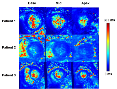 |
35 |
Accelerated, Free-Breathing 3D Cardiac T1ρ Mapping Pulse
Sequence with XD-GRASP Reconstruction
Suvai Gunasekaran1,
KyungPyo Hong1,
Roberta Catania1,
Joshua Robinson2,
Rod Passman3,
Aggelos Katsaggelos4,
Cynthia Rigsby5,
Walter Witschey6,
and Daniel Kim1
1Radiology, Northwestern University, Feinberg School of Medicine, Chicago, IL, United States, 2Cardiology, Lurie Children’s Hospital, Chicago, IL, United States, 3Cardiology, Northwestern University, Feinberg School of Medicine, Chicago, IL, United States, 4Electrical and Computer Engineering, Northwestern University, Evanston, IL, United States, 5Radiology, Lurie Children’s Hospital, Chicago, IL, United States, 6Radiology, University of Pennsylvania Perelman School of Medicine, Philadelphia, PA, United States Keywords: Myocardium, Quantitative Imaging, Fibrosis T1ρ mapping is an emerging non-contrast pulse sequence for measuring cardiac fibrosis. Unfortunately, current T1ρ techniques suffer from lack of coverage, poor spatial resolution, and long scan time. Thus, we developed an accelerated, free-breathing 3D cardiac T1ρ mapping pulse sequence using XD-GRASP reconstruction including both respiratory and contrast dimensions; additionally, view sharing and KWIC filtering were incorporated to improve spatial resolution. This sequence was tested in 8 patients undergoing clinically indicated cardiac MRI, resulting in robust image quality and T1ρ values that are in agreement with literature. |
|
4982.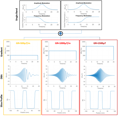 |
36 |
Dual-Band Adiabatic Pulses in cardiac MRI: Application in Black
Blood Imaging at 3T
Ayda Arami1,
Christal van de Steeg-Henzen2,
Hildo J.Lamb3,
and Sebastian Weingärtner1
1TU Delft, Delft, Netherlands, 2HollandPTC, Delft, Netherlands, 3LUMC, Leiden, Netherlands Keywords: Myocardium, RF Pulse Design & Fields Black-Blood imaging is an integral part of clinical cardiac MRI, due to it’s clear depiction of the cardiac morphology. Double inversion recovery, is most commonly used for black-blood contrast, by successively applying a non-selective inversion and a slice-selective reinversion. In this work, we investigate dual band adiabatic inversion pulses, as an alternative, to achieve nulling of the blood signal outside of the imaging slice. Thorough blood suppression and comparable noise performance to double inversion recovery were demonstrated in vivo while achieving an 11% reduction in SAR and a 10% reduction in pulse duration. |
|
4983.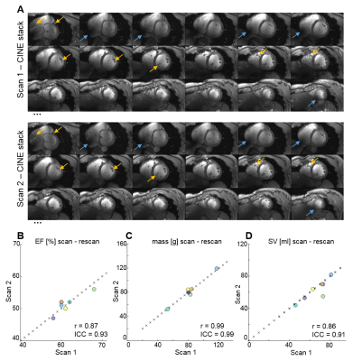 |
37 |
Ultrahigh Field CMR for the Assessment of Cardiac Function –
Scan Rescan Reproducibility and Shim Limitations
David Lohr1,
Wiebke Schlötelburg2,
Maxim Terekhov1,
and Laura Maria Schreiber1
1Chair of Molecular and Cellular Imaging, Comprehensive Heart Failure Center (CHFC), University Hospital Würzburg, Würzburg, Germany, 2Department of Nuclear Medicine, University Hospital Würzburg, Würzburg, Germany Keywords: Myocardium, High-Field MRI In this study we aim to analyze blood tissue contrast (BTC), SNR, and reproducibility in measurements of cardiac function at 7T. Furthermore, we assess shim quality and its impact on image quality as well as T2*. Measurements are performed in n=7 healthy volunteers using a 7T system. Diastolic BTC and SNR in scan1 and scan2 were 2.14±0.24 and 2.22±0.33 as well as 96±35 and 105±29, respectively. Reproducibility assessed via intra class correlation coefficient was found to be excellent for ejection fraction (0.93), myocardial mass (0.99), stroke volume (0.91), end-systolic volume (0.94), and end-diastolic volume (0.93). |
|
4984.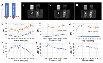 |
38 |
Off-resonance encoded fat suppression methods for 5D whole-heart
free-running cardiac MRI at 1.5T
Yasaman Safarkhanlo1,2,
Jerome Yerly3,4,
Mariana Falcao3,
Adèle LC Mackowiak2,3,5,
Davide Piccini3,6,
Matthias Stuber3,4,
Bernd Jung2,5,
Christoph Gräni1,2,
and Jessica AM Bastiaansen2,5
1Department of Cardiology, University Hospital Bern, Inselspital, Bern, Switzerland, 2Translation Imaging Center (TIC), Swiss Institute for Translational and Entrepreneurial Medicine, Bern, Switzerland, 3Department of Diagnostic and Interventional Radiology, Lausanne University Hospital (CHUV), Lausanne, Switzerland, 4Center for Biomedical Imaging, Lausanne, Switzerland, 5Diagnostic, Interventional and Pediatric Radiology (DIPR), University Hospital Bern, Inselspital, Bern, Switzerland, 6Advanced Clinical Imaging Technology, Siemens Healthineers International AG, Lausanne, Switzerland Keywords: Heart, Fat, data acquisition, data analysis, image reconstruction, low-field MRI The presence of fat signals around the heart can affect the diagnostic quality of cardiovascular MR images. There are multiple fat-suppression pulses, such as FISS, and off-resonance water-excitation (WE) pulses, such as BORR, LIBRE, and LIBOR, that have been developed for the free-running balanced Steady-State Free-Precession (bSSFP) sequences at low-field MRI (1.5T). These fat-suppression pulses have never been thoroughly compared to each other, therefore, in this work, we implemented four different fat-suppression pulses and validated their performance on phantoms and healthy volunteers. Our results indicated LIBOR provided better fat suppression compared to BORR and LIBRE, while having fast acquisition time. |
|
4985. |
39 |
Recovering image orientation for unconventional acquisition
plane with deep learning algorithm: cardiac magnetic resonance
case.
Habib Rebbah1 and
Timothé Boutelier1
1Research & Innovation, Olea Medical, La Ciotat, France Keywords: Myocardium, Segmentation Deducing the orientation main view of an image from the DICOM information could be complicated especially for unconventional image plane as for cardiac MR. We explore here the feasibility of deducing the orientation of the image based on CNN approach. |
|
4986.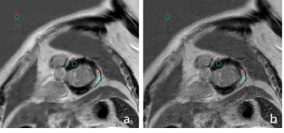 |
40 |
A clinical application of deep learning reconstructed myocardial
late gadolinium enhancement on short-breath-hold patients
Xuefang Lu1,
Weiyin Vivian Liu2,
Yuchen Yan1,
Changsheng Liu1,
Wei Gong1,
Yan Wang1,
Yilin Zhao1,
Guangnan Quan3,
and Yunfei Zha1
1Department of Radiology, Renmin Hospital of Wuhan University, Wuhan, China, 2GE Healthcare, MR Research China, Beijing, China, 3General Electric Medical (China) Co, Beijing, China Keywords: Myocardium, Image Reconstruction SNR and CNR are essential for radiologists to precisely assess the signal enhancement in myocardia tissues. High-resolution late gadolinium enhancement cardiac magnetic resonance (LGE-CMR) is important but often possess low SNR and takes long scan time. Compared with original PSMDE (PSMDEO), AIRTM Recon DL-based PSMDE (PSMDEDL) effectively and significantly improved SNR, CNR and image quality without extra scan time. High-resolution PSMDEDL images also accelerated diagnossis speed of identifying defected tissues from noisy but normal myocardial tissues and elevated the diagnosis confidence despite no statistically different diagnostic performance between PSMDEDL and PSMDEO images. |
|
 Back to Meeting Home
 Back to the Program-at-a-Glance
The International Society for Magnetic Resonance in Medicine is accredited by the Accreditation Council for Continuing Medical Education to provide continuing medical education for physicians.
 Back to Meeting Home
 Back to the Program-at-a-Glance
 View Presentation Video