Digital Poster
Advanced Cardiac Tissue Characterization & Applications of Novel Techniques I
ISMRM & ISMRT Annual Meeting & Exhibition • 03-08 June 2023 • Toronto, ON, Canada

Digital Poster
Advanced Cardiac Tissue Characterization & Applications of Novel Techniques I
| Computer # | |||
|---|---|---|---|
4283.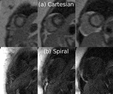 |
21 |
Opportunities for improved myocardial first-pass perfusion
imaging at 0.55T
Ye Tian1,
Sophia X. Cui2,
Parveen Garg3,
and Krishna S. Nayak1
1Ming Hsieh Department of Electrical and Computer Engineering, University of Southern California, Los Angeles, CA, United States, 2Siemens Medical Solutions, Los Angeles, CA, United States, 3Division of Cardiovascular Medicine, Keck School of Medicine, University of Southern California, Los Angeles, CA, United States Keywords: Myocardium, Perfusion Myocardial first-pass perfusion (FPP) MRI is used in the clinic to diagnose coronary artery disease but has limited spatial resolution and coverage. Contemporary low-field scanners have the potential to provide FPP imaging with improved resolution and coverage by leveraging different acquisitions, such as spiral bSSFP. In this study, we demonstrate a time-efficient spiral bSSFP FPP pulse sequence at 0.55T and compare it with the standard Cartesian FPP in 4 healthy volunteers. Measured SNR, CNR, and myocardial boundary sharpness scores are all higher in the spiral images than in Cartesian images. |
|
4284.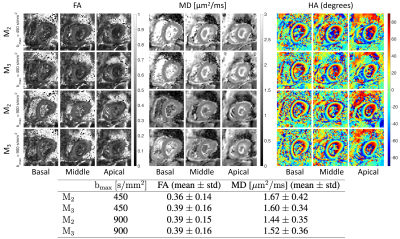 |
22 |
Cardiac diffusion MRI using Connectom scanner
Maryam Afzali1,2,3,
Lars Mueller1,3,
Sam Coveney1,
Fabrizio Fasano4,5,
John Evans2,
Maria Engel2,
Filip Szczepankiewicz6,
Irvin Teh1,
Erica Dall'Armellina1,
Derek K Jones2,
and Jürgen E Schneider1
1Leeds Institute of Cardiovascular and Metabolic Medicine, Leeds, United Kingdom, 2Cardiff University Brain Research Imaging Centre (CUBRIC), School of Psychology, Cardiff University, Cardiff, United Kingdom, 3These authors contributed equally to this work, Leeds, United Kingdom, 4Siemens Healthcare Ltd, Camberly, United Kingdom, 5Siemens Healthcare GmbH, Erlangen, Germany, 6Medical Radiation Physics, Clinical Sciences Lund, Lund University, Lund, Sweden Keywords: Heart, Diffusion Tensor Imaging, Strong gradients Cardiac diffusion Magnetic Resonance Imaging (cardiac dMRI) allows to non-invasively assess the microstructure of the heart. Cardiac motion typically necessitates the use of motion-compensated diffusion gradients. Here we report for the first time on the application of cardiac dMRI on a Connectom MR scanner with a gradient strength of 300 mT/m using a spin echo (SE) sequence with EPI readout and up to third order (M3) motion-compensated diffusion gradients. The results show the benefits of applying dMRI with higher order motion compensation in the human heart using a Connectom scanner. |
|
4285.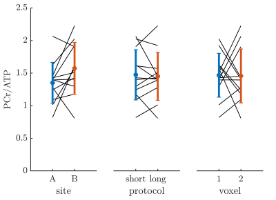 |
23 |
Reproducibility of Cardiac 31P MRS at 7 T – Initial Results of a
Multi-Center and Longitudinal Study
Stefan Wampl1,
Ladislav Valkovic2,
Ferenc Mozes2,
Martin Meyerspeer1,
and Albrecht Ingo Schmid1
1High Field MR Center, Center for Medical Physics and Biomedical Engineering, Medical University of Vienna, Vienna, Austria, 2Oxford Centre for Clinical MR Research, RDM Cardiovascular Medicine, University of Oxford, Oxford, United Kingdom Keywords: Heart, Spectroscopy, 31P MRS To demonstrate reproducibility of cardiac 31P MR spectroscopy at 7T, three subjects were scanned on two sites in two different countries. The two sites are equipped with similar MR scanners but scanner platform versions, RF coil hardware and operators differed. The 16-channel receive array at site A provided 50% higher SNR than the 14cm single loop coil at site B, while site B achieved better linewidths, resulting in similar quality of spectral fitting. Two scan protocols were compared between the sites, both provided good reproducibility of cardiac PCr/ATP. This demonstrates the feasibility of larger multi-centre trials of cardiac MR spectroscopy. |
|
4286.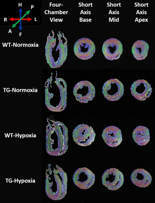 |
24 |
Structural alterations in right ventricle of hypoxic pulmonary
hypertensive rats assessed by high-resolution DTI
Dounia El Hamrani1,
Emma Le Nezet1,
Marilyne Campagnac2,
Céline Ayez1,
Benjamin Pere2,
Christelle Guibert2,
David Benoist1,
and Bruno Quesson3
1IHU Liryc, Bordeaux, France, 2CRCTB U1045, Bordeaux, France, 3CRMSB UMR 5536, Bordeaux, France Keywords: Cardiomyopathy, Hypertension Transgenic rats with Bmpr2 mutation is a model of heritable pulmonary arterial hypertension. This study aims to evaluate structural alterations of Bmpr2-mutants associated with a risk factor (chronic hypoxia) by high-resolution diffusion tensor imaging (DTI). Chronic increases of pressure-loading conditions appears to be poorly tolerated by the right ventricle (RV). Indeed, chronic hypoxia groups showed a hypertrophy in RV. Moreover, DTI analysis showed disturbances in RV (myofibers and sheetlets) and this more prominently at basal level in transgenic rats. Consequently, hypoxic pulmonary hypertension can induce microstructural changes and electrophysiological remodeling, which are potentiated by Bmpr2 mutation. |
|
4287.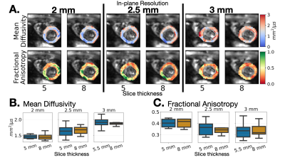 |
25 |
The Effect of Resolution and Voxel Size on SNR for Cardiac
Diffusion Tensor Imaging
Ariel J Hannum1,2,3,
Tyler E Cork1,2,3,
Kawin Setsompop1,4,
and Daniel B Ennis1,2
1Department of Radiology, Stanford University, Stanford, CA, United States, 2Division of Radiology, Veterans Administration Health Care System, Palo Alto, CA, United States, 3Department of Bioengineering, Stanford University, Stanford, CA, United States, 4Department of Electrical Engineering, Stanford University, Stanford, CA, United States Keywords: Heart, Diffusion Tensor Imaging The purpose of this work is to characterize how SNR varies with slice thickness for a given in-plane resolution for cDTI. We observed that noise in cDTI is largely physiological dominated such that increasing voxel volume yields smaller gains in SNR than what is observed in a phantom. More specifically, increased slice thickness did not significantly improve cDTI metric maps for a given in-plane resolution. However, higher in-plane resolution improved MD and FA maps. Therefore, future work will evaluate the optimal in-plane resolution and slice thickness. |
|
4288.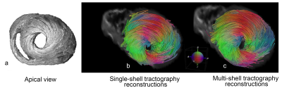 |
26 |
Multi-shell versus single-shell cardiac diffusion imaging
Nahla M H Elsaid1,
Dana C Peters1,
Gigi Galiana1,
and Albert J Sinusas2
1Radiology and Biomedical Imaging, Yale University, New Haven, CT, United States, 2Medicine (Cardiology), Yale University, New Haven, CT, United States Keywords: Heart, Diffusion/other diffusion imaging techniques Myocardial infarction (MI) remains a leading cause of morbidity and death in the Western world. MI causes regional dysfunction, which places remote areas of the heart at a mechanical disadvantage resulting in long-term adverse left ventricular (LV) remodeling and congestive heart failure (CHF). While the cardiac fiber structure has been the topic of study for decades, to this day, it has not been fully understood. There is a need for a deeper understanding of the normal and pathological myocardial structure. Standard techniques using diffusion tensor imaging (DTI) which typically needs one b-value (single shell) set of data, yield poor quality data, as DTI is incapable of delineating fibers that form torsions and complex interdigitation. However, multi-shell diffusion magnetic resonance imaging (dMRI) can delineate these complex fibers. This research investigates the difference between multi-shell versus single shell on the quality of the resulting cardiac tractography applied to an ex vivo normal porcine heart. |
|
4289.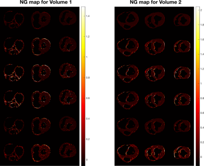 |
27 |
Applying diffusion signal representation further than DTI
improves cardiac dMRI insights
Justino Rafael Rodríguez-Galván1,
Susana Merino-Caviedes1,
David Filgueiras-Rama2,
Javier Sánchez-González3,
Antonio Tristán-Vega1,
and Carlos Alberola-López1
1Laboratorio de Procesado de Imagen (LPI) Universidad de Valladolid, Valladolid, Spain, 2Centro Nacional de Investigaciones Cardiovasculares, Madrid, Spain, 3Philips Ibérica, Madrid, Spain Keywords: Myocardium, Diffusion/other diffusion imaging techniques Diffusion MRI in the brain has undergone tremendous advances in the last two decades while cardiac and general diffusion in the body show a lower speed. As for modelling, either isotropic or diffusion tensor imaging (DTI) are, by far, the methods used outside the brain. In this abstract we intend to quantify the better modeling capabilities of higher order (HO) methods than DTI, i.e., than Gaussian distributions. We show, by means of ex-vivo diffusion in pig hearts, that non-Gaussianity is observable and that diffusion weighted images (DWI) generated out of a HO model show more closeness to reality than DTI. |
|
4290.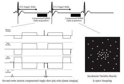 |
28 |
Second-order motion compensated single-shot spin echo planar
imaging sequence using Compressed SENSE: a pilot study
Rui Chen1,
Wei Luo1,
Zhigang Wu2,
Yongzhou Xu2,
Hui Liu1,
and Zaiyi Liu1
1Radiology, Guangdong Provincial People’s Hospital, Guangzhou, China, 2Philips Healthcare China, Shenzhen, China Keywords: Heart, Diffusion/other diffusion imaging techniques Cardiac diffusion weighted imaging (DWI) is a novel tool that could provide non-invasive in-vivo microstructural assessment. However, cardiac DWI remains a challenging task due to low signal-to-noise-ratio (SNR) and motion artifacts. The second-order motion compensated (M2C) could be used to decrease the motion artifacts, and Compressed SENSE could improve the SNR of the echo planar imaging (EPI) based sequences. Hence, the combination of M2C DWI and Compressed SENSE was firstly proposed in the present study. We found that M2C DWI using Compressed SENSE showed better overall image quality and higher SNR. |
|
4291.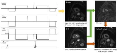 |
29 |
Computed DWI with 2nd Order motion Compensated DWI for Diagnosis
of the Myocardial Infarction without Contrast Agents
Wei Luo1,
Rui Chen1,
Zhigang Wu2,
Hui Liu1,
and Zaiyi Liu1
1Department of Radiology, Guangdong Provincial People’s Hospital, Guangzhou, China, 2Philips Healthcare, Shenzhen, Ltd., Shenzhen, China Keywords: Heart, Diffusion/other diffusion imaging techniques, Computed DWI Diagnosis of myocardial infarction (MI) remains challenge since it’s still difficult to quantify the infarction area, and to assess prognosis. Cardiac DWI could provide essential information for diagnosis of MI without exogenous contrast agents. Higher b-value DWI could improve the detection rate of myocardial infarction, but it is challenging for image acquisition in cardiac DWI due to low SNR and motion. The present study aimed to combine the 2nd order motion compensated diffusion with computed DWI to overcome these challenges. Results of this study indicates the combination is potentially a promising and valuable non-invasive method in detection of MI. |
|
4292.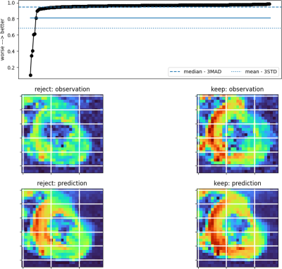 |
30 |
Semi-Automated Rejection of Corrupted Images in Cardiac
Diffusion Tensor Imaging
Sam Coveney1,
Chris Kelly1,
Irvin Teh1,
Maryam Afzali1,2,
Lars Mueller1,
Arka Das3,
Filip Szczepankiewicz4,
Derek K Jones2,
Erica Dall’Armellina1,
and Jurgen E Schneider 1
1Leeds Institute of Cardiovascular and Metabolic Medicine, University of Leeds, Leeds, United Kingdom, 2Cardiff University Brain Research Imaging Centre (CUBRIC), Cardiff University, Cardiff, United Kingdom, 3Leeds Teaching Hospitals NHS Trust, Leeds, United Kingdom, 4Medical Radiation Physics, Clinical Sciences Lund, Lund University, Lund, Sweden Keywords: Heart, Diffusion Tensor Imaging Cardiac Diffusion Tensor Imaging (cDTI) is prone to imaging artefacts including distortion, signal dropout, and misregistration even after post-processing. We developed a method for image rejection based on a comparison between the observed images and the corresponding set of predicted images generated by tensor models fit to the observed data. A rejection threshold to exclude images from subsequent refitting of the tensor model can be chosen by the user with a simple graphical method. |
|
4293.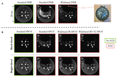 |
31 |
Wideband bright- and black-blood late gadolinium enhancement
imaging for patients with cardiac implantable electronic device
Pauline Gut1,2,
Hubert Cochet1,3,
Frederic Sacher1,4,
Pierre Jaïs1,4,
Matthias Stuber1,2,5,
and Aurélien Bustin1,2,3
1IHU LIRYC, Electrophysiology and Heart Modeling Institute, Université de Bordeaux – INSERM U1045, Pessac, France, 2Department of Diagnostic and Interventional Radiology, Lausanne University Hospital and University of Lausanne, Lausanne, Switzerland, 3Department of Cardiovascular Imaging, Hôpital Cardiologique du Haut-Lévêque, CHU de Bordeaux, Pessac, France, 4Department of Cardiac Pacing and Electrophysiology, Hôpital Cardiologique du Haut-Lévêque, CHU de Bordeaux, Pessac, France, 5CIBM Center for Biomedical Imaging, Lausanne, Switzerland Keywords: Myocardium, Tissue Characterization, Metallic device Conventional LGE PSIR provides very good contrast between healthy and scar tissue, but scar patterns may be confused with blood signal. Joint LGE bright- and black-blood imaging allows for improved scar contrast and detailed cardiac anatomy. However, many patients with cardiac implantable electronic device (CIED) do not undergo cardiac imaging due to severe hyperintensity artefacts. Here we propose a technology to image myocardial scars with unprecedent scar contrast in patients with CIED by combining bright- and black-blood SPOT imaging with wideband MRI. We showed that wideband SPOT can suppress CIED-related hyperintensity artefacts, while maintaining improved scar contrast and localization. |
|
4294.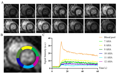 |
32 |
Low-dose Dobutamine Stress CMR perfusion for the Evaluation of
Myocardial Microcirculation Induced by Chronic High-Altitude
Hypoxia in Rats
Bo He1
1Radiological department, Sichuan Academy of Medical Sciences – Sichuan Provincial People's Hospital(SAMSPH), Chengdu, China Keywords: Heart, Perfusion This study investigated the cardiovascular effects of altitude exposure, and determined the feasibility of stress first-pass perfusion CMR to objectively and noninvasively diagnose chronic HAH-induced microvascular changes. The results showed a significant decreased in LVEF as compared with NC animals at two time points. Our study has shown rest_RU and stress_RU were significantly lower and rest_MPI, stress_MPI and MPR were significantly higher. The pilot testing demonstrated that the feasibility of stress first-pass perfusion CMR to objectively and noninvasively diagnose chronic HAH-induced microvascular changes. |
|
4295.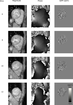 |
33 |
Characterization of Cardiac Quantitative Susceptibility Mapping
(QSM)
Andrew Tyler1,
Li Huang1,
Radhouene Neji1,2,
Ronald Mooiweer1,2,
Pier-Giorgio Masci1,
and Sébastien Roujol1
1School of Biomedical Engineering and Imaging Sciences, King's College London, London, United Kingdom, 2MR Research Collaborations, Siemens Healthcare Limited, Camberley, United Kingdom Keywords: Heart, Susceptibility Cardiac Quantitative Susceptibility Mapping (QSM) is a promising technique for the evaluation of iron levels in the myocardium, particularly after a hemorrhagic infarction. In this abstract, we validate the accuracy of our QSM acquisition and reconstruction procedure in phantom experiments, and characterize its precision and repeatability in the myocardium in a healthy volunteer cohort. We found a strong linear relationship between the gadolinium concentration and measured susceptibility in the phantom. In vivo QSM precision and repeatability in the myocardium were 0.11±0.05 ppm and 0.02±0.02 ppm, respectively. |
|
4296.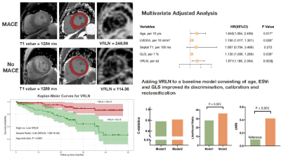 |
34 |
Left Ventricular Vertical Run-Length Non-Uniformity Adds
Prognostic Value to MACE in Patients with ESRD
Tian-yi Zhang1,
Peng Wu2,
Lian-Ming Wu3,
and Shan Mou1
1Nephrology, Shanghai Jiaotong University School of Medicine Affiliated Renji Hospital, Shanghai, China, 2Philips Healthcare, Shanghai, China, Shanghai, China, 3Radiology, Shanghai Jiaotong University School of Medicine Affiliated Renji Hospital, Shanghai, China Keywords: Myocardium, Tissue Characterization, End-stage renal disease Left ventricular vertical run-length non-uniformity (VRLN) represents heterogeneity within native T1 images and reflects the extent of cardiac fibrosis. In uremic cardiomyopathy, interstitial fibrosis was found to be the major histological alteration. The aim of this study was to evaluate the prognostic value of VRLN in patients with ESRD. The primary endpoint was major adverse cardiac events (MACE). Survival analysis was calculated by Kaplan-Meier curves with log-rank test. VRLN remained associated with MACE in the multivariable model. Adding VRLN to a model containing clinical and CMR parameters significantly improved the accuracy and reclassification of the predictive model. |
|
4297.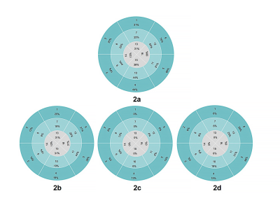 |
35 |
Global, segmental and layer specific analysis of myocardial
involvement in Duchenne muscular dystrophy by LGE
Xuelin Fang1,2,
Longwei Sun1,
Yizhen Luo1,
Guohua Liang1,
Liqi Yang1,
Kan Deng3,
and Zhiyong Li1
1Shenzhen Children’s Hospital, shenzhen, China, 2Shantou University Medical College, shantou, China, 3Philips Healthcare, guangzhou, China Keywords: Myocardium, Tissue Characterization To provide more imaging basis for early identification and prognosis evaluation of DMD-associated cardiomyopathy(DMD-CM), we analyzed the distribution characteristics of LGE in global and regional left ventricle and the correlation between myocardial fibrosis index and cardiac function indexes in 29 boys with DMD. The range of LGE and the distribution pattern of interventricular septal involvement are related to the decrease of cardiac function. LGE is a reliable tool to identify subclinical DMD-CM and evaluate the severity of the disease. |
|
4298.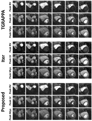 |
36 |
Fast reconstruction of SMS bSSFP myocardial perfusion images
using a combination of parallel imaging and AI-based denoising
Naledi Lenah Adam1,
Kowalik Tomasz Grzegorz 1,
Andrew Tyler1,
Ronald Mooiweer1,2,
Alexander Paul Neofytou1,
Sarah McElroy1,
Karl Kunze1,2,
Peter Speier3,
Daniel Stäb4,
Radhouene Neji1,2,
Muhummad Sohaib Nazir1,
Reza Razavi1,
Amedeo Chiribiri1,
and Sébastien Roujol1
1School of Biomedical Engineering and Imaging Sciences, Faculty of Life Sciences and Medicine, King's College London, London, United Kingdom, 2MR Research Collaborations, Siemens Healthcare Limited, Camberley, United Kingdom, 3Cardiovascular Predevelopment, Siemens Healthcare GmbH, Erlangen, Germany, 4MR Research Collaborations, Siemens Healthcare Limited, Melbourne, Australia Keywords: Heart, Perfusion Simultaneous multi-slice (SMS) imaging enables high spatial coverage and resolution for myocardial perfusion imaging. We developed and evaluated a fast, noise-removing and signal-preserving SMS reconstruction approach combining parallel imaging and deep-learning and evaluated its benefit for myocardial perfusion imaging. In comparison to conventional parallel imaging reconstruction, the proposed SMS reconstruction provides improved perceived SNR and image quality (p<0.008), preserved sharpness and signal fidelity (p>0.05) in a short computation time (+20s for an entire dataset). |
|
4299.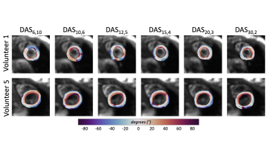 |
37 |
Trading Off Averages and Directions for Cardiac Diffusion Tensor
Imaging
Tyler E. Cork1,2,3,
Ariel J. Hannum1,2,3,
Matthew J. Middione1,2,
and Daniel B. Ennis1,2
1Radiological Sciences Laboratory, Stanford University, Stanford, CA, United States, 2Division of Radiology, Veterans Administration Health Care System, Palo Alto, CA, United States, 3Bioengineering, Stanford University, Stanford, CA, United States Keywords: Myocardium, Microstructure The objective of this work was to control the total number of collected diffusion weighted images (DWI) across several different acquisition schemes to evaluate which combination of directions and averages provides the highest quality data. In vivo cDTI data was collected in volunteers (N=5) using a series of fixed duration protocols that span a range of diffusion directions and signal averages. We found that balancing the number of diffusion directions (12-20) and averages (5-3) allows for robust quantification of mean diffusivity, fractional anisotropy, and helix measurements in a fixed scan time. |
|
4300.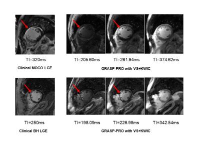 |
38 |
Single-shot, Multi-TI Late Gadolinium Enhancement MRI using
GRASP-Pro Reconstruction with View-Sharing and KWIC Filtering
Mingyue Zhao1,2,
Daming Shen1,2,
Lexiaozi Fan1,2,
Kyungpyo Hong2,
Li Feng3,
Bradley D Allen2,
Daniel C Lee4,
and Daniel Kim1,2
1Biomedical Engineering, Northwestern University, Evanston, IL, United States, 2Radiology, Northwestern University Feinberg School of Medicine, Chicago, IL, United States, 3BioMedical Engineering and Imaging Institute(BMEII), Icahn School of Medicine at Mount Sinai, New York, NY, United States, 4Division of Cardiology, Internal Medicine, Northwestern University Feinberg School of Medicine, Chicago, IL, United States Keywords: Myocardium, Tissue Characterization Late gadolinium enhancement (LGE) is the clinical standard for assessment of myocardial scarring. Current limitations of standard LGE are lengthy scan time, sensitivity to arrhythmia and/or dyspnea, and reliance of optimal inversion time (TI), which may be difficult to identify for patients with subendocardial scarring. We propose a free-breathing, single-short, multi-TI LGE approach to address the aforementioned challenges. Our results show that GRASP-Pro reconstruction with view-sharing (VS) and k-space weighted image contrast (KWIC) filtering produces multi-TI LGE images (i.e., no need for TI scout) with relatively high spatial resolution. |
|
4301.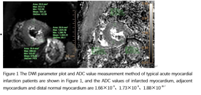 |
39 |
Application of cardiac diffusion imaging with second-order
motion compensation and breath hold in the diagnosis of acute
myocardial infarction
Xiaowei Ruan1,
yanbing yang1,
yishi wang2,
and Xiuzheng yue2
1Peoples hospital of ningxia hui autonomous region, Yinchuan, China, 2Philips Healthcare, Beijing, China Keywords: Heart, Cardiovascular, Heart; magnetic resonance imaging; myocardial diffusion imaging; myocardial infarction Cardiac magnetic resonance imaging can non-invasively evaluate the anatomy and function of the heart, and has been widely used in various fields of cardiovascular diseases. This study explores the diagnostic value of DWI in the evaluation of acute myocardial infarction. The preliminary results of 17 patients showed that the ADC value of infarcted myocardium was significantly reduced, and DWI had the ability to diagnose acute myocardial infarction. |
|
4302.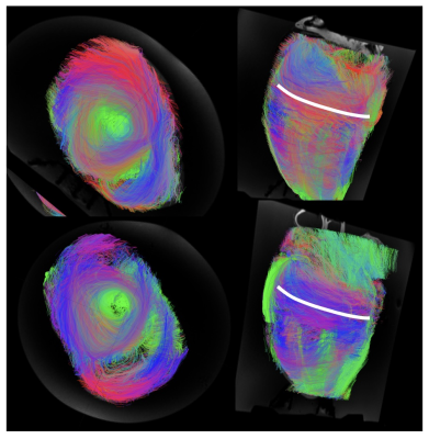 |
40 |
Evaluating the myocardial fibre pattern in the equine heart
using DTI: Protocol development for very a large ex-vivo sample
Lucy Elizabeth Kershaw1,
Rachel Jago2,
Jenny Reed2,
and John Keen2
1Edinburgh Imaging, University of Edinburgh, Edinburgh, United Kingdom, 2The Royal (Dick) School of Veterinary Studies and The Roslin Institute, University of Edinburgh, Edinburgh, United Kingdom Keywords: Myocardium, Ex-Vivo Applications We aim to develop a protocol suitable for ex-vivo DTI of the equine heart. DTI on a large sample is challenging because this is already a low SNR technique. The main requirement for good-quality data in a formalin-fixed heart is to reduce TE as much as possible. The protocol recommendations presented here are largely common sense, but the long acquisitions required make troubleshooting extremely time consuming. We thought it important to share these findings for the benefit of other investigators |
|
 Back to Meeting Home
 Back to the Program-at-a-Glance
The International Society for Magnetic Resonance in Medicine is accredited by the Accreditation Council for Continuing Medical Education to provide continuing medical education for physicians.
 Back to Meeting Home
 Back to the Program-at-a-Glance
 View Presentation Video