Digital Poster
Cardiac Function II
ISMRM & ISMRT Annual Meeting & Exhibition • 03-08 June 2023 • Toronto, ON, Canada

| Computer # | |||
|---|---|---|---|
4850.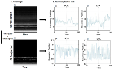 |
81 |
Improved self-navigation of respiratory motion in XD-GRASP
reconstruction using exploratory factor analysis on 3D late
gadolinium enhancement
Dima Bishara1,2,
Kyungpyo Hong1,
Suvai Gunasekaran1,
Daniel C Lee1,3,
Bradley D Allen1,
Michael Markl1,2,
Rod Passman3,
and Daniel Kim1,2
1Department of Radiology, Northwestern University Feinberg School of Medicine, Chicago, IL, United States, 2Department of Biomedical Engineering, Northwestern University, Evanston, IL, United States, 3Department of Medicine, Division of Cardiology, Northwestern University Feinberg School of Medicine, Chicago, IL, United States Keywords: Arrhythmia, Image Reconstruction Signal processing of self-navigation in 3D late gadolinium enhancement (LGE) with XD-GRASP reconstruction often leads to variable results due to a variety of factors including arrhythmia (variations in inversion-recovery of self-navigated signal) and bright signals around the body that are projected onto the edge profile used for tracking respiratory motion. Principal component analysis (PCA) is the conventional approach to process navigation signal. We hypothesize that exploratory factor analysis (EFA) produces better extraction of respiratory motion. Our results show that EFA improves respiratory motion tracking in 3D LA LGE obtained b-SSFP readout, but not in gradient echo [GRE] readout. |
|
4851.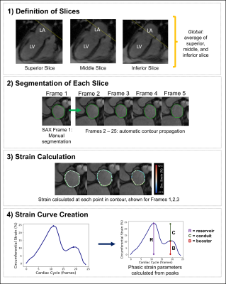 |
82 |
Left Atrial Circumferential Strain for Evaluation of Mitral
Regurgitation: A Feasibility and Reproducibility Study
Siva Sreedhar1,
Aakash Gupta2,
Teodora Chitiboi3,
Maurice Pradella4,
and Mohammed S.M. Elbaz1
1Northwestern University Feinberg School of Medicine, Chicago, IL, United States, 2Radiology, Stanford University, Stanford, CA, United States, 3Digital Technology and Innovation, Siemens Healthineers, Erlangen, Germany, 4Cardiothoracic Radiology, University Hospital of Basel, Basel, Switzerland Keywords: Myocardium, Valves, strain Short-axis CMR left atrial (LA) circumferential strain has not yet been investigated as a cardiac biomarker. This study assessed feasibility and reproducibility of LA circumferential strain in a healthy cohort, examined changes in strain within mitral regurgitation (MR) patients, and assessed correlations between reproducible strain measures and MR severity and cardiac function. Reservoir strain globally and at superior and mid LA regions was found to be reproducible, was significantly different between healthy volunteers and MR patients, and was correlated with cardiac function. CMR LA circumferential strain is reproducible with potential utility in evaluating impaired LA mechanics in MR patients. |
|
4852.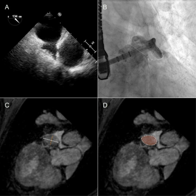 |
83 |
Cardiac magnetic resonance imaging for preprocedural planning of
percutaneous left atrial appendage closure
Dagmar Bertsche1,
Patrick Metze1,
Erfei Luo1,
Tillman Dahme1,
Birgit Gonzka1,
Wolfgang Rottbauer1,
Ina Vernikouskaya1,
Leonhard Moritz Schneider1,
and Volker Rasche1
1Internal Medicine II, Ulm University Medical Center, Ulm, Germany Keywords: Arrhythmia, MR-Guided Interventions, LAA closere, preprocedural planning An isotropic 2D six-point Dixon method was applied for assessment of the left arterial appendix (LAA) morphology prior to LAA closure intervention. Sufficient image quality was achieved even in highly arrhythmic patients allowing for the preinterventional quantification of the LAA dimensions as required for proper closure device selection. MR derived diameters were compared to periprocedurally TEE and XR derived values, which were in good agreement. Further, optimal XR angulation providing projections perpendicular to the envisaged landing zone could be identified. |
|
4853.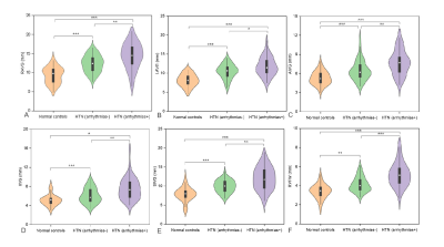 |
84 |
The relationship of epicardial adipose tissue thickness with
arrhythmias in patients with hypertension: a 3.0 T cardiac
magnetic resonance study
Zhaoxia Yang1,
Dazong Tang1,
Yi Luo2,
Chunlin Xiang1,
Lu Huang1,
and Liming Xia1
1Tongji Hospital,Huazhong University of Science and Technology, wuhan, China, 2Tongji Hospital,Huazhong University of Science and Technology, Wuhan, China Keywords: Heart, Hypertension, epicardial adipose tissue Cardiac MRI enables quantification of EAT thickness with good to excellent reproducibility for differentiating the hypertensive patients with arrhythmias from those without arrhythmias and normal controls. Additionally, EAT thickness was correlated with native T1 and LVMi, which demonstrated EAT might induce cardiac remodeling and promote myocardial fibrosis. Cardiac MRI-derived EAT thickness metrics has great potential to further explore the mechanism of EAT with arrhythmias and guide an appropriate treatment in patients of hypertension. |
|
4854.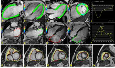 |
85 |
Impact of Epicardial Adipose Tissue Assessed on Early Left
Atrial Dysfunction in Heart Failure with Preserved Ejection
Fraction
Yunling Li1,
Shengliang Liu1,
Jianxiu Lian2,
Goukun Wang1,
Xia Gu1,
Yong Sun1,
and Bo Yu1
1Department of Cardiology, The Second Affiliated Hospital of Harbin Medical University, Harbin, China, HarBin, China, 2Phillips Healthcare, Beijing, China Keywords: Heart, Cardiovascular, Heart failure Heart failure with preserved ejection fraction (HFpEF) characterized by heart diastolic dysfunction. Epicardial adipose tissue (EAT) has been speculated to play an important role in HFpEF pathophysiological processes. In present study, EAT volume was significantly increased in patients with HFpEF and patients with high risk for developing HFpEF compared with healthy controls, and it was correlated with CMR parameters indicative of diastolic dysfunction, particularly GLS and LA reservoir, conduit, and pump strains. These findings provided compelling evidence for the potential role of EAT in adverse myocardial remodeling and the pathophysiology of HFpEF. |
|
4855.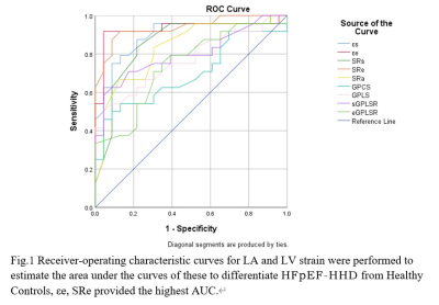 |
86 |
Association between HFpEF and Impaired Left Heart strain in
Hypertensive Heart Disease: Evaluation by Cardiac MRI Feature
Tracking
Tianyu Ke1,
Xiaolin Mu2,
Jing Chen1,
and Yang Song2
1Graduate School of Dalian Medical University, Dalian Municipal Central Hospital, Dalian, China, 2The Affiliated Hospital of Dalian University of Technology, Dalian Municipal Central Hospital, Dalian, China Keywords: Heart, Hypertension, Heart failure LA/LV strain The application value of left atrial and left ventricular strain parameters to heart failure with preserved left ventricular ejection fraction in hypertensive heart disease was evaluated by cardiac magnetic resonance feature tracking technology. We found that the left atrial function, strain, and left ventricular strain parameters were impaired to varying degrees in the HFpEF-HHD group, and the left atrial ejection fraction was significantly correlated with some feature parameters of left atrial and left ventricular strain. However, NT-proBNP is only significantly correlated with LAεa.In the identification of the HFpEF-HHD population, the LASRe parameter has shown optimal diagnostic value. |
|
4856.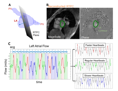 |
87 |
Left Atrial Hemodynamics Assessed with 2D Real Time Phase
Contrast MRI are Associated with Prior Stroke History in AF
Patients
Justin J Baraboo1,
Maurice Pradella1,
Anthony Maroun1,
Elizabeth Weiss1,
Amanda Dicarlo1,
Suvai Gunasekaran1,
Daniel Lee1,
Rod Passman1,
Daniel Kim1,
and Michael Markl1
1Northwestern, Chicago, IL, United States Keywords: Arrhythmia, Velocity & Flow 2D real time phase contrast was utilized in atrial fibrillation patients with and without a prior history of stroke. To account for within patient heartbeat variability, heartbeats were aggregated within each patient according to ECG trace. Heartbeat durations (RR-intervals) were sorted from fast to slow for each patient. Three groups were formed from the tertiles of the RR-intervals (faster, regular, and slower heartbeats). Blood stasis and peak velocity were compared between the stoke subgroups for each grouping. Patients with prior stroke had significantly higher stasis which remained significant in multivariable modeling using demographic variables as well. |
|
4857.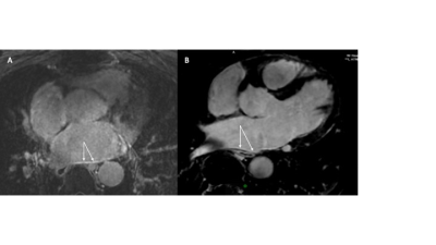 |
88 |
High-resolution, free-breathing, fat-suppressed 3D Late
Gadolinium Enhancement to define atrial wall scar after atrial
fibrillation ablation
Rajesh Dash1,2,
Henry Chubb3,
Xianglun Mao4,
Fara Nikbeh4,
Pingni Wang4,
Yuko Tada1,
Gaspar Delso5,
Martin Janich6,
Sanjiv Narayan1,
Daniel Ennis1,
and Phillip C Yang1
1Stanford University, Stanford, CA, United States, 2Cardiovascular Institute, Stanford, CA, United States, 3Stanford Lucille Packard Childrens Hospital, Stanford, CA, United States, 4GE Healthcare, Inc., Menlo Park, CA, United States, 5GE Healthcare, Inc., Barcelona, Spain, 6GE Healthcare, Inc., Munich, Germany Keywords: Myocardium, Arrhythmia, atrial fibrillation, ablation, atrial wall, scar, delayed gadolinium enhancement Atrial fibrillation (AF) ablation procedures still face challenges to achieve durable restoration of sinus rhythm. Advanced cardiac MRI delayed enhancement can delineate atrial wall scar to aid in planning AF ablation. However, thin atrial walls demand more advanced sequence development. In this study, we report protocol optimizations for high-resolution, free-breathing, fat-suppressed 3D LGE to detect atrial wall scar. In three AF patients to date, we found progressive improvements in CNR and detailed the sequence modification that led to these improvements. Future iterations will be used to assess atrial scar in patients, pre- and post-AF ablation. |
|
4858.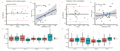 |
89 |
Magnetic resonance multitasking T1 mapping in atrial
fibrillation
Chengchen Zhao1,2,
Chunna Jin1,2,
Xiaopeng He1,2,
Simin Meng1,2,
Qi Liu3,
Zhongqi Zhang3,
Jianzhong Sun4,
and Meixiang Xiang4
1Department of Cardiology, The Second Affiliated Hospital, Zhejiang University School of Medicine, Hangzhou, China, 2Key Lab of Cardiovascular Disease of Zhejiang Province, Hangzhou, China, 3UIH America, Inc., Houston, TX, United States, 4Department of Radiology, The Second Affiliated Hospital, Zhejiang University School of Medicine, Hangzhou, China Keywords: Arrhythmia, Arrhythmia CMR multitasking T1 mapping, a recently proposed free-breathing, non-ECG technique, was evaluated in atrial fibrillation patients. It demonstrated good image quality, differentiated patients from healthy participants, and correlated with important cardiac function biomarkers. It is a promising T1 mapping tool in atrial fibrillation patients. |
|
4859.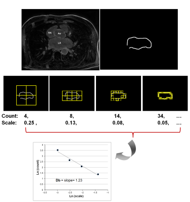 |
90 |
Characterization of left atrial remodelling and atrial
fibrillation subtype using fractal analysis of late gadolinium
enhanced MRI
Rebecca E Thornhill1,2,
Jessie Kang1,2,
Pablo B Nery3,4,
Calum Redpath3,4,
David Birnie3,4,
Sophie Chenier3,
and Elena Peña1,2
1Medical Imaging, The Ottawa Hospital, Ottawa, ON, Canada, 2Radiology, Radiation Oncology, and Medical Physics, University of Ottawa, Ottawa, ON, Canada, 3University of Ottawa Heart Institute, Ottawa, ON, Canada, 4Medicine, University of Ottawa, Ottawa, ON, Canada Keywords: Arrhythmia, Heart Atrial Fibrillation (AF) is a heart rhythm disorder of the left atrium (LA) that is associated with an increased risk of stroke and death. AF has been known to induce morphological changes of the LA that may be detectable with fractal analysis. The purpose of this study was to assess whether there are differences in fractal dimension of the LA based on AF severity or subtype. The paroxysmal AF groups demonstrated increased fractal dimension compared to persistent or permanent groups. LA dilatation associated with persistent or permanent AF may explain this result, but this requires validation in a larger cohort. |
|
4860.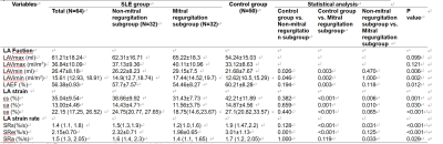 |
91 |
Cardiac magnetic resonance imaging with feature tracking to
evaluate left atrial strain for patients with systemic lupus
erythematosus
huang wei wei1,
luo song1,
zhang jun1,
zhang ling yan1,
and dou wei qiang2
1Jinling Hospital, Medical School of Nanjing University, nanjing, China, 2MR Research, GE Healthcare, beijing, China Keywords: Cardiomyopathy, MR Value This study aimed to detect left atrial (LA) strain and left ventricular (LV) stain by cardiac magnetic resonance imaging feature tracking (CMR-FT) in patients with systemic lupus erythematosus (SLE). SLE patients showed lower LVEF and multiple LV strain data than control group. Furthermore, significantly different LA reservoir, conduit and bump function were found in SLE patients than healthy controls. With these findings, CMR-FT has proven as an effective method in the diagnosis of cardiac injury in SLE patients. |
|
4861.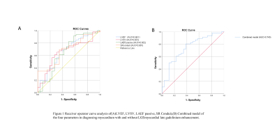 |
92 |
Cardiac MRI left ventricular and left atrial parameters predict
myocardial late gadolinium enhancement in hypertrophic
cardiomyopathy
Di Tian1,
Ziqi Xiong1,
Yifan He1,
Jingyu Zhang1,
and Zhiyong Li1
1Department of Radiology, The First Affiliated Hospital of Dalian Medical University, Dalian, China, Dalian, China Keywords: Cardiomyopathy, Cardiomyopathy Myocardial fibrosis causes functional impairment in patients with hypertrophic cardiomyopathy (HCM). The clinical gold standard for imaging myocardial fibrosis is cardiac magnetic resonance imaging (MRI) with late gadolinium enhancement (LGE), which is commonly used for the diagnosis and prognostic evaluation of patients with HCM. However, LGE imaging is an invasive test and some patients have adverse reactions to contrast agents. The aim of this study is to predict the presence of fibrosis in the left ventricular myocardium by cardiac magnetic resonance (CMR) non-invasive assessment of left atrial and left ventricular parameters in patients with HCM. |
|
4862.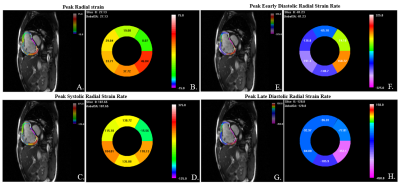 |
93 |
DRA-CMR based layer-specific right ventricle strain predicts
pulmonary arterial hypertension prognosis: results from a
Chinese registry study
Wen Li1,
Xianchang Zhang2,
Xiaoyue Zhou3,
Jens Wetzl4,
Qing Gu5,
and Jianguo He6
1State Key Laboratory of Cardiovascular Disease, Fuwai Hospital, National Center for Cardiovascular Diseases, Chinese Academy of Medical Sciences and Peking Union Medical College, Beijing, China, 2MR Collaboration, Siemens Healthineers Ltd, Beijing, China, 3MR Collaboration, Siemens Healthineers Ltd, Shanghai, China, 4Magnetic Resonance, Siemens Healthcare, Erlangen, Germany, 5Emergency Center, State Key Laboratory of Cardiovascular Disease, Key Laboratory of Pulmonary Vascular Medicine, Fuwai Hospital, National Center for Cardiovascular Diseases, Chinese Academy of Medical Sciences and Peking Union Medical College, Beijing, China, 6Center of Pulmonary Vascular Disease, State Key Laboratory of Cardiovascular Disease, Fuwai Hospital, National Center for Cardiovascular Diseases, Chinese Academy of Medical Sciences and Peking Union Medical College, Beijing, China Keywords: Heart, Heart, Strain, Right ventricle This is the first study to demonstrate that right ventricle (RV) layer-specific strain rate measured by deformation registration algorithm based cardiovascular magnetic resonance had the long-term and 3-year mortality predictive values in pulmonary arterial hypertension (PAH) patients. Our findings indicated that RV layer-specific strain rate may be sensitive indicators of RV dysfunction in PAH. |
|
4863.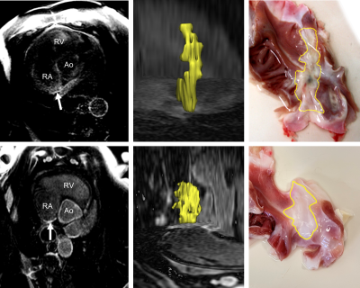 |
94 |
3D LGE MRI at 3T for the Detection of Chronic Scars after RF
Ablation in the Right Atrium: Assessment of Precision in a
Porcine Study
Simon Reiss1,
Enaam Chleilat2,
Thomas Lottner1,
Peter Kohl2,
and Michael Bock1
1Division of Medical Physics, Department of Radiology, University Medical Center Freiburg, Freiburg, Germany, 2Institute for Experimental Cardiovascular Medicine, University Heart Center Freiburg Bad Krozingen, and Faculty of Medicine, University of Freiburg, Freiburg, Germany Keywords: Heart, Arrhythmia RF ablation is an established treatment option for cardiac arrhythmia. After RF ablation the scar can be assessed with LGE MRI. However, imaging of scars in the right atrium (RA) remains difficult due to the small geometry. In this animal study, we assessed the conspicuity of chronic RA scars by comparing scar extent on MRI to ex vivo anatomical dissected preparations. We show, the detection of chronic RF ablation scars in the RA is feasible but with limited precision. The presented comparison to gross histology provides and important evaluation of RF ablation scar conspicuity in LGE MRI. |
|
4864.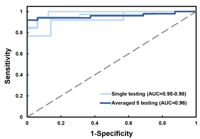 |
95 |
Radiomics-Based Classification of Patients with Repaired
Tetralogy of Fallot by Cardiac Magnetic Resonance
Jo-Hua Peng1,
Ming-Ting Wu2,
Nai-Yu Pan3,
Teng-Yi Huang3,
Yi-Jui Liu4,
Ken-Pen Weng5,6,
and Hsu-Hsia Peng1
1Department of Biomedical Engineering and Environmental Sciences, National Tsing Hua University, Hsinchu, Taiwan, 2Department of Radiology, Kaohsiung Veterans General Hospital, Kaohsiung, Taiwan, 3Department of Electrical Engineering, National Taiwan University of Science and Technology, Taipei, Taiwan, 4Department of Automatic Control Engineering, Feng Chia University, Taichung, Taiwan, 5Department of Pediatrics, Kaohsiung Veterans General Hospital, Kaohsiung, Taiwan, 6Dpartment of Pediatrics, National Yang Ming Chiao Tung University, Taipei, Taiwan Keywords: Heart, Radiomics Radiomics is a novel technique of advanced cardiac magnetic resonance (CMR) phenotyping by analyzing a couple of variables of cardiac shape and tissue texture. This study aimed to develop the radiomics-based classification model to differentiate rTOF patients from normal controls and evaluate the representative features of ventricular and myocardial abnormalities in rTOF patients. In conclusion, the radiomics-based classification model can successfully differentiate rTOF patients from normal controls with routine CMR cine images. The selected features underlined the right ventricular remodeling and left ventricular myocardium fibrosis in rTOF patients. |
|
4865.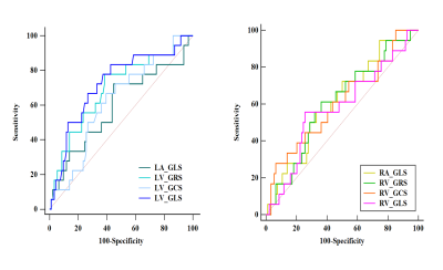 |
96 |
Analysis of Biventricular and Biatrial Strain by Cardiovascular
Magnetic Resonance in Dilated Cardiomyopathy
Shengliang Liu1,
Yunling Li1,
Jianxiu Lian2,
Guokun Wang1,
Xueying Wang1,
Ye Li1,
Yanming Zhao1,
and Bo Yu1
1The Second Affiliated Hospital of Harbin Medical University, Harbin, China, 2Philips Healthcare, Beijing, China Keywords: Cardiomyopathy, Myocardium, strain, feature tracking, fast long-axis Dilated cardiomyopathy (DCM) remains to attract worldwide attention for its poor prognosis and high mortality. The interaction of biatrial and biventricular deformation capacity in DCM patients is unclear. In this study, feature tracking and fast long-axis method were used to evaluate myocardial strain. We found that biventricular and biatrial strain were significantly impaired in DCM group than control group. LV GLS was obviously impaired in event group than no event group, and it showed significantly prognostic value in predicting cardiovascular events. Comprehensive CMR analysis should be performed to improve the diagnosis ability for DCM patients. |
|
4866. |
97 |
Left ventricular layer-specific strain predicts pulmonary
arterial hypertension long-term prognosis
Wen Li1,
Xianchang Zhang2,
Jens Wetzl3,
Qing Gu4,
and Jianguo He5
1State Key Laboratory of Cardiovascular Disease, Fuwai Hospital, National Center for Cardiovascular Diseases, Chinese Academy of Medical Sciences and Peking Union Medical College, Beijing, China, 2MR Collaboration, Siemens Healthineers Ltd, Beijing, China, 3Magnetic Resonance, Siemens Healthcare, Erlangen, Germany, 4Emergency Center, State Key Laboratory of Cardiovascular Disease, Key Laboratory of Pulmonary Vascular Medicine, Fuwai Hospital, National Center for Cardiovascular Diseases, Chinese Academy of Medical Sciences and Peking Union Medical College, Beijing, China, 5Center of Pulmonary Vascular Disease, State Key Laboratory of Cardiovascular Disease, Fuwai Hospital, National Center for Cardiovascular Diseases, Chinese Academy of Medical Sciences and Peking Union Medical College, Beijing, China Keywords: Heart, Data Processing, Strain Studies have reported left ventricular (LV) dysfunction was associated with more severe disease and poorer prognosis in pulmonary arterial hypertension (PAH). However, there are only few studies that evaluated LV strain abnormalities using cardiovascular magnetic resonance (CMR) in PAH patients. This is the first study to describe the distribution pattern and prognostic values of LV layer-specific strain/strain rate in PAH patients using a deformation registration algorithm (DRA) based on CMR. It’s found that CMR derived LV layer-specific strain could predict long-term prognosis in PAH patients. |
|
4867. |
98 |
Cine MRI-derived radiomics features of the blood pool in
pulmonary hypertension–heart failure with preserved ejection
fraction (PH-HFpEF)
Kai Lin1,
Roberto Sarnari1,
Daniel Z Gordon1,
Michael Markl1,
and James C Carr1
1Northwestern University, Chicago, IL, United States Keywords: Heart, Radiomics, blood pool Radiomics features acquired from the blood pool in different chambers (LV/RV/LA/RA) at various periods within a cardiac cycle present diverse property in characterizing PH-HFpEF. Multiple blood pool radiomics features were significantly related to PCWP and mPAP (r: 0.4 – 0.643). Compared to the LV wall, the blood pools provide more efficient features (individual AUC: 0.7 – 0.821) to discriminate PH-HFpEF from controls (p < 0.05). Cine MRI-derived radiomics features of the blood pool can be used to characterize PH-HFpEF. Radiomics features of the blood pool have the potential to become novel quantitative imaging biomarkers for presenting cardiovascular pathology. |
|
4868.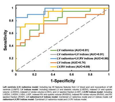 |
99 |
Can Radiomics Identify Severe Right Ventricular Dilation in
Patients with Repaired Tetralogy of Fallot by LV Cardiac
Magnetic Resonance Images?
Jo-Hua Peng1,
Ming-Ting Wu2,
Nai-Yu Pan3,
Teng-Yi Huang3,
Yi-Jui Liu4,
Ken-Pen Weng5,6,
and Hsu-Hsia Peng1
1Department of Biomedical Engineering and Environmental Sciences, National Tsing Hua University, Hsinchu, Taiwan, 2Department of Radiology, Kaohsiung Veterans General Hospital, Kaohsiung, Taiwan, 3Department of Electrical Engineering, National Taiwan University of Science and Technology, Taipei, Taiwan, 4Department of Automatic Control Engineering, Feng Chia University, Taichung, Taiwan, 5Department of Pediatrics, Kaohsiung Veterans General Hospital, Kaohsiung, Taiwan, 6Dpartment of Pediatrics, National Yang Ming Chiao Tung University, Taipei, Taiwan Keywords: Heart, Radiomics Radiomics analysis of cardiac magnetic resonance (CMR) may provide new insights into the quantitative analysis of cardiac imaging by extracting many computational quantitative metrics. This study aimed to develop a radiomics-based classification model by left ventricular (LV) CMR images to identify repaired tetralogy of Fallot (rTOF) patients with severe right ventricular (RV) dilation. In our results, the AUC were 0.81 and 0.91 for LV radiomics and LV radiomics+LV indices models, respectively. The extracted features underlined the LV intracardiac flow alteration due to RV dilation and the potential LV remodeling in rTOF patients with severe RV dilation. |
|
4869. |
100 |
Effect of Left Ventricular Myocardial Strain on Right Ventricle
in Hypertrophic Cardiomyopathy
Tingting Liu1,
Weibin Dai1,
Yueyou Peng1,
Qianyu Hu1,
Kunkun Liu1,
Ningning Song1,
Tianfeng Shi1,
Donglian Du1,
Xiaoqiong Li1,
Yueluan Jiang2,
and Yanfeng Meng1
1Taiyuan Central Hospital of Shanxi Medical University, Taiyuan, China, 2Department of MR Scientific Marketing, Siemens Healthineers Ltd., Beijing, China Keywords: Myocardium, Cardiomyopathy, Hypertrophic cardiomyopathy, Biventricular function This study evaluated the right ventricular (RV) myocardial strain of Hypertrophic Cardiomyopathy (HCM) patients with preserved ejection fraction (EF) and without myocardial hypertrophy in RV based on Cardiovascular magnetic resonance feature tracking technique (CMR-FT), and further explored the effect of left ventricular (LV) myocardial mechanics on the RV. The results showed that the global longitudinal strain (GLS) of the RV has been impaired, although the structure and EF of the RV were normal, so myocardial strain can be more sensitive and earlier to identify cardiac impairment. The systole function of the LV has a great influence on the RV function. |
|
 Back to Meeting Home
 Back to the Program-at-a-Glance
The International Society for Magnetic Resonance in Medicine is accredited by the Accreditation Council for Continuing Medical Education to provide continuing medical education for physicians.
 Back to Meeting Home
 Back to the Program-at-a-Glance
 View Presentation Video