Digital Poster
Advanced Cardiac Tissue Characterization & Applications of Novel Techniques II
ISMRM & ISMRT Annual Meeting & Exhibition • 03-08 June 2023 • Toronto, ON, Canada

Digital Poster
Advanced Cardiac Tissue Characterization & Applications of Novel Techniques II
| Computer # | |||
|---|---|---|---|
4455.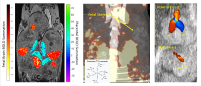 |
21 |
The Brain-Heart-Placenta Connection: Multimodal Investigation of
Neurodevelopment in Mice with Congenital Heart Disease
Margaret Stapleton1,2,
Devin Raine Everaldo Cortes2,3,4,
Samuel Wyman1,2,
Cody Ruck1,2,
Shanim Manzoor1,2,
Gabriella M Saladino1,2,
George C Gabriel1,
Tuan Tuan Tan1,
Jiuann-Huey Lin3,5,
Sebastian Ho1,
Sivakama S. Bharathi6,
Eric Goetzman6,
Cecilia W. Y. Lo1,
Anthony G Christodoulou7,
and Yijen L. Wu1,2
1Department of Developmental Biology, University of Pittsburgh School of Medicine, Pittsbrugh, PA, United States, 2Rangos Research Center Animal Imaging Core, Children's Hospital of Pittsburgh, Pittsburgh, PA, United States, 3Department of Developmental Biology, University of Pittsburgh School of Medicine, Pittsburgh, PA, United States, 4Department of Bioengineering, University of Pittsburgh Swanson School of Engineering, Pittsburgh, PA, United States, 5Department of Critical Care Medicine, University of Pittsburgh School of Medicine, Pittsburgh, PA, United States, 6Department of Pediatrics, University of Pittsburgh School of Medicine, Pittsburgh, PA, United States, 7Biomedical Imaging Research Institute, Cedars-Sinai Medical Center, Los Angeles, CA, United States Keywords: Heart, Fetus, Congenital Heart Disease Neurodevelopmental deficits (NDD) are a prevalent debilitating factor in patients with congenital heart disease (CHD) even after successful surgical palliation, but the etiology for NDD is not understood. Our study in a transgenic mouse model carrying causative genetic mutations found that NDD associated with CHD is not merely the consequence of compromised fetal hemodynamics, but primarily driven by intrinsic genetic factors and modulated by the placenta. Our study suggests that placental functions might potentially be a possible intervention strategy to overcome intrinsic genetic disadvantages. |
|
4456.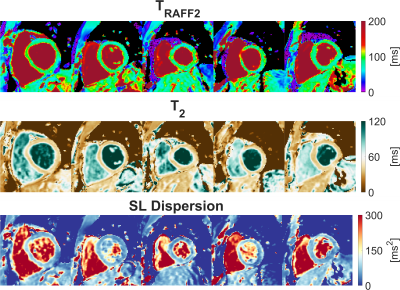 |
22 |
Intra-subject repeatability and initial clinical evaluation of
approximate myocardial spin-lock dispersion mapping
Joao Tourais1,
Yidong Zhao1,
Iain Pierce2,
Christian Nitsche2,
George D Thornton2,
Mehmet Akcakaya3,
Qian Tao1,
Thomas A Treibel2,
and Sebastian Weingartner1
1Imaging Physics, Delft University of Technology, Delft, Netherlands, 2Barts Heart Centre and Institute for Cardiovascular Science, University College London, London, United Kingdom, 3Department of Electrical and Computer Engineering and Center for Magnetic Resonance Research, University of Minnesota, Minneapolis, MN, United States Keywords: Myocardium, Quantitative Imaging Approximate myocardial spin-lock (SL) dispersion mapping was obtained using simultaneous TRAFF2 and T2 mapping with good image quality and consistent repeatability among healthy subjects at 3T. Initial patient data indicates clinical feasibility and sensitivity to scar tissue, as identified by LGE. T2 and TRAFF2 are sensitive to distinct macromolecular interactions, showing differential contrast in disease. SL dispersion may exploit this and allow for isolating contrast components (e.g. chemical exchange effects). Our results suggest that these may allow for robust, complementary, and comprehensive clinical sensitivity to pathological remodeling. Future evaluation in a larger patient cohort with different myocardial injuries is warranted. |
|
4457.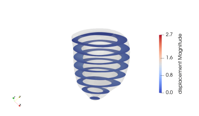 |
23 |
Framework for Characterizing in vivo Passive Myocardial
Stiffness using in vivo MRI
Fikunwa O Kolawole1,2,3,
Tyler E Cork1,2,4,
Michael Loecher1,2,
Vicky Y Wang1,2,
Ellen Kuhl3,
and Daniel B Ennis1,2
1Radiology, Stanford University, Stanford, CA, United States, 2Radiology, Veterans Administration Health Care System, Palo Alto, CA, United States, 3Mechanical Engineering, Stanford University, Stanford, CA, United States, 4Bioengineering, Stanford University, Stanford, CA, United States Keywords: Myocardium, In Silico, computational modeling; inverse finite element modeling As increased passive myocardial stiffness is implicated in the etiology of many cardiac diseases, its in vivo estimation can improve management of heart disease. MRI-driven computational constitutive modeling can be used to obtain subject-specific passive myocardial stiffness. We present a method for building an in silico model to estimate subject-specific passive myocardial stiffness by combining LV geometric data derived from cine bSSFP, regional kinematics extracted from tagged MRI, and myocardial microstructure measured using in vivo cDTI. This project aims to develop a clinically translatable in vivo passive myocardial stiffness evaluation framework by integrating cardiac MRI and computational modeling. |
|
4458.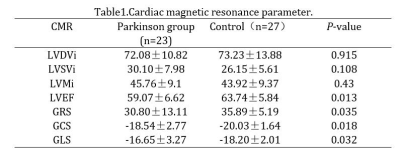 |
24 |
Preliminary Study of CMR to Evaluate Structural and Functional
Alterations of the Left Ventricle in Patients with Parkinson's
Disease
Xun Yue1,
Pengfei Peng1,
and Jiayu Sun1
1Radiology Department, West China Hospital, Sichuan University, Chengdu, China Keywords: Cardiomyopathy, Parkinson's Disease, Cardiac magnetic resonance,Cardiac function,Myocardial strain Parkinson's disease (PD) is the second most frequent neurodegenerative disease. Likewise, heart failure (HF) is one of the most common causes of mortality and morbidity in the world. The prevalence of HF has been found to be twice among PD patients compared with overall population regardless any other heart diseases and cardiovascular disease risk factors. Cardiac MRI is the clinically recognised gold standard for the assessment of ventricular functional parameters and myocardial histology. We have designed a study to assess the cardiac function and structure in patients with PD compared with a control group by means of cardiac magnetic resonance. |
|
4459.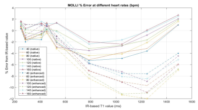 |
25 |
No separate acquisition timing scheme is necessary for the MOLLI
technique when used for the post contrast cardiac T1 mapping
Seonghwan Yee1
1Radiology, Massachusetts General Hospital, Boston, MA, United States Keywords: Heart, Relaxometry, MOLLI The modified Look-Locker imaging (MOLLI) technique, widely used in cardiac T1 mapping, requires specification of an acquisition timing scheme. For better quantification of wide range of T1 values, different acquisition schemes have been used for the prior- and post-contrast conditions. However, the strategy of having two different acquisition schemes might add complexity to the consistent management of clinical protocols, especially in a clinical environment with multiple MRI systems. Hence, a phantom experiment was performed to examine the necessity of this strategy. The results here suggest that having a separate acquisition scheme for the post-contrast condition would not be necessary. |
|
4460. |
26 |
Influence of Temporal Sampling on Reproducibility of Radiomics
Features in Cardiac Cine MRI
Ann Laube1,2,
Matthias Ivantsits1,
Markus Hüllebrand1,3,
Lennart Tautz3,
Léa Ter-Minassian4,
Hannu Zhang4,
Patrick Doeblin2,5,
Sebastian Kelle2,5,
and Anja Hennemuth1,2,3,6
1Institute for Computer-assisted Cardiovascular Medicine, Charité - Universitätsmedizin Berlin, Berlin, Germany, 2DZHK (German Centre for Cardiovascular Research), Partner Site Berlin, Berlin, Germany, 3Fraunhofer MEVIS, Bremen, Germany, 4Charité - Universitätsmedizin Berlin, Berlin, Germany, 5Deutsches Herzzentrum Berlin, Berlin, Germany, 6University Medical Center Hamburg-Eppendorf, Hamburg, Germany Keywords: Heart, Radiomics, Reproducibility Radiomics has been applied in cardiovascular imaging with promising results. Still, the effect of imaging parameters on the reproducibility of radiomics features needs to be well understood for these to be used in clinical diagnostics. We conduct a retrospective study on short-axis cine CMR images of healthy volunteers to assess the impact of different temporal sampling parameters on the reproducibility of shape, first-order, texture, and deformation field-based features, and contextualize the findings with inter- and intra-observer variability. We find that the reproducibility of features is dependent on temporal sampling, warranting the introduction of parameter standardization for radiomics analyses. |
|
4461. |
27 |
Intravoxel Incoherent Motion Diffusion Weighted MR Imaging to
Evaluate Myocardial Microvascular Dysfunction of Exertional Heat
Illness
Zhang Jun1,
Luo Song1,
and Dou Weiqiang2
12. Department of Radiology, Jinling Hospital, Medical School of Nanjing University, Nanjing, China, 24. MR Research, GE Healthcare, Beijing, China Keywords: Heart, Myocardium Purpose: To investigate the feasibility of assessing myocardial microvascular perfusion of patients with exertional heat illness (EHI) by intravoxel incoherent motion imaging. Methods: CMR-IVIM image quality was evaluated with a 3-point scale. CMR-IVIM derived parameters were analyzed and compared between EHI and HC. Receiver operating characteristic curve was used. Results: CMR-IVIM image quality were significantly improved. EHI patients showed decreased D*, decreased f values and higher D values compared to HC. f showed the most robust efficacy for detecting EHI related myocardial injury with the highest area under the curve. Conclusion: CMR-IVIM can evaluate myocardial injury of EHI patients. |
|
4462.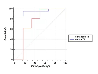 |
28 |
Cardiac MRI Quantitative Analysis of Native T1 and Enhanced T1
Maps in Patients with Acute Heart Failure
Yanhui Hao1,
Jianxin Guo2,
Yi Zhu3,
Rui Zhang1,
Lihong Chen2,
and Jiantao Liu4
1THE FIRST AFFILIATED OF XI'AN JIAOTONG UNIVERSITY, Xi'an, China, 2THE FIRST AFFILIATED OF XI'AN JIAOTONG UNIVERSITY, XI'an, China, 3Philips Healthcare, Beijing, China, 4Philips Healthcare, XI'an, China Keywords: Heart, Body The purpose of this study was to investigate the predictive value of native T1 and enhanced T1 in the diagnosis of acute heart failure. Using echocardiographic ejection fraction and clinical symptoms as reference standards, The statistical results of our data showed that the native T1 value was found to have high sensitivity and specificity in predicting acute heart failure. |
|
4463.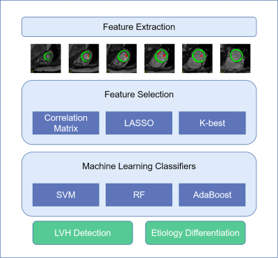 |
29 |
Papillary Muscle Derived Radiomic Features Facilitate LVH
Detection and Improve LVH Etiology Classification
Qifan Lu1,
Qiming Liu1,
Yezi Chai1,
and Meng Jiang1
1Renji Hospital,Shanghai Jiao Tong University School of Medicine, Shanghai, China Keywords: Cardiomyopathy, Radiomics Detecting left ventricular hypertrophy(LVH) is common in a clinical scenario, while a precise classification of LVH etiology is quite challenging. In this study, we applied radiomics methods to LVH detection and LVH etiology classification. Innovatively, we extracted radiomic features not only from the myocardium(MYO) but also includes features from the papillary muscle(PM). After strict feature selection and analysis, we found the incorporation of PM features improves our model performance compared with models that simply use MYO features. |
|
4464.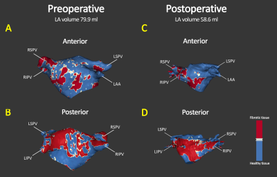 |
30 |
Impact of mitral valve repair on left atrial fibrosis in severe
mitral valve regurgitation patients: a 3D LGE pilot follow-up
study.
Sulayman el Mathari1,
Jolanda Kluin1,
Luuk Hopman2,
Pranav Bhagirath2,
Maurice Oudeman1,
Alexander Vonk1,
Steven Chamuleau2,
Robert Klautz1,3,
Aart Nederveen4,
Pim van Ooij4,
and Marco Götte2
1Cardiothoracic Surgery, AmsterdamUMC, Amsterdam, Netherlands, 2Cardiology, AmsterdamUMC, Amsterdam, Netherlands, 3Cardiothoracic Surgery, LUMC, Leiden, Netherlands, 4Radiology and Nuclear Medicine, AmsterdamUMC, Amsterdam, Netherlands Keywords: Valves, Valves, Fibrosis Timing of mitral valve repair (MVR) surgery in mitral valve regurgitation (MR) patients remains a subject of debate. Assessment of left atrial (LA) fibrosis might aid to improve clinical management of these patients. We assessed LA fibrosis extent using 3-dimensional late gadolinium enhanced cardiac magnetic resonance imaging prior to and after MVR surgery in MR patients (n=6). Our data demonstrates a decrease of LA volume, while a concomitant increase in LA fibrosis was observed. Further study is required to determine the potential relation between LA fibrosis, pre- and post-operative changes and long-term clinical outcome. |
|
4465.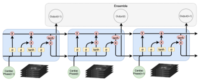 |
31 |
Deep Learning Algorithm Based on LTSM for Automated Bi-Ventricle
Segmentation of CMR Images
Lu Lin1,
Difei Jiang2,
Yueting Xiao2,
and Yining Wang1
1Department of Radiology, Peking Union Medical College Hospital, Chinese Academy of Medical Sciences and Peking Union Medical College, Beijing, China, 2Shukun (Beijing) Technology Co., Ltd, Beijing, China Keywords: Heart, Heart Cardiac Magnetic Resonance(CMR) imaging is an advanced cardiovascular imaging modality to evaluate cardiac structure and function. Therefore, the accuracy of segmentation directly affects the clinical evaluation and diagnosis. In this study, we used Long Short-Term Memory(LSTM) network, proposing a novel deep learning-based model for accurate automated bi-ventricle segmentation of CMR images. |
|
4466.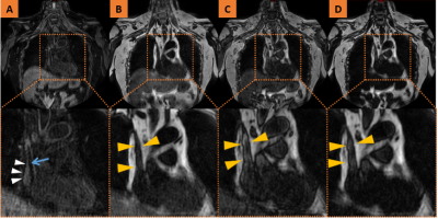 |
32 |
Magnetic Resonance Imaging of Central Vein in the diagnosis of
Stuck Hemodialysis Central Venous Catheter: A Case report
Zhe Jin1,
Ruimeng Yang1,
Yongzhou Xu2,
Peng Wu3,
Faith Zeng4,
and Xinhua Wei1
1Guangzhou First People's Hospital, Guangzhou, China, 2Philips Healthcare, Guangzhou, China, 3Philips Healthcare, Shanghai, China, 4Philips Healthcare, Nanchang, China Keywords: Vessels, Rare disease, Stuck Catheter;Central Venous;Atrial Laceration The stuck central venous catheter (CVC) results from fibrin sheaths that firmly adhere the catheter to the wall of the superior vena cava or right atrium. Here, we report a patient with stuck CVC. The patient underwent preoperative magnetic resonance imaging (MRI) of the central vein to remove the catheter and then suffered intraoperative cardiac arrest with a bedside ultrasound suggesting cardiac tamponade and considered a right atrial laceration. The patient was then transferred to ICU, where he was discharged over a week later with stable vital signs, pending further surgery. The preoperative MRI features were analyzed retrospectively. |
|
4467. |
33 |
Utility of single breath-hold compressed sensing cine imaging in
optimizing clinical cardiac magnetic resonance scanning
procedure
Fuyan Wang1,
Cailing Pu1,
and Hongjie Hu1
1Department of Radiology, Sir Run Run Shaw Hospital, Zhejiang University School of Medicine, Hangzhou, China Keywords: Quantitative Imaging, Cardiovascular, compressed sensing The main goal of this study is to evaluate the practicability of a single breath-hold whole-heart coverage compressed sensing cine sequence in daily CMR examination, moreover, to optimize the scanning procedure for patients with different kinds of cardiac dysfunction. For patients, especially those are diagnosed with chronic heart failure, the single breath-hold CS cine can substantially shorten scanning time and provide reliable cardiac function parameters comparing with routine bSSFP cine. This single breath-hold CS cine sequences is recommended to be used before contrast injection because of gadolinium agent induced reduction of end-systolic/diastolic volume and image contrast. |
|
4468.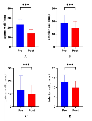 |
34 |
Myocardial changes evaluation based on CMR imaging in patients
with obstructive hypertrophic cardiomyopathy after Liwen
procedure
Minwen Zheng1 and
shuangxin li2
1Department of Radiology, Xijing Hospital, Fourth Military Medical University, Xian, China, 2Department of Radiology, Xijing Hospital, Fourth Military Medical University, xinan, China Keywords: Heart, Cardiovascular This study evaluated the structural, functional and tissue characteristic changes of myocardium in patients with hypertrophic obstructive cardiomyopathy after treated with the Liwen procedure using cardiac magnetic resonance (CMR) techniques. Compared with pre-operation, one year after treated with Liwen procedure (a new effective procedure), patients’ septal and left ventricular free wall thickness and left ventricular mass (LVM) were significantly reduced. There was no significant change in left ventricular ejection fraction (LVEF) (P>0.05). |
|
4469.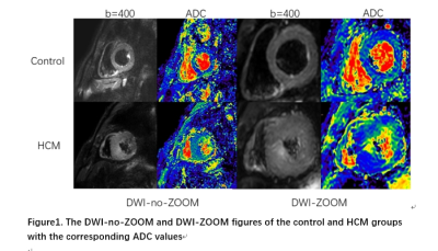 |
35 |
Comparison of cardiac DWI with and without ZOOM by using
second-order motion compensated spin echo for healthy control
and HCM patients
Jianmin Zheng1,
Yishi Wang2,
Jianxiu Lian2,
Yijun Li1,
Ping Tian1,
Ying Liu1,
and Minwen Zheng1
1Xijing Hospital, Fourth Military Medical University, Xi 'an, China, 2Philips Healthcare,, Beijing, China Keywords: Myocardium, Diffusion/other diffusion imaging techniques, Hypertrophic Cardiomyopathy(HCM) Hypertrophic cardiomyopathy (HCM) is a hereditary cardiomyopathy with a relatively high rate of sudden death. Diffusion-weighted imaging (DWI) is widely used for evaluating motion of water protons as one of the functional magnetic resonance imaging methods. Second-order motion-compensated spin echo (M2SE) has been applied for cardiac DWI, but there are few reports of M2SE combined with ZOOM for HCM. The results showed that both signal-to-noise ratio (SNR) and contrast-to-noise ratio (CNR) of ZOOM-M2SE were significantly higher than those of M2SE only, and the ADC value of hypertrophic cardiomyopathy was also significantly higher than that of healthy controls. Cardiac DWI using ZOOM-M2SE would be a more appropriate method for investigating the myocardial microenvironment in healthy and HCM subjects. |
|
4470.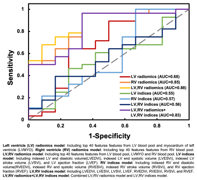 |
36 |
Radiomics-Based Classification of Severe Pulmonary Regurgitation
in Patients with Repaired Tetralogy of Fallot by Cardiac
Magnetic Resonance
Jo-Hua Peng1,
Ming-Ting Wu2,
Nai-Yu Pan3,
Teng-Yi Huang3,
Yi-Jui Liu4,
Ken-Pen Weng5,6,
and Hsu-Hsia Peng1
1Department of Biomedical Engineering and Environmental Sciences, National Tsing Hua University, Hsinchu, Taiwan, 2Department of Radiology, Kaohsiung Veterans General Hospital, Kaohsiung, Taiwan, 3Department of Electrical Engineering, National Taiwan University of Science and Technology, Taipei, Taiwan, 4Department of Automatic Control Engineering, Feng Chia University, Taichung, Taiwan, 5Department of Pediatrics, Kaohsiung Veterans General Hospital, Kaohsiung, Taiwan, 6Dpartment of Pediatrics, National Yang Ming Chiao Tung University, Taipei, Taiwan Keywords: Heart, Radiomics Cardiac magnetic resonance (CMR) radiomics is a novel technique for advanced cardiac image phenotyping. This study aimed to develop a radiomics-based classification model by CMR images and thereby to identify rTOF patients with severe pulmonary regurgitation (PR). In our results, the radiomics-based classification model can successfully identify rTOF patients with PR ≥25% by routine CMR cine images. The extracted features underlined the RV intracardiac flow alteration due to PR and the potential right ventricular remodeling in rTOF patients with severe PR. This radiomics-based information may be helpful in determining the appropriate timing for pulmonary valve replacement in the future. |
|
4471.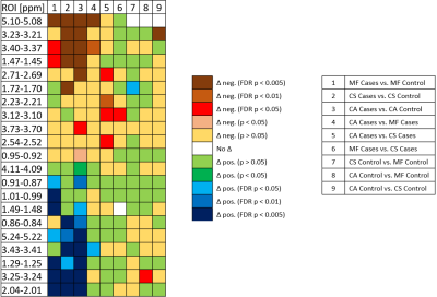 |
37 |
Using Magnetic Resonance Spectroscopy for Metabolomic
Characterization of Blood Serum in Patients with Myocardial
Fibrosis
Leo L. Cheng1,
Mark V. Füzesi1,2,
Xiaoyu Wang1,3,
Antonia Leist1,4,
Anna-Laura M. Hasubek1,4,
and Paul B. Yu5
1Molecular Pathology, Massachusetts General Hospital, Boston, MA, United States, 2Charité - Berlin University of Medicine, Berlin, Germany, 3Nanjing University, Nanjing, China, 4Julius-Maximilians-University of Würzburg, Würzburg, Germany, 5Cardiovascular Research Center, Massachusetts General Hospital, Boston, MA, United States Keywords: Myocardium, Spectroscopy Myocardial fibrosis plays a key role in the pathological remodeling of the diseased heart. Yet, there is currently no routine screening method for it. The gold standard, histological examination, is highly invasive. Other alternatives are either costly (e.g. PET-CT, MRI) or subjective (echocardiography). Using HRMAS MRS, we analyzed blood samples from subjects diagnosed with common myocardial fibrosis (MF), amyloidosis (CA), or sarcoidosis (CS), plus controls. The identified regions of interest (ROI) and principal components (PC) enabled us to distinguish between diseased and not-diseased, and between the different disease groups. This may contribute to earlier diagnosis and increased success of treatment. |
|
4472.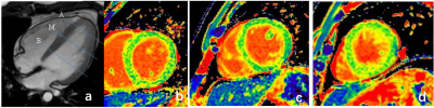 |
38 |
A priliminary evaluation of myocardialgadolinium deposition in
apical hypertrophic cardiomyopathy with T1mapping
Hongzhi Yang1,
shaoyu wang2,
and Ruwu Yang1
1Xi dian group hospital, Xi'an, China, 2DI MR SMK, Siemens Healthineers, Shanghai, China Keywords: Myocardium, Cardiovascular, Gd deposition In this study, we used the CMR T1mapping technique was to evaluate the correlation between T1R and gadolinium deposition in the myocardium of patients with apical hypertrophic cardiomyopathy after gadolinium enhancement. |
|
4473.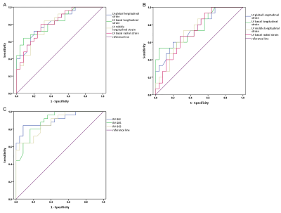 |
39 |
Early detection of left ventricular involvement in
arrhythmogenic right ventricular cardiomyopathy by Feature
Tracking
Xuan Li1 and
Jiang Wu1
1Shanxi Cardiovascular Hospital, Taiyuan, China Keywords: Cardiomyopathy, Quantitative Imaging This retrospective study aimed to detect left ventricular myocardial strain by CMR-FT in ARVC patients. The 25 ARVC patients were divided into 2 subgroups: the preserved LVEF group (LVEF ≥50%, n=15) and the reduced LVEF group (LVEF <50%, n=10). The biventricular global and regional myocardial strain parameters in the radial, circumferential, and longitudinal directions were compared between ARVC and control group. ROC curve analysis showed the left ventricular basal longitudinal strain exhibited good performance in differential diagnosis between the LVEF ≥50% ARVC and the control group. In conclusion, CMR-FT can early detect left ventricular myocardial motion changes in ARVC patients. |
|
4474.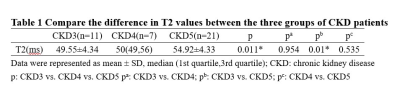 |
40 |
Cardiac magnetic resonance T2 mapping for evaluating the degree
of myocardial edema in patients with chronic kidney disease
Jingyu Zhang1,
Di Tian1,
Ziqi Xiong1,
Yifan He1,
and Zhiyong Li1
1The First Affiliated Hospital of Dalian Medical University, Dalian, China Keywords: Heart, Kidney T2 mapping is used to assess the degree of myocardial damage in CKD patients, and T2 values increase as the glomerular filtration rate decreases in CKD patients. |
|
 Back to Meeting Home
 Back to the Program-at-a-Glance
The International Society for Magnetic Resonance in Medicine is accredited by the Accreditation Council for Continuing Medical Education to provide continuing medical education for physicians.
 Back to Meeting Home
 Back to the Program-at-a-Glance
 View Presentation Video