Digital Poster
Parametric Mapping of the Heart II
ISMRM & ISMRT Annual Meeting & Exhibition • 03-08 June 2023 • Toronto, ON, Canada

Digital Poster
Parametric Mapping of the Heart II
| Computer # | |||
|---|---|---|---|
4130.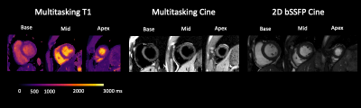 |
41 |
Free-breathing, ECG-Free, Myocardial T1 Mapping: Initial
Feasibility Experiments of Cardiac MR Multitasking on GE Systems
Xianglun Mao1,
Hsu-Lei Lee2,
Katerina Eyre3,
Gaspar Delso4,
Debiao Li2,5,
Anthony G Christodoulou2,5,
Matthias G Friedrich3,6,
Michael Salerno7,
and Martin A Janich8
1GE HealthCare, Menlo Park, CA, United States, 2Biomedical Imaging Research Institute, Cedars-Sinai Medical Center, Los Angeles, CA, United States, 3Department of Radiology, McGill University Health Centre, Montreal, QC, Canada, 4GE HealthCare, Barcelona, Spain, 5Department of Bioengineering, University of California in Los Angeles, Los Angeles, CA, United States, 6Area 19, Montreal, QC, Canada, 7Departments of Medicine and Radiology, Stanford University, Stanford, CA, United States, 8GE HealthCare, Munich, Germany Keywords: Myocardium, Quantitative Imaging, T1 Mapping Quantitative imaging biomarkers such as T1 are promising for assessing focal and diffuse myocardial pathologies across different sites and scanners. However, the lack of reliable T1 mapping methods across different scanner platforms reduces patient access to robust quantitative imaging. CMR Multitasking has shown promise for non-ECG and free-breathing quantitative imaging in the heart, primarily at 3T. This work is an initial feasibility study implementing the CMR Multitasking on GE 1.5T MR scanners. Radial trajectories, self-gating/training data, and the CMR Multitasking framework together provided co-registered T1 and cine maps in a 1-min, free-breathing, non-ECG CMR scan. |
|
4131.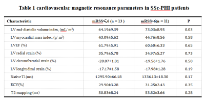 |
42 |
SSc primary heart involvement assessed by cardiovascular
magnetic resonance imaging and explore its relationship with the
skin involvement
Yanan Zhao1,
Jianing Cui1,
Xiuzheng Yue2,
and Tao Li1
1Department of Radiology, The First Medical Center of PLA General Hospital, Beijing, China, 2Philips Healthcare(Beijing), Beijing, China Keywords: Heart, Heart, SSc primary heart involvement, cardiovascular magnetic resonance,extracellular-volume fraction, modified Rodnan skin score Cardiac involvement is a significant cause of morbidity and mortality in subclinical systemic sclerosis (SSc). There are, however, no effective methods for detecting cardiac involvement in a general, asymptomatic SSc cohort. We aim to characterize SSc-primary heart involvement (SSc-pHI) through cardiovascular magnetic resonance (CMR), exploring the relationship between SSc-pHI and skin involvement by modified Rodnan skin score (mRSS). We found that 71% of SSC patients had Late gadolinium enhancement (LGE) fibrosis, and 75% and 46% had anomalous native T1 and extracellular-volume fraction (ECV). In addition, SSc-pHI is related to the severity of skin involvement. |
|
4132.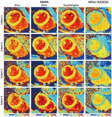 |
43 |
Improving SASHA T1 precision via a deep-learning approach
Xiaofeng Qian1,
Yingwei Fan1,
Dongyue Si2,
Yongsheng Jin3,
Haiyan Ding2,
and Rui Guo1
1Shool of Medical Technology, Beijing Institute of Technology, Beijing, China, 2Center for Biomedical Imaging Research, Department of Biomedical Engineering, School of Medicine, Tsinghua University, Beijing, China, 3Department of Infectious Diseases, The Affiliated Hospital of Yan’an University, Shanxi, China Keywords: Myocardium, Machine Learning/Artificial Intelligence, SASHA SASHA with a 3-parameter fitting model has high T1 accuracy but low precision due to low SNR in saturation-recovery T1-weighted images. Alternatively, a two-parameter model could improve the precision with the penalty of losing accuracy. In this study, we developed a 1D neural network (DeepFittingNet) to predict SASHA T1 and alleviate the impaction from noise. We trained DeepFittingNet using simulation of SASHA with different Rician noise levels and tested it using in-vivo MR data. Results showed that T1 from DeepFittingNet had high precision and comparable accuracy to that of the 3-parameter fitting. |
|
4133.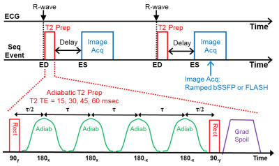 |
44 |
Improved 3T Adiabatic T2-Prep for Cardiac T2-Mapping
Ronald J. Beyers1 and
Thomas S. Denney1
1MRI Research Center, Auburn University, Auburn University, AL, United States Keywords: Myocardium, Cardiomyopathy, T2prep, Mapping, Adiabatic Cardiac T2-mapping allows quantifying myocardial edema resulting from events, such as, acute myocardial infarction, myocarditis and tako-tsubo cardiomyopathy. Many T2-mapping sequences exist in literature, however, most have design shortcuts that degrade their consistency, accuracy and prognostic value. We previously reported our sequence that T2-maps at end-systole (ES) for maximum wall thickness, minimized artifacts, and had four-point T2 curve-fits3. Here we present an improved 3T T2prep with four adiabatic pulses to eliminate prep inhomogeneity. This new 3T T2-mapping sequence produces T2maps at ES within 22 seconds breath hold. Pre-testing on phantoms and validation on healthy human volunteers demonstrated superior results. |
|
4134.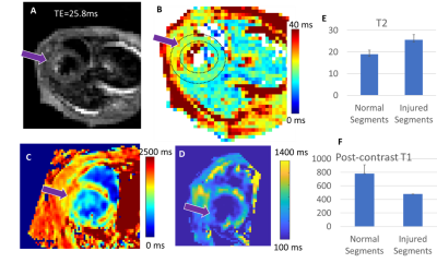 |
45 |
Cardiac MR evaluation of a myocarditis mouse model
Dana C Peters1,
Daniel Coman1,
Peter Herman1,
Jennifer M Kwan2,
Hanming Zhang2,
Raja Chakraborty2,
Jeacy Espinoza2,
Hyung Chun2,
Saejong Park2,
and Lauren A Baldassarre2
1Radiology and Biomedical Imaging, Yale University, New Haven, CT, United States, 2Cardiovascular Medicine, Yale University, New Haven, CT, United States Keywords: Inflammation, Contrast Mechanisms Tools were developed to evaluate mice models of myocarditis with cardiac MRI. Cine imaging with self-gating, T2 mapping using ECG-gated multi-spin echo acquisition, and pre- and post-contrast injection T1 mapping were employed. In twelve mice, including 4 mice models of myocarditis, normal T2 values were 19ms±2ms at 11.7T, and regions of higher T2 (25±2ms) were observed in some myocarditis mice. T1 mapping showed reduced T1 after injection of gadolinium, and regions of lower T1 in myocarditis vs. normal regions. These tools will be useful in improved characterization of myocarditis in mice. |
|
4135.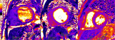 |
46 |
Dark right ventricular blood pool on myocardial T2 mapping in
dilated cardiomyopathy: a predictor of cardiac function
Yajuan Li1,
Hui Zhou1,
and Huiting Zhang2
1Department of Radiology, Xiangya Hospital Central South University, Changsha, Hunan, China, 2Scientific Marketing, Siemens Healthineers Ltd., Wuhan, Hubei, China Keywords: Cardiomyopathy, Cardiovascular Although the T2 mapping is mainly used to evaluate myocardial water content, it is also sensitive to blood oxygenation levels, we found that in dilated cardiomyopathy the right ventricular dark right ventricular blood pool may affect the accurate quantification of myocardial T2 values. This study investigated the relationship between right ventricular blood pool on myocardial T2 mapping and indicators of cardiac function. |
|
4136.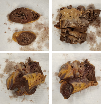 |
47 |
MRI relaxometry mapping of hearts extracted post-mortem from
acute COVID-19 patients
El-Sayed H. Ibrahim1,
Andrii Puzyrenko1,
and Ivor Benjamin1
1Medical College of Wisconsin, Milwaukee, WI, United States Keywords: Myocardium, COVID-19 Cardiac MRI is capable of myocardial tissue characterization using parametric mapping techniques such as T1 and T2 mappings, which are sensitive for detecting diffuse inflammation, fibrosis and edema. These techniques seem adequate for the detection of myocardial changes in COVID-19. In this study, we demonstrated the use of ex vivo MRI mapping techniques for noninvasive myocardial tissue characterization in COVID-19 patients and compared the MRI results to findings from autopsy reports. The proposed approach would allow for better understanding of COVID-19 pathophysiological effects on the myocardium through longitudinal studies that could be conducted without the need for invasive endomyocardial biopsy. |
|
4137.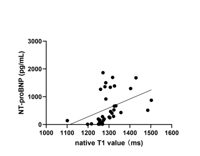 |
48 |
Relation of N-Terminal Pro–B-Type Natriuretic Peptide to Native
T1 at Cardiac MRI
Rui Zhang1,
Jianxin Guo2,
Lihong Chen2,
Yi Zhu3,
Yanhui Hao1,
and Kai Ai4
1THE FIRST AFFILIATED OF XI'AN JIAOTONG UNIVERSITY, XI'AN, China, 2THE FIRST AFFILIATED OF XI'AN JIAOTONG UNIVERSITY, XI'an, China, 3Philips Healthcare, Beijing, China, 4Philips Healthcare, Xi'an, China Keywords: Heart, Body The purpose of this study was to explore the relationship and closeness between N-Terminal pro–B-Type natriuretic peptide (NT-proBNP) and native T1, so as to predict the occurrence of heart failure(HF). This retrospective study included 43 participants. Native T1 value were measured on T1 mapping images. The result reflects native T1 values correlated positively with NT-proBNP, and had high sensitivity and specificity. Native T1 has high predictive performance for HF. |
|
4138.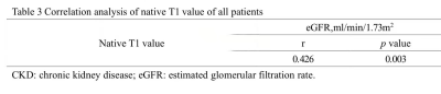 |
49 |
Cardiac magnetic resonance native T1 mapping assessed different
CKD stages, a polit study
Yating Chen1,
Di Tian1,
Jingyu Zhang1,
and Zhiyong Li1
1Department of radiology, The first affiliated hospital of dalian medical university, Dalian, China Keywords: Myocardium, Cardiovascular, Chronic kidney disease,T1mapping, cardiac magnetic resonance Chronic kidney disease (CKD) is a condition in which kidney damage or decreased kidney function persists for more than three months. Moreover, toxins inhibit myocardium leads to myocardial dysfunction and heart failure. T1 mapping, also known as longitudinal relaxation time quantitative imaging technology, is a new cardiac magnetic resonance (CMR) scanning technology, which can quantitatively evaluate myocardial T1 time and reflect changes in myocardial fibrosis.The purpose of this study is to analyze the difference and correlation between T1 value and CKD stage. |
|
4139.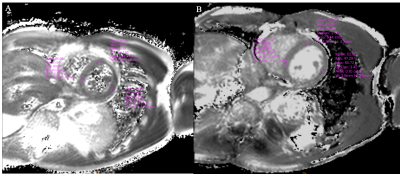 |
50 |
A preliminary comparison of multi-echo GraSE and T2
preparation-based myocardium T2 mapping
Yang Wu1,
Peng Sun2,
Zhigang Wu2,
Xiaoxiao Zhang2,
Jing Zhang2,
and Jiazheng Wang2
1Department of Medical Imaging, Wuhan Asia General Hosipital, Wuhan, China, 2Philips Healthcare, Beijing, China Keywords: Myocardium, Cardiovascular T2 mapping is a quantitative technique for measuring T2 in tissues, which is gaining popularity in the MR community, particularly for the detection of cardiac edema. There are several representative T2 mapping methods, including GraSE and T2 preparation-based schemes. However, there is still a lack of clinical experience with these T2 mapping methods. In this study, we tentatively compared the GraSE-based and T2 preparation-based methods for myocardium imaging. |
|
4140.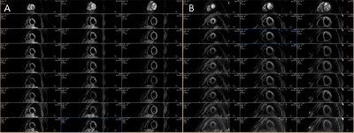 |
51 |
Evaluation of right ventricular myocardial microcirculation in
diabetes patients by using optimized intravoxel incoherent
motion imaging
Shilan Li1,
Jiahuan Tao1,
Jing Zhang1,
Kai Ai2,
and Jiantao Liu2
1Department of Magnetic Resonance, Lanzhou University Second Hospital, Lanzhou, China, 2Philips Healthcare, Xi’an, China Keywords: Myocardium, Diffusion/other diffusion imaging techniques, IVIM, diabetes, myocardial microcirculation, right ventricular myocardial Intra-voxel incoherent motion imaging (IVIM) can noninvasively and quantitatively evaluate myocardial microcirculation. At present, some scholars have demonstrated that IVIM can observe left ventricular myocardial microcirculation. However, due to the thickness of the right ventricular wall, IVIM imaging is difficult to assess it. This study proposed an optimized navigator-based and free-breathing acquisition scheme for cardiac IVIM. The results found that right ventricular myocardial microcirculation was impaired, pseudo-diffusion coefficient (Dfast) were significantly lower than healthy volunteers. It is helpful to monitor cardiovascular risk and evaluate prognosis. |
|
| 4141. | 52 |
The preliminary application of fully quantitative CMR myocardial
perfusion in ST elevation myocardial infarction
Shi-hai Zhao1 and
Wei-bo Chen2
1Radiology, Zhongshan Hospital Fudan University, Shanghai, China, 2Philips Healthcare, Shanghai, China Keywords: Myocardium, Perfusion Fully quantitative CMR-MPI was preliminarily applied for evaluating the area at risk in patients with acute ST elevation myocardial infarction. As a result, the fully quantitative CMR-MPI provided quantitative myocardial blood flow of the infarction territories, which was correlated with the extent of Salvageable zone. |
|
4142.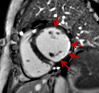 |
53 |
A clinical study using T1-mapping and ECV and late gadolinium
enhancement to detect early myocardium involoved in
rheumatologic diseases
Ying Liu1,
Ping Tian1,
Jianmin Zheng1,
and Minwen Zheng1
1Xijing Hospital, Xi'an, China Keywords: Myocardium, Quantitative Imaging Myocardial involvement is frequently observed in rheumatologic diseases but typically remains subclinical. This study aimed to detect early subclinical cardiac involvement and to investigate characteristics of myocardial involvement by cardiac MR (CMR) imaging. In 75 of 83 patients with rheumatologic diseases, elevated global native T1 and ECV values were observed. Furthermore, 51 patients showed positive segment distribution of late gadolinium enhancement, including subendocardial contrast, intramural contrast and transmural contrast. Thus, T1-mapping, ECV and late gadolinium enhancement could detect early myocardial involvement in rheumatologic diseases patients without overt LV dysfunction. |
|
4143.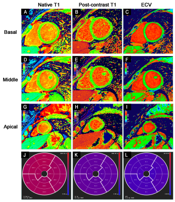 |
54 |
Association of CMR-verified diffuse myocardial fibrosis with
depressed myocardial strain in HFpEF
Yi Zhang1 and
Xiance Zhao2
1Shanghai General Hospital, Shang Hai, China, 2Philips Healthcare,, Shang Hai, China Keywords: Myocardium, Quantitative Imaging HFpEF was seen approximately in half of all hospitalized patients for heart failure and is associated with a poor prognosis. Using CMR, the present study shows that the increased diffuse myocardial fibrosis was associated with the degree of impaired myocardial strain in patients with HFpEF. As diffuse fibrosis is recognized as prognostic markers and reversible. Our findings demonstrate the utility of myocardial strain in assessing patients with HFpEF. The measurement of myocardial strain could be used to inform clinicians about whether HF medications could improve the degree of diffuse myocardial fibrosis in HFpEF. |
|
4144.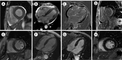 |
55 |
Predict late gadolinium enhancement using left ventricular
long-axis strain in multiple myeloma with secondary cardiac
amyloidosis patients
Mengyao Hu1,
Yipei Song1,
Ao Kan1,
Jiankun Dai2,
and Lianggeng Gong1
1the Second Affiliated Hospital of Nanchang University, Nanchang, China, 2GE Healthcare, MR Research China, Beijing, China Keywords: Cardiomyopathy, MR Value, Multiple myeloma; Cardiac amyloidosis; Long-axis strain The purpose of this study was to evaluate the function of left ventricular (LV) in multiple myeloma (MM) with secondary cardiac amyloidosis (MM-CA) patients by cardiac magnetic resonance feature tracking (CMR-FT), and investigate the role of simple and fast CMR-FT long-axis strain (LAS) analysis in predicting late gadolinium enhancement (LGE). The results showed the LV function was more impaired in enhanced (LGE+) than in no-enhanced (LGE-) MM-CA patients. Among the investigated CMR-FT parameters, the LV LAS presented with best prediction efficacy of LGE. Our study indicated the simple and fast LAS may be beneficial for MM-CA patients with renal failure. |
|
4145.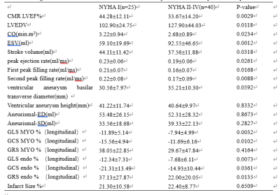 |
56 |
Global Longitudinal and Global Circumferential Strain by
Speckle-Tracking Echocardiography and Feature-Tracking CMRI:
Comparison with LVEF
Jing Liang1,
Dan Mu1,
Xiance Zhao2,
and Xiuzheng Yue3
1Department of Radiology, Affiliated Nanjing Drum Tower Hospital of Nanjing University Medical School, nanjing, China, 2Philips Healthcare, Shanghai, China, 3Philips Healthcare, beijing, China Keywords: Atherosclerosis, Atherosclerosis Understanding the relationship between left ventricular function and ventricular aneurysmafter myocardial infarction is crucial for surgical treatment and drug therapy. The CMR feature-tracking technique can quantitatively evaluate strain changes of LV and detect myocardial dysfunction. The study aims to use CMR-FT in patients with left VA after MI to assess strain changes and evaluate the clinical value for predicting patient prognosis. We found that the LV GLS, GCS, and GRS could be used for predicting the NYHA class after MI. |
|
4146.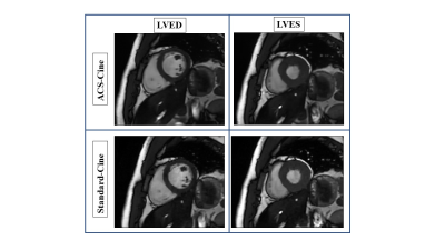 |
57 |
Applications of artificial intelligence-assisted compressed
sensing cardiac cine in left heart function imaging: Quality and
Efficiency
Zijing Zhang1,
Fei FENG1,
Yulong Qi1,
Hui Zhang1,
and Guanxun Cheng1
1Peking University Shenzhen Hospital, ShenZhen, China Keywords: Heart, Cardiomyopathy Cardiac magnetic resonance (CMR) imaging is the gold standard for non-invasive evaluation of cardiac structure and function. Conventional 2D segmented steady-state free precession cine imaging(Standrad-Cine)is widely used but the scanning time is long. Artificial intelligence-assisted compressed sensing(ACS)is a new technology that can reduce the scanning time and improve image quality. By comparing the image quality and left ventricular function (LVF) of CMR sequence based on Standrad-Cine and ACS-Cine, we found that the use of ACS-Cine can significantly reduce the scanning time while obtaining high-quality and accurate parameters images. |
|
4147.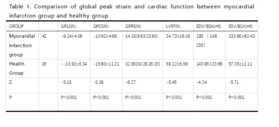 |
58 |
The predictive value of left ventricular strain analysis in
myocardial infarction with ventricular remodeling
Jing Chen1,
Xiaolin Mu2,
Tianyu Ke3,
and Yang Song2
1Graduate School of Dalian Medical University, Dalian Municipal Central Hospital, Dalian, China, 2The Affiliated Hospital of Dalian University of Technology, Dalian Municipal Central Hospital, Dalian, China, 3Graduate School of Dalian Medical University, Dalian Municipal Central Hospita, Dalian, China Keywords: Cardiomyopathy, Ischemia, CMR-feature tracking The subtle changes of myocardial strain in patients with myocardial infarction complicated with ventricular remodeling remain unclear. We used the CMR-FT technique to investigate the degree of strain damage, the degree of scar tissue penetration and the long-term prognostic value after ventricular remodeling in 41 patients with old myocardial infarction.The results showed that the myocardial strain was of great value in quantitatively assessing the degree of myocardial damage, identifying the degree of scar tissue penetration in infarcted myocardium and predicting the long-term end events, especially GLS, which was the best predictor |
|
| 4148. | 59 |
Developing and Evaluating a Chronic Ischemic Cardiomyopathy in
Swine Model by Rest and Stress CMR
baiyan zhuang1,
Minjie Lu2,
jian he3,
and jing xu2
1Department of Magnetic Resonance Imaging, Key Laboratory of Cardiovascular Imaging(cultivation) , Cardiovascular imaging and intervention Center, Fuwai Hospital, State Key Laboratory of Cardiovascular Disease, National Center for Cardiovascular Diseases, Chinese Academy of Medical Sciences and Peking Union Medical College, Beijing, China, 2Department of Magnetic Resonance Imaging, Key Laboratory of Cardiovascular Imaging(cultivation) , Cardiovascular imaging and intervention Center, Fuwai Hospital, State Key Laboratory of Cardiovascular Disease, National Center for Cardiovascular Diseases, Chinese Academy of Medical Sciences and Peking Union Medical College, beijing, China, 3Department of Magnetic Resonance Imaging, Key Laboratory of Cardiovascular Imaging(cultivation) , Cardiovascular imaging and intervention Center, Fuwai Hospital, State Key Laboratory of Cardiovascular Disease, National Center for Cardiovascular Diseases, Chinese Academy of Medical Sciences and Peking Union Medical College, beijjing, China Keywords: Myocardium, MR Value This preclinical study has established that chronic coronary artery stenosis can induce significant myocyte loss with modest global replacement fibrosis that leads to global LV dysfunction and varying degrees of congestive heart failure. |
|
 Back to Meeting Home
 Back to the Program-at-a-Glance
The International Society for Magnetic Resonance in Medicine is accredited by the Accreditation Council for Continuing Medical Education to provide continuing medical education for physicians.
 Back to Meeting Home
 Back to the Program-at-a-Glance
 View Presentation Video