Digital Poster
Brain Microstructure I
ISMRM & ISMRT Annual Meeting & Exhibition • 03-08 June 2023 • Toronto, ON, Canada

| Computer # | |||
|---|---|---|---|
4303.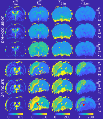 |
41 |
Multi-echo NODDI for the study of tissue compartmentalisation in
transient ischaemic stroke: initial insights
Ezequiel Farrher1,
Chia-Wen Chiang2,
Chang-Hoon Choi1,
Kuan-Hung Cho2,
Sheng-Min Huang2,
Ming-Jye Chen2,
Li-Wei Kuo2,3,
and N. Jon Shah1,4,5,6
1Institute of Neuroscience and Medicine 4, Forschungszentrum Jülich, Jülich, Germany, 2Institute of Biomedical Engineering and Nanomedicine, National Health Research Institutes, Miaoli, Taiwan, 3Institute of Medical Device and Imaging, National Taiwan University College of Medicine, Taipei, Taiwan, 4Department of Neurology, Faculty of Medicine, RWTH Aachen University, Aachen, Germany, 5JARA – BRAIN – Translational Medicine, RWTH Aachen University, Aachen, Germany, 6Institute of Neuroscience and Medicine – 11, Forschungszentrum Jülich, Jülich, Germany Keywords: Stroke, Diffusion/other diffusion imaging techniques, NODDI, ischemia, multi-echo, transverse relaxation, intra-neurite, extra-neurite Neurite orientation dispersion and density imaging (NODDI) has been broadly used in diffusion MRI for the characterisation of tissue microstructure in healthy ageing and the diseased brain. However, the compartment-specific volume fractions provided by NODDI suffer from echo-time (TE) dependence due to differences in the compartment-specific transverse relaxation times. The recently proposed multi-TE NODDI (MTE-NODDI) model provides both TE-independent compartmental volume fractions and compartment-specific transverse relaxation times. Here we aim to assess the benefits and limitations of using MTE-NODDI for studying the brain tissue microstructure affected by transient ischaemic stroke in MCAo animal models. |
|
4304.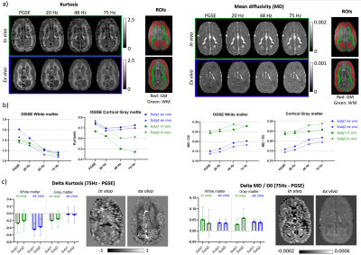 |
42 |
Evidence of axon beading and loss of extracellular fluid
following perfusion and fixation of the marmoset brain
Tales Santini1,
Naila Rahman1,
Alyson Shim1,
Matthew Budde2,
Stefan Everling1,
and Corey Baron1
1Western University, London, ON, Canada, 2Medical College of Wisconsin, Milwaukee, WI, United States Keywords: Microstructure, Microstructure Previous work showed that advanced diffusion metrics (OGSE and μFA) are sensitive to microstructural changes between in vivo and ex vivo marmoset brain tissues. Here, we utilized Monte Carlo simulations to investigate potential microstructure changes that could be occurring during tissue perfusion and fixation. A comparison between simulations and experiments showed that beading and decreased extracellular space are in agreement with the experimental trends observed. Changing microstructural properties during these processes has implications for studies that compare ex vivo tissue samples with in vivo findings. |
|
4305.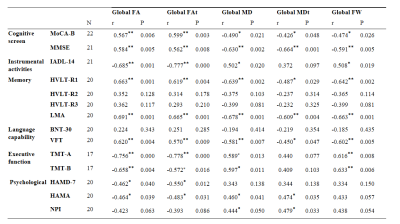 |
43 |
Free-water DTI and cognitive performance in patients with
intracranial vascular occlusion disease and transient ischemic
attack
Siyuan Fang1,
Jian Hai2,
Gaiying Li1,
Yupeng Wu1,
Weiwei Zhao1,
and Jianqi Li1
1Shanghai Key Laboratory of Magnetic Resonance, School of Physics and Electronic Science, East China Normal University, Shanghai, China, 2Department of Neurosurgery, Tong Ji Hospital, School of Medicine, Tong Ji University,, Shanghai, China Keywords: Data Analysis, Diffusion Tensor Imaging, free-water elimination DTI The objective of this study was to explore the relationship between cognitive impairment and free-water elimination DTI (FWE-DTI) metrics in patients with intracranial vascular occlusion disease (IVOD) and transient ischemic attack (TIA). 22 IVOD with TIA patients were tested with cognitive scales and scanned using a 3T echo-planar imaging sequence. The results show global FWE-DTI metrics were associated with patients' cognitive performance in some domains, which indicated the potential of FWE-DTI as new indices for assessing global brain changes in this disease. |
|
4306.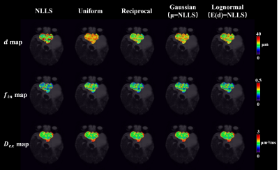 |
44 |
Improving microstructural estimation in time-dependent diffusion
MRI model with a Bayesian method
Kuiyuan Liu1,
Tianshu Zheng1,
Ruicheng Ba1,
Hongxi Zhang2,
and Dan Wu1
1Department of Biomedical Engineering, College of Biomedical Engineering & Instrument Science, Zhejiang University, Hangzhou, China, 2Department of Radiology, Children's Hospital, Zhejiang University School of Medicine, Hangzhou, China Keywords: Signal Modeling, Microstructure In this study, we proposed Bayesian estimation of tissue microstructures in td-dMRI model, and compared its performance with the traditional non-linear least square fitting method in simulation data and glioma patient data. We found that the performance of Bayesian fitting was dependent on the prior distribution and choice of hyperparameters, and the combination of Bayesian and least-square fitting could achieve reasonable performance without prior information. |
|
4307.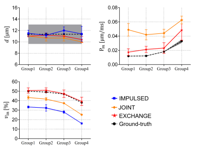 |
45 |
A general quantitative diffusion MRI model of water exchange,
confinement, and hindrance with arbitrary gradient waveform
encoding
Sisi Li1,
Diwei Shi2,
Xiaoyu Jiang3,4,
Li Chen2,
Quanshui Zheng2,
Hua Guo1,
and Junzhong Xu3,4,5,6
1Center for Biomedical Imaging Research, Tsinghua University, Beijing, China, 2Center for Nano and Micro Mechanics, Department of Engineering Mechanics, Tsinghua University, Beijing, China, 3Institute of Imaging Science, Vanderbilt University Medical Center, Nashville, TN, United States, 4Department of Radiology and Radiological Sciences, Vanderbilt University Medical Center, Nashville, TN, United States, 5Department of Biomedical Engineering, Vanderbilt University, Nashville, TN, United States, 6Department of Physics and Astronomy, Vanderbilt University, Nashville, TN, United States Keywords: Signal Modeling, Microstructure This study proposed and validated a general diffusion MRI biophysical model enabling simultaneous estimation of cell size, intracellular volume fraction, and transcytolemmal water exchange. The model introduces two-mode intracellular diffusion, corrects for restriction-induced “edge-enhancement” effect, and handles arbitrary diffusion gradient waveforms. The results of both in silico and in vitro experiments suggest this model not only improves the accuracy of estimated microstructural parameters such as cell size but also provides reasonable estimates of water exchange rate constant that is usually ignored previously. Such a comprehensive model may have potential to probe tumor status more precisely which is feasible in clinics. |
|
4308. |
46 |
Evaluation of reliability and self-consistency of compartment
size-distribution estimations by oscillating gradient spin-echo
sequences
Melisa L. Gimenez1,2,
Leonardo A. Pedraza Pérez1,2,
and Gonzalo A. Álvarez1,2,3
1Instituto Balseiro, CNEA, Universidad Nacional de Cuyo, San Carlos de Bariloche, Argentina, 2Centro Atómico Bariloche, CNEA, CONICET, San Carlos de Bariloche, Argentina, 3Instituto de Nanociencia y Nanotecnologia, CNEA, CONICET, San Carlos de Bariloche, Argentina Keywords: Microstructure, Quantitative Imaging Changes in tissue microstructure are promising biomarkers of neurological diseases. However, achieving robust and reliable, non-invasive diagnostic tools to estimate microstructural features is a major challenge. We perform a systematic analysis using Non-uniform Oscillating Gradient Spin Echo (NOGSE) sequences to estimate size distributions of white-matter phantoms. We evaluate the reliability and self-consistency of the inference method. We find that estimation of the distribution’s mode is very robust and reliable for sharp and smooth gradient modulations. These results contribute to developing reliable diagnostic tools based on quantitative images of tissue microstructure. |
|
4309. |
47 |
Mapping Myelin Volume Fraction using Multiple Echo Gradient Echo
and Dictionary Matching
Mert Şişman1,2,
Dominick J. Romano2,3,
Alexey V. Dimov2,
Ilhami Kovanlikaya2,
Pascal Spincemaille2,
Thanh D. Nguyen2,
and Yi Wang2,3
1Electrical and Computer Engineering, Cornell University, Ithaca, NY, United States, 2Department of Radiology, Weill Cornell Medicine, New York, NY, United States, 3Biomedical Engineering, Cornell University, Ithaca, NY, United States Keywords: Microstructure, White Matter Myelin Volume Fraction (MVF) is an important biomarker of demyelination various diseases Multiple Sclerosis. In this study, we propose a method that provides quantitative MVF maps from routine multiple echo gradient echo acquisitions and dictionary matching. The dictionary is generated using the Hollow Cylindrical Fiber Model (HCFM) and employs both the magnitude signal decay and QSM obtained from the mGRE phase. The obtained maps show both qualitative and quantitative superiority over the standard multiexponential fitting-based myelin water fraction (MWF) maps. |
|
4310.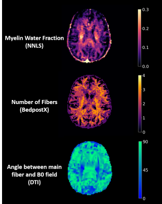 |
48 |
The effect of inter-individual differences and number of fiber
directions on possible orientation dependence of myelin water
imaging
Tigris Joseph1,2,
Sarah Morris1,2,3,
Shannon H. Kolind1,2,3,4,
Alex MacKay1,3,
Irene M. Vavasour2,3,
and Cornelia Laule1,2,3,5
1Physics and Astronomy, University of British Columbia, Vancouver, BC, Canada, 2International Collaboration on Repair Discoveries, University of British Columbia, Vancouver, BC, Canada, 3Radiology, University of British Columbia, Vancouver, BC, Canada, 4Medicine, University of British Columbia, Vancouver, BC, Canada, 5Pathology & Laboratory Medicine, University of British Columbia, Vancouver, BC, Canada Keywords: Microstructure, Relaxometry, myelin, white matter, orientation dependence, fiber direction, BedpostX, myelin water fraction, myelin water imaging Previous work suggests myelin water fraction (MWF) may be fibre orientation dependent. We compared MWF to fiber angle (from diffusion tensor imaging (DTI)) in white matter (WM) from 16 healthy participants using BedpostX to estimate the number of fiber directions. MWF vs. fiber angle graphs showed trends which varied between participants, suggesting that real myelination differences may be at least partly responsible for apparent orientation dependence of MWF. There were little obvious differences between trends with 1, 2, or 3+ fiber orientations in WM, suggesting number of fibers directions within a voxel does not impact orientation dependence. |
|
4311. |
49 |
A new qMRI approach for mapping microscopic water populations
and tissue relaxivity in the in-vivo human brain
Shir Filo1 and
Aviv A Mezer1
1The Edmond and Lily Safra Center for Brain Science, The Hebrew University of Jerusalem, Jerusalem, Israel Keywords: Microstructure, Magnetization transfer We present an array of new quantitative maps, highlighting different aspects of the tissue’s water. Furthermore, our technique allows to calculate the voxel-wise tissue relaxivity, associated with the molecular composition of the brain. Our approach is based on the pioneering two-site exchange model. We implemented it for magnetization-transfer weighted water content mapping of brain tissue for the first time. The new in-vivo MRI measurements conform well with the theory. They can be easily acquired with the standard quantitative MRI protocol and do not require complex data fitting. Therefore, the proposed technique may further advance human brain research and diagnosis. |
|
4312.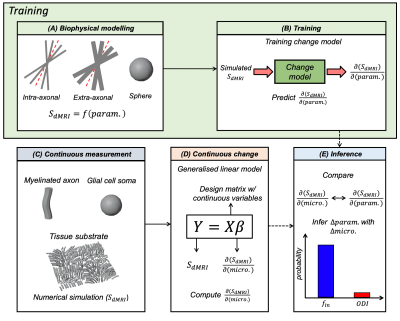 |
50 |
Parameter inference using continuous change in degenerate
biophysical diffusion models
Daniel Z.L. Kor1,
Hossein Rafipoor1,
Michiel Cottaar1,
Saad Jbabdi1,
Karla L. Miller1,
and Amy F.D. Howard1
1Wellcome Centre for Integrative Neuroimaging, FMRIB, University of Oxford, Oxford, United Kingdom Keywords: Diffusion/other diffusion imaging techniques, Microstructure, biophysical diffusion modelling Biophysical modelling of diffusion MRI (dMRI) may elucidate key microstructural features. However, most models include many input parameters, making simultaneous estimations of all parameters ill-posed. To overcome this, the recently published Bayesian framework EstimatioN for CHange (BENCH) characterises changes (variation) in parameters across multiple measurements/samples, rather than inferring the actual parameters from a single measurement/sample. BENCH has been previously applied to understand group-wise changes (e.g., patients vs. controls) in biophysical parameters. Here, we adapted BENCH to interpret situations of continuous change and validate its behaviour using synthetic dMRI data from numerical simulations. |
|
4313.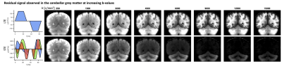 |
51 |
Diffusion MRI with spherical tensor encoding at high b-values
reveals cerebellar grey matter abnormalities in movement
disorders
Chantal Tax1,2,
Sila Genc3,
Claire L MacIver1,4,
Markus Nilsson5,
Mark Wardle6,
Filip Szczepankiewicz5,
Derek K Jones1,
and Kathryn Peall4
1CUBRIC, Cardiff University, Cardiff, United Kingdom, 2Image Sciences Institute, University Medical Center Utrecht, Utrecht, Netherlands, 3Department of Neurosurgery, The Royal Children's Hospital, Parkville, Australia, 4Neuroscience and Mental Health Research Institute, Division of Psychological Medicine and Clinical Neurosciences, Cardiff University, Cardiff, United Kingdom, 5Diagnostic Radiology, Clinical Sciences Lund, Lund Unversity, Lund, Sweden, 6Cardiff and Vale University Health Board, University Hospital of Wales Cardiff, Cardiff, United Kingdom Keywords: Diffusion/other diffusion imaging techniques, Microstructure, Movement disorders Most work on understanding movement disorder pathophysiology has focused on grey/white matter volumetric (macrostructural) and white matter microstructural effects, limiting understanding of frequently implicated grey matter microstructural differences. Using ultra-strong diffusion MRI with spherical tensor encoding, a persistent MRI signal was seen in healthy cerebellar grey matter at ultra-high diffusion-weightings. This work quantifies the proportion of this signal, previously ascertained to originate from small spherical spaces, in a clinical cohort, including patients with diagnosed movement disorders where the cerebellum has been implicated in symptom pathophysiology. Significant differences were found in individuals diagnosed with SCA6 and dystonia compared to age-matched controls. |
|
4314.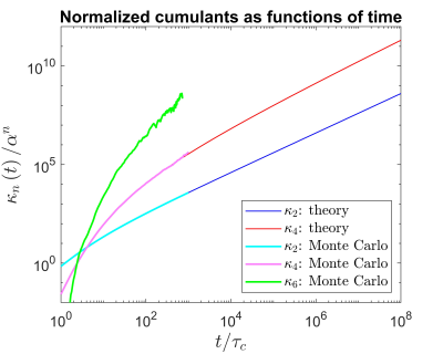 |
52 |
Twinkle, twinkle, T2*, still we wonder what you are
Pippa Storey1 and
Dmitry S. Novikov1
1Department of Radiology, New York University School of Medicine, New York, NY, United States Keywords: Microstructure, Relaxometry, T2* decay In the absence of macroscopic magnetic field inhomogeneity, the decay of gradient-echo signals is generally observed to be approximately monoexponential, with time constant $$$T_2^*$$$. The monoexponential behavior is typically attributed to diffusion, which averages out the dephasing of spins due to microstructural sources of magnetic susceptibility. The rationale is that, over times long enough for spins to have explored the microstructure, the cumulants of the spins’ phase distribution should increase linearly with time. By means of a simple counterexample, we demonstrate that this assumption is incorrect, thereby calling into question the mechanism underlying the monoexponential decay of gradient-echo signals. |
|
4315.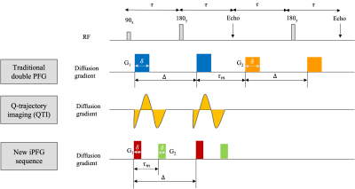 |
53 |
High Resolution In Vivo Diffusion Tensor Distribution MRI of the
Whole Human Brain with 300 mT/m Gradients
Kulam Najmudeen Magdoom1,2,
Alexandru V. Avram1,2,
Dario Gasbarra3,
Thomas Witzel4,
Susie Yi Huang4,
and Peter J Basser2
1The Henry M. Jackson Foundation for the Advancement of Military Medicine (HJF) Inc., Bethesda, MD, United States, 2National Institute of Health, Bethesda, MD, United States, 3University of Helsinki, Helsinki, Finland, 4Athinoula A. Martinos Center for Biomedical Imaging, Massachusetts General Hospital, Charlestown, MA, United States Keywords: Diffusion/other diffusion imaging techniques, Microstructure, Multiple diffusion encoding Whole brain DTD MRI was performed at 1.2 mm isotropic resolution in-vivo using 300 mT/m gradients. An efficient and easy to implement interfused-PFG (iPFG) sequence was used to acquire multiple diffusion encoded images. Size, shape and orientation heterogeneity are measured and mapped from the estimated DTD along with a new form of tractography. The results show varying heterogeneity in different areas of the cerebral cortex, and subcortical and white matter regions. DTD tractography captured complex fiber configurations absent in DTI tractography. |
|
4316.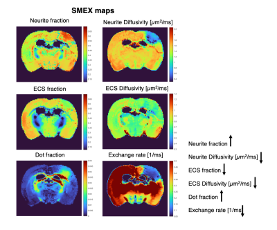 |
54 |
Mapping biophysical changes in stroked tissue via the Standard
Model with Exchange (SMEX) diffusion model
Rita Alves1,
Rafael Neto Henriques1,
Jonas Olesen2,3,
Sune Nørhøj Jespersen2,3,
and Noam Shemesh1
1Champalimaud Research, Champalimaud Foundation, Lisbon, Portugal, 2Center of Functionally Integrative Neuroscience (CFIN) and MINDLab, Department of Clinical Medicine, Aarhus University, Aarhus, Denmark, 3Department of Physics and Astronomy, Aarhus University, Aarhus, Denmark Keywords: Diffusion/other diffusion imaging techniques, Microstructure, Exchange Apparent diffusion coefficient (ADC) contrasts in stroke are still debated over 30 years after their discovery, mainly because of the nonspecific nature of ADC. Here, we harness Standard Model with Exchange (SMEX) measurements in ex-vivo stroked brains, to investigate how tissue microstructure is changed with ischemia. SMEX provides insight into neurite density, diffusivity and – importantly – exchange between neurite and extracellular environments. Using an extensive dataset, we find changes in neurite density and diffusivity that are consistent with neurite beading effects, which were further confirmed histologically. Dramatic reductions in exchange rates in the ischemic core were also observed. |
|
4317.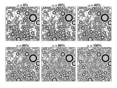 |
55 |
On the practical influence of extra-axonal heterogeneity in a
numerically informed model of extra-axonal water diffusion
Kevin D Harkins1,2,3,
Junzhong Xu1,2,
John C Gore1,2,3,
and Mark D Does2,3
1Radiology and Radiological Sciences, Vanderbilt University Medical Center, Nashville, TN, United States, 2Institute of Imaging Science, Vanderbilt University Medical Center, Nashville, TN, United States, 3Biomedical Engineering, Vanderbilt University, Nashville, TN, United States Keywords: Simulations, Microstructure This study explores the role of extra-axonal environment on the time dependent diffusion coefficient of extra-axonal water in white matter. Monte Carlo simulations of water diffusion in geometries with varying extra-axonal heterogeneity were compared with a proposed analytic model. The results agree with previously published power-law relationships that depend on structural order, but also uncovers trends between model parameters with characteristics of extra-axonal space. |
|
4318.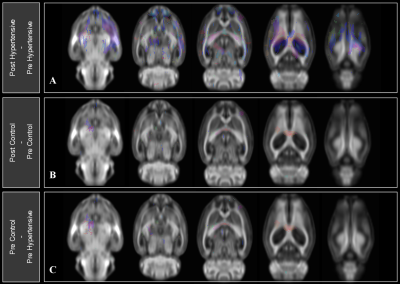 |
56 |
MULTI-SHELL DIFFUSION MRI TO INVESTIGATE THE EFFECTS OF
HYPERTENSION ON RAT BRAIN
Haley Elizabeth Wiskoski1,2,
Loi Do1,
Marc Zempare3,
Natalie Carey3,
Amy Delmendray3,
Kimberly Young3,
Kimberly Bohne3,
Monica Chawla3,
Pradyumna Bharadwaj4,
Kenneth Mitchell5,
Gene Alexander3,4,6,
Carol Barnes3,4,
and Theodore Trouard1,3,7
1Department of Biomedical Engineering, The University of Arizona, Tucson, AZ, United States, 2James C. Wyant College of Optical Sciences, The University of Arizona, Tucson, AZ, United States, 3Evelyn F. McKnight Brain Institute, The University of Arizona, Tucson, AZ, United States, 4Department of Psychology, Neurology, and Neuroscience, The University of Arizona, Tucson, AZ, United States, 5Health Sciences Center, Tulane University, New Orleans, LA, United States, 6Division of Neural Systems, Memory, and Aging, The University of Arizona, Tucson, AZ, United States, 7Department of Medical Imaging, The University of Arizona, Tucson, AZ, United States Keywords: Microstructure, Diffusion/other diffusion imaging techniques, DTI, fixels, Tractography & Fibre Modeling, Software Tools, Hypertension Hypertension is associated with an increased risk of cardiovascular disease and cognitive decline in aging humans. This study investigated longitudinal effects of induced HTN in regional and microstructural neuroanatomy of F344 rats using noninvasive diffusion-weighted MRI and fixel-based analysis. Single-shot spin-echo EPI along 64 diffusion directions and three shells (b=1000, 2000, and 3000 s/mm2) was performed. Microstructural changes in the brain appeared in certain regions after 10 weeks of hypertension, yet, most regions remained unaffected, contrasting peripheral organs which showed dramatic fibrosis due to the HTN. This likely demonstrates a robust protective mechanism of the central nervous system. |
|
4319.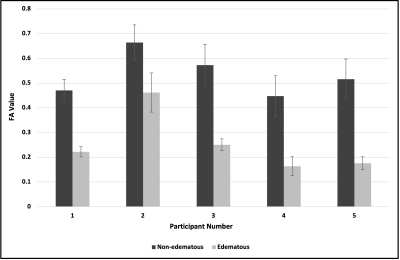 |
57 |
NODDI-based identification of white matter tracts in regions of
peritumoral edema: a validation study in brains bearing
meningioma tumors.
Sasha Hakhu1,
Jennapher Lingo VanGilder1,
Lucas Paulson1,
Leland Hu2,
Yuxiang Zhou2,
Kurt Schilling3,
and Scott Beeman1
1School of Biological and Health Systems Engineering, Arizona State University, Tempe, AZ, United States, 2Mayo Clinic, Scottsdale, AZ, United States, 3Vanderbilt University, Nashville, TN, United States Keywords: White Matter, Diffusion/other diffusion imaging techniques White matter (WM) tract detection proximal to brain tumors, surrounded by vasogenic edema during tumor resection is critical. Previous work has shown that NODDI presents a more accurate quantification of WM in edema as compared to DTI. However, these studies included gliomas and metastases, which are known to infiltrate WM. Here, we focus on validating the use of the NODDI model for WM identification in edema only in non-invasive meningiomas. Our results showed that DTI-derived FA of WM near edematous regions were lower than contralateral WM in non-edematous regions; in contrast, NODDI-derived ODI values for the same regions remained comparable. |
|
4320.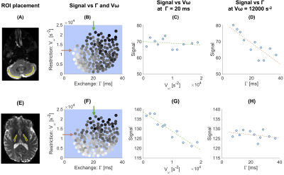 |
58 |
Probing restriction and exchange in the human brain using free
waveforms on a high-performance gradient system
Arthur Chakwizira1,
Ante Zhu2,
Thomas Foo2,
Carl-Fredrik Westin3,
Filip Szczepankiewicz1,
and Markus Nilsson4
1Medical Radiation Physics, Lund, Lund University, Lund, Sweden, 2GE Research, Niskayuna, New York, NY, United States, 3Department of Radiology, Brigham and Women's Hospital, Harvard Medical School, Boston, MA, United States, 4Clinical Sciences Lund, Lund University, Lund, Sweden Keywords: Diffusion/other diffusion imaging techniques, Microstructure Monitoring time dependence with diffusion MRI provides observables sensitive to restricted diffusion and exchange. Probing these phenomena by simply varying the diffusion time is a challenge because they have opposite effects on the diffusion-weighted signal and may cancel each other. A theoretical framework was recently proposed for disentangling the effects using free gradient waveforms. Here we explore the potential of the approach for neuroimaging in vivo using a high-performance gradient system. Results demonstrate unprecedented ability to disentangle the effects of restriction and exchange. Maps of the exchange rate show plausible contrast in both cortical and subcortical regions of the brain. |
|
4321.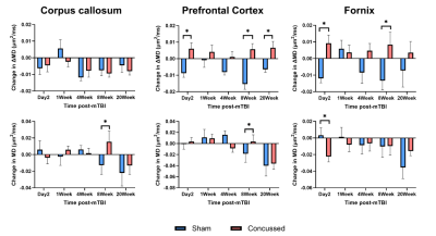 |
59 |
Evolution of Microstructural Changes in a Mouse Model of Mild
Traumatic Brain Injury
Naila Rahman1,
Kathy Xu1,
Matthew Budde2,
Arthur Brown1,
and Corey Baron1
1Robarts Research Institute, London, ON, Canada, 2Medical College of Wisconsin, Milwaukee, WI, United States Keywords: Diffusion/other diffusion imaging techniques, Microstructure, Brain Injury Single mild concussive impacts remain sparsely explored by in vivo neuroimaging techniques. Multimodal microstructural MRI has shown increased sensitivity and specificity to microstructural changes in various disease and injury models. In this work, we apply oscillating gradient spin-echo (OGSE) diffusion MRI, microscopic anisotropy (µA) diffusion MRI, and magnetization transfer (MT) MRI longitudinally to increase sensitivity to smaller spatial scales, disentangle fiber orientation dispersion from true microstructural changes, and acquire myelin sensitivity, respectively. We demonstrate that multimodal microstructural MRI provides sensitivity to evolving changes following a mild impact in both acute and chronic regimes, with OGSE demonstrating higher sensitivity than µA. |
|
4322.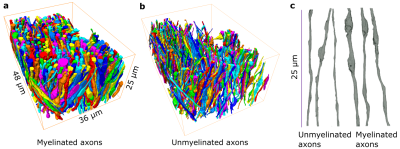 |
60 |
Characterization of white matter myelinated and unmyelinated
axons from diffusion MRI perspective
Ali Abdollahzadeh1,
Ricardo Coronado-Leija1,
Subah Mehrin1,
Hong-Hsi Lee2,
Alejandra Sierra3,
Els Fieremans1,
and Dmitry S. Novikov1
1Radiology, New York University School of Medicine, New York, NY, United States, 2Radiology, Harvard Medical School, Boston, MA, United States, 3A.I. Virtanen Institute for Molecular Sciences, University of Eastern Finland, Kuopio, Finland Keywords: Diffusion/other diffusion imaging techniques, Simulations, Time-dependence diffusion We segment all myelinated and unmyelinated axons from electron microscopy (EM) images of brain white matter (WM) of a mouse and a rat and quantify their cross-sectional morphology. We provide the exact relation between the long-time limit of the along-axon diffusion coefficients and the geometry of segmented intra-axonal spaces (IAS). By performing Monte Carlo (MC) simulations of diffusion in the IASs at varying diffusion times, we establish the time-dependence of diffusion along segmented myelinated and unmyelinated axons and validate the relation between axonal geometry and the long-time behavior of the longitudinal diffusion coefficient. |
|
 Back to Meeting Home
 Back to the Program-at-a-Glance
The International Society for Magnetic Resonance in Medicine is accredited by the Accreditation Council for Continuing Medical Education to provide continuing medical education for physicians.
 Back to Meeting Home
 Back to the Program-at-a-Glance
 View Presentation Video