Digital Poster
Brain Microstructure II
ISMRM & ISMRT Annual Meeting & Exhibition • 03-08 June 2023 • Toronto, ON, Canada

| Computer # | |||
|---|---|---|---|
4475.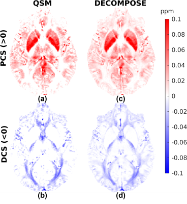 |
41 |
RECOMPOSE – Reproducing DECOMPOSE Using Susceptibility Maps
Acquired for Clinical Research
Patrick Fuchs1,
Jingjia Chen2,3,
Oliver C Kiersnowski1,
Russell Murdoch1,
Chunlei Liu2,3,
and Karin Shmueli1
1Medical Physics and Biomedical Engineering, University College London, London, United Kingdom, 2Electrical Engineering and Computer Sciences, University of California, Berkeley, Berkeley, CA, United States, 3Helen Wills Neuroscience Institute, University of California, Berkeley, Berkeley, CA, United States Keywords: Susceptibility, Quantitative Susceptibility mapping Here, we reproduced the results of the DECOMPOSE susceptibility separation model using new $$$T_2^*$$$-weighted data independently acquired using three clinically applicable sequences and processed with different QSM pipelines. This allowed us to investigate the sensitivity of DECOMPOSE to various dipole inversion algorithms. Good susceptibility source separation results were achieved using a 5-echo GRE acquisition, but maps of diamagnetic and paramagnetic sources from a highly accelerated 5-echo EPI sequence were noisy. When the input susceptibility maps exhibited artefacts, these were exacerbated by DECOMPOSE. Care must be taken not to lose local structural information when using (highly) regularised input susceptibility maps. |
|
4476.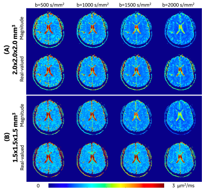 |
42 |
Evaluating High-Resolution High-b-value OGSE Time-dependent
Diffusion in Human Brain at Ultra-high-gradient 3.0T
Ante Zhu1,
Nastaren Abad1,
Robert Y Shih2,3,
Raymond Huang4,
Chitresh Bhushan1,
Tim Sprenger5,
Jennifer A McNab6,
J Kevin DeMarco2,3,
Vincent Ho2,3,
and Thomas K.F. Foo1,2
1GE Research, Niskayuna, NY, United States, 2Uniformed Services University of the Health Sciences, Bethesda, MD, United States, 3Walter Reed National Military Medical Center, Bethesda, MD, United States, 4Brigham and Women's Hospital, Boston, MA, United States, 5GE Healthcare, Stockholm, Sweden, 6Stanford University, Palo Alto, CA, United States Keywords: Microstructure, Diffusion/other diffusion imaging techniques Oscillating gradient spin echo (OGSE) time-dependent diffusion MRI has been shown to improve assessment of intra-cranial disease progression including glioma and multiple sclerosis. In human brain studies, however, high-resolution and high-b-value OGSE diffusion measurements suffer from low signal-to-noise ratio and Rician noise bias. In our preliminary studies with ultra-high-performance gradient 3.0T system, apparent diffusivity coefficient (ADC) of white matter at OGSE 35 Hz showed lower values when ADC was estimated from high b-values ≥1000 s/mm2 with 1.5-mm isotropic resolution, due to Rician noise bias. The effect of Rician noise bias on OGSE ADC measurement was reduced by using real-valued signals. |
|
4477.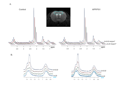 |
43 |
Investigating changes in brain lactate compartmentation using
diffusion-weighted MRS in APP/PS1 mice
Sophie Malaquin1,
Eloïse Mougel1,
Rodrigo Lerchundi1,
and Julien Valette1
1Université Paris-Saclay, CEA, CNRS, MIRCen, Laboratoire des Maladies Neurodégénératives, Fontenay-aux-Roses, France Keywords: Diffusion/other diffusion imaging techniques, Microstructure Diffusion-weighted MR spectroscopy yields information about the microstructure of the environment where brain metabolites are diffusing. Here, we measure the diffusion of two purely intracellular metabolites (astrocytic myo-inositol and neuronal N-acetyl aspartate) and of lactate, which is also present in the extracellular space, in the cortex of 12-month-old APP/PS1 and control mice. While astrocytic and neuronal microstructures appear unchanged in APP/PS1 mice, lactate diffusion appears slower. Modeling DW-signals suggests decreased extracellular lactate fraction. |
|
4478.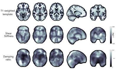 |
44 |
Estimating brain tissue stiffness from cardiac-induced 7T MRI
displacement measurements
Marius Burman Ingeberg1,
Elijah van Houten2,
and Jaco Zwanenburg1
1UMC Utrecht, Utrecht, Netherlands, 2University of Sherbrooke, Sherbrooke, QC, Canada Keywords: Image Reconstruction, Brain, Shear Stiffness, Tissue properties, Elastography, DENSE, 7T MRI Mechanical properties of the human brain can be altered by a variety of pathologies. These properties can be estimated from in vivo MRI measurements of brain tissue displacements as induced by the heartbeat. Previously obtained 7T MRI displacements measurements were used to reconstruct stiffness parameters using a subzone-based non-linear inversion scheme. Various structures of the brain can be observed in the reconstructed parameter distributions, and the results show good test-retest reliability. The preliminary results show promise of this approach to yield non-invasive assessment of brain tissue microstructure as a tool to investigate brain disease. |
|
4479.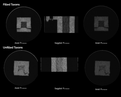 |
45 |
Diffusion spectrum imaging at 7T for probing Restricted
compartment of Textile-based Phantom
Sudhir Kumar Pathak1,
Yijen Wu2,3,
Tony Zuccolotto4,
and Walter Schneider1,5,6,7,8
1Learning Research and Development Center, University of Pittsburgh, Pittsburgh, PA, United States, 2Department of Developmental Biology, University of Pittsburgh, Pittsburgh, PA, United States, 3Rangos Research Center Animal Imaging Core, University of Pittsburgh, Pittsburgh, PA, United States, 4Psychology Software Tools, Pittsburgh, PA, United States, 5Psychology, University of Pittsburgh, Pittsburgh, PA, United States, 6Bioengineering, University of Pittsburgh, Pittsburgh, PA, United States, 7Neurosurgery, University of Pittsburgh, Pittsburgh, PA, United States, 8Radiology, University of Pittsburgh, Pittsburgh, PA, United States Keywords: Phantoms, Quantitative Imaging, Diffusion MRI, microstructural imaging Validating mathematical models that describe the diffusion in the restricted compartment in biological tissue is an active research area. Techniques like NODDI, SMT, and CHARMED models claim to quantify restricted, hindered, and free water compartment but lacks validation on a typical clinical scanner. We present a 3D-printed phantom that uses (non)-water-filled textile-based hollow fiber with a 0.9μ diameter to validate restricted and hindered compartments. This study used a DSI-based scanning protocol and reconstruction method (GDSI and RDI) to probe the restricted compartment at a 2-6μ length scale. Scalar metrics derived from reconstruction methods show a significant difference between water-(un)filled taxons. |
|
4480.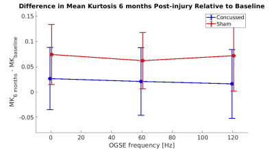 |
46 |
Investigating Diffusion Time Dependent Kurtosis Evolution in
Repeated Mild Traumatic Brain Injury in Mice
Jake Hamilton1,2,
Naila Rahman1,2,
Kathy Xu3,
Arthur Brown3,4,
and Corey Baron1,2
1Department of Medical Biophysics, Schulich School of Medicine and Dentistry, University of Western Ontario, London, ON, Canada, 2Centre for Functional and Metabolic Mapping (CFMM), Robarts Research Institute, University of Western Ontario, London, ON, Canada, 3Translational Neuroscience Group, Robarts Research Institute, Schulich School of Medicine and Dentistry, University of Western Ontario, London, ON, Canada, 4Department of Anatomy and Cell Biology, University of Western Ontario, London, ON, Canada Keywords: Traumatic brain injury, Microstructure, mild Traumatic Brain Injury (mTBI); Concussion Probing the diffusion time dependence of diffusional kurtosis within brain microstructure is being recognized as a valuable method to study various neurological pathologies, however, its use in the study of repeated mild traumatic brain injury (mTBI) has not been explored. In this work, we investigated differences in the time-dependence of diffusional kurtosis in injured and sham mice using oscillating gradient spin echo (OGSE) diffusion kurtosis imaging (DKI). The results of this work show promising differences in kurtosis within the hippocampus of injured and sham mice, illustrating the sensitivity of OGSE DKI in detecting long-lasting pathological microstructural changes following brain injury. |
|
4481.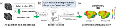 |
47 |
Best Response Constraint Generative Adversarial Network for
Diffusion MRI-based Estimation of Cortical micro-Architecture
Tianjia Zhu1,2,
Minhui Ouyang1,3,
Xuan Liu4,
Risheng Liu4,
and Hao Huang1,3
1Department of Radiology, Children's Hospital of Philadelphia, Philadelphia, PA, United States, 2Department of Bioengineering, University of Pennsylvania, Philadelphia, PA, United States, 3Department of Radiology, University of Pennsylvania, Philadelphia, PA, United States, 4School of Information Science and Engineering, Dalian University of Technology, Dalian, China Keywords: Diffusion/other diffusion imaging techniques, Microstructure, machine learning/artificial intelligence, neuro Advanced diffusion MRI (dMRI) has enabled noninvasive assessment of conventional cortical histological measures. However, analytical models are limited by their restrictive model assumptions and lack of validation from quantitative histology. We have developed a Diffusion-MRI based Estimation of Cortical micro-Architecture (DECAM) method using a novel deep learning technique Best Response Constraint Generative Adversarial Network (BRC-GAN) for accurately estimating cortical soma density (SD) leveraging rich dMRI data information. By providing high-fidelity, reproducible whole-brain estimated SD maps validated with histology, DECAM paves the way for data-driven noninvasive virtual histology for potential applications such as Alzheimer’s diseases. |
|
4482.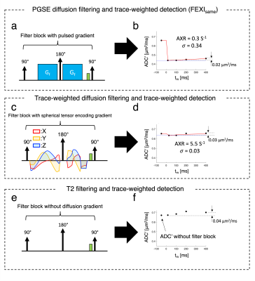 |
48 |
Diffusion filters, T2-filters, diffusion tensor component
diffusivities, and compartmental diffusivities. What is
happening in FEXI?
Hyeong-Geol Shin1,2,
Xu Li1,2,
Hye-Young Heo1,
Linda Knutsson1,2,3,
Filip Szczepankiewicz3,
Markus Nilsson4,
and Peter van Zijl1,2
1Department of Radiology and Radiological Science, Johns Hopkins University School of Medicine, Baltimore, MD, United States, 2F.M. Kirby Research Center for Functional Brain Imaging, Kennedy Krieger Research Institute, Baltimore, MD, United States, 3Department of Medical Radiation Physics, Lund University, Lund, Sweden, 4Deparment of Radiology, Clinical Sciences Lund, Lund University, Lund, Sweden Keywords: Diffusion/other diffusion imaging techniques, Diffusion/other diffusion imaging techniques, FEXI, trans-membrane exchange We explored how compartmental anisotropy affects filter-exchange imaging (FEXI) data in white matter by applying different types of tissue water magnetization filters, including multi-orientation and spherical tensor encoded diffusion filters, as well as a T2 filter. The results show that, depending on the mutual orientation of the filter and detection gradient pairs relative to fiber orientation, filtering efficiencies can be positive or negative. We conclude that the fast compartment reduced during diffusional filtering need not be extracellular, but can also reflect intra-axonal signal losses with effect size determined by the diffusion tensor components perpendicular and parallel to the fibers. |
|
4483.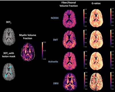 |
49 |
G-ratio Calculation in Multiple Sclerosis: The Impact of
Diffusion Modelling for Axonal and Fiber Volume Fraction
Determination
Tigris Joseph1,2,
Shannon H. Kolind1,2,3,4,
Guojun Zhao3,
Peng Sun5,
Robert Carruthers3,
Alice Schabas3,
Ana-Luiza Sayao3,
Virginia Devonshire3,
Roger Tam4,6,
G. R. Wayne Moore2,3,7,
David K. B. Li3,4,
Sheng-Kwei Song8,
Anthony Traboulsee3,
Irene M. Vavasour2,4,
and Cornelia Laule1,2,4,7
1Physics and Astronomy, University of British Columbia, Vancouver, BC, Canada, 2International Collaboration on Repair Discoveries, University of British Columbia, Vancouver, BC, Canada, 3Medicine, University of British Columbia, Vancouver, BC, Canada, 4Radiology, University of British Columbia, Vancouver, BC, Canada, 5Imaging Physics, MD Anderson Cancer Center, Houston, TX, United States, 6School of Biomedical Engineering, University of British Columbia, Vancouver, BC, Canada, 7Pathology & Laboratory Medicine, University of British Columbia, Vancouver, BC, Canada, 8Radiology, Washington University, St. Louis, MO, United States Keywords: Multiple Sclerosis, White Matter, myelin, axons, g-ratio, brain, microstructure, lesions, normal appearing white matter, multiple sclerosis G-ratio is the ratio of the inner axonal diameter to the total outer diameter, including myelin. MRI-derived g-ratios in multiple sclerosis (MS) may convey microstructural tissue abnormalities. G-ratios were estimated using myelin water fraction scaled to myelin volume fraction, and axonal metrics from Diffusion Basis Spectrum Imaging (DBSI), Neurite Orientation Dispersion and Density Imaging (NODDI), Spherical Mean Technique (SMT), and ActiveAx for axon/fiber volume fraction in 122 MS patients. DBSI and ActiveAx derived g-ratios were higher in lesions than normal appearing white matter (NAWM), reflecting MS pathology. NODDI and SMT derived g-ratios were unexpectedly lower in lesions than NAWM. |
|
4484.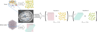 |
50 |
A multi-compartment fingerprinting model for non-invasive tumor
cell characterization via diffusion MRI
Manon Dausort1,
Nicolas Delinte1,2,
Quentin Dessain1,
Colin Vanden Bulcke1,2,
and Benoit Macq1
1ICTEAM, UCLouvain, Louvain-la-neuve, Belgium, 2IoNS, UCLouvain, Louvain-la-neuve, Belgium Keywords: Microstructure, Tissue Characterization Brain tumor tissue characteristics are important for treatment planning but are nowadays often recovered via invasive biopsy analysis. This work attempts to characterize the microstructural properties of tumor cells using diffusion MRI and Monte Carlo simulations to build a dictionary composed of several fingerprints, combining both a representation of axonal fibers and tumor cells. We demonstrate the use of our method on in-vivo brain data as a reliable estimation of tumor cell properties. |
|
4485.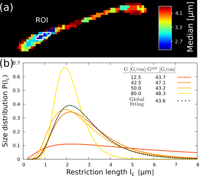 |
51 |
Optimization method for estimating tissue microstructure
size-distributions with diffusion weighted imaging
Pablo Javier Jimenez1,2,
Analia Zwick1,2,3,
and Gonzalo Alvarez1,2,3
1Centro Atómico Bariloche, CONICET, CNEA, Bariloche, Argentina, San Carlos de Bariloche, Argentina, 2Instituto Balseiro, CNEA, Universidad Nacional de Cuyo, Bariloche, Argentina, San Carlos de Bariloche, Argentina, 3Instituto de Nanociencia y Nanotecnologia,CNEA, CONICET, Bariloche, Argentina, San Carlos de Bariloche, Argentina Keywords: Diffusion/other diffusion imaging techniques, Microstructure Characterization of microstructures in living-tissues by non-invasive imaging is one of the key and outstanding challenges for diagnosing early stages of pathologies and understanding disease mechanisms. Here we implement an optimal control strategy for obtaining microstructure details by Diffusion-Weighted Imaging sequences. We estimate microstructure parameters of size-distribution models for axon bundles, attaining the ultimate precision limits predicted by quantum-information tools. We performed proof-of-principle experiments of the optimization protocol with an ex-vivo mouse-brain contrasted with simulations. These results might drastically reduce the total acquisition time compared to the present state-of-the-art, opening alternative avenues towards unraveling diagnostic information by quantitative MRI. |
|
4486.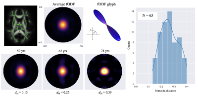 |
52 |
Inter-subject variability in fiber orientation density function
increases with aging
Hunter Moss1,
Andreana Benitez2,
and Jens Jensen1
1Neuroscience, Medical University of South Carolina, Charleston, SC, United States, 2Neurology, Medical University of South Carolina, Charleston, SC, United States Keywords: Tractography & Fibre Modelling, Microstructure The fiber orientation density function (fODF) estimated from diffusion MRI is typically used as an input for fiber tractography or microstructural modeling. However, since it encodes the fine details of the intra-voxel distribution of axon orientations, it can also be regarded as a quantity of physical interest that is potentially sensitive to subtle white matter changes. Here we demonstrate a method for quantifying inter-subject fODF differences in individual voxels within a white matter skeleton for a cohort of healthy older adults. We find that inter-subject fODF variability increases with age suggesting that diverse processes affect axons in healthy aging. |
|
4487.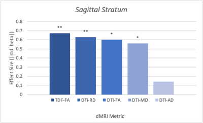 |
53 |
Assessing Sex Differences in the Brain’s White Matter
Microstructure during Development using the Tensor Distribution
Function
Sebastian M. Benavidez1,
Katherine E. Lawrence1,
Emily Laltoo1,
James T. McCracken2,
and Paul M. Thompson1
1Imaging Genetics Center, Mark and Mary Stevens Neuroimaging & Informatics Institute, University of Southern California, Marina del Rey, CA, United States, 2Department of Psychiatry and Biobehavioral Sciences, University of California Los Angeles, Los Angeles, CA, United States Keywords: Signal Modeling, Microstructure Diffusion magnetic resonance imaging (dMRI) can detect sex differences in developmental populations, as shown previously using the conventional diffusion tensor imaging (DTI) model. However, DTI cannot model complex brain fiber orientations, which the advanced tensor distribution function (TDF) model does. Here we compared the two models’ sensitivity in detecting sex differences, with the goal of improving our understanding of WM sex differences during development. We discovered WM microstructure sex effects, with the TDF model detecting more regions with significant differences and yielding larger effect sizes. Our results suggest that TDF is better able to detect developmental sex effects on WM. |
|
4488.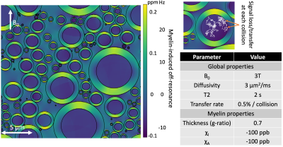 |
54 |
Modelling transverse relaxometry using myelin-induced
off-resonance fields and magnetisation transfer
Michiel Cottaar1 and
Saad Jbabdi1
1Wellcome Center for Integrative Neuroimaging (WIN), University of Oxford, Oxford, United Kingdom Keywords: Microstructure, Microstructure We present a new Monte Carlo MR simulator, which in addition to simulating the random diffusion of water restricted by any obstacles also simulates (1) the off-resonance field due to myelin and (2) signal loss due to magnetisation transfer. We use this simulator to study the interaction of diffusion, relaxation, and magnetisation transfer. We simulate gradient and spin echo measurements in white matter. The myelin off-resonance field is found to greatly reduce T2* due to dephasing of the signal between compartments, but barely affects T2. Magnetisation transfer at tissue boundaries explains T2 differences between intra-, extra-axonal, and myelin water. |
|
4489.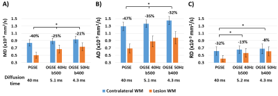 |
55 |
Exploring the oscillating gradient waveform frequency dependence
of diffusivity changes in human acute ischemic stroke
Mi Zhou1,
Robert Stobbe1,
Brian Buck2,
Mahesh Kate2,
Paige Fairall2,
Derek Emery3,
Thorsten Feiweier4,
and Christian Beaulieu1
1Biomedical Engineering, University of Alberta, Edmonton, AB, Canada, 2Neurology, University of Alberta, Edmonton, AB, Canada, 3Radiology and Diagnostic Imaging, University of Alberta, Edmonton, AB, Canada, 4Siemens Healthcare GmbH, Erlangen, Germany Keywords: Diffusion/other diffusion imaging techniques, Microstructure Two previous oscillating-gradient-spin-echo (OGSE) studies of human acute ischemic stroke using different diffusion waveforms (40 Hz, one-period; 50 Hz, two-period) have reported considerably different mean diffusivity (MD) changes in lesion white matter relative to long diffusion time (“0 Hz”) pulsed-gradient-spin-echo (PGSE). Here both OGSE waveforms were acquired in 8 stroke patients. Lesions showed marked diffusion time dependencies with lesion MD reduction of 40% for PGSE compared to 25% for OGSE 40Hz, and yet OGSE 50Hz was only a bit less (21%). Large heterogeneity between patients (+6% to -43% of OGSE 50Hz MD lesion changes) may explain earlier study differences. |
|
4490.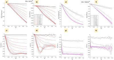 |
56 |
Evaluating frequency offsets due to local dephasing in
magnetically inhomogeneous tissue
Ross Shaw1,
Richard Bowtell1,
and Penny Gowland1
1Physics, University of Nottingham, Nottingham, United Kingdom Keywords: Simulations, Microstructure Through simulation the frequency offsets due to signal dephasing around biologically relevant local regions of high magnetic susceptibility are explored. Monte Carlo simulations are used to characterise the frequency offsets produced by spherical inclusions of varying radius and susceptibility in the presence of diffusion. The results indicate that heterogeneity of non-haeme iron concentration produce $$$\delta\Omega R^{2}/D$$$ values associated with local frequency offsets of up to $$$ -0.053 \gamma B_{0}\chi_{av}$$$: such offsets would be a confound for QSM measurements that merits further investigation. |
|
4491.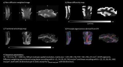 |
57 |
Investigating Multi-Dimensional Diffusion-Weighted MRI for
High-Resolution Post-Mortem Brain Acquisitions at 9.4 T
Hannah L. Gerbeth1,2,
Michael Paquette1,
Carsten Jäger3,4,
Patricia Wenk5,
Eike Budinger5,
Markus Morawski3,4,
and Cornelius Eichner1
1Department of Neuropsychology, Max Planck Institute for Human Cognitive and Brain Sciences, Leipzig, Germany, 2Translational Molecular Imaging, German Cancer Research Centre, Heidelberg, Germany, 3Department of Neurophysics, Max Planck Institute for Human Cognitive and Brain Sciences, Leipzig, Germany, 4Paul Flechsig Institute - Center of Neuropathology and Brain Research, Medical Faculty, University of Leipzig, Leipzig, Germany, 5Combinatorial Neuroimaging Core Facility, Leibniz Institute for Neurobiology, Magdeburg, Germany Keywords: Diffusion/other diffusion imaging techniques, Diffusion/other diffusion imaging techniques, ex vivo, human, brain Diffusion-weighted MRI (dMRI) is a widely used tool to non-invasively study the microstructure of the human brain. However, conventionally employed diffusion tensor imaging (DTI) applications only provide limited information. In contrast, Multi-Dimensional Diffusion Imaging, MD-dMRI, aims to provide a framework for further discrimination between highly complex biological structures. In this context, post-mortem MD-dMRI data play an important role to validate in vivo imaging results. Because the fixation process modifies tissue properties, adapted and optimized acquisition methods are required. Here, we investigate the feasibility to acquire and analyse high-resolution MD-dMRI data from a post-mortem brain tissue sample. |
|
4492.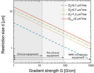 |
58 |
Optimization of non-uniform oscillating gradient spin echo
sequences for selective microstructure-size imaging
Milena Capiglioni1,2,3,
Analía Zwick1,2,4,
and Gonzalo A. Álvarez1,2,4
1Instituto Balseiro, CNEA, Universidad Nacional de Cuyo, Bariloche, Argentina, 2Centro Atómico Bariloche, CONICET, CNEA, Bariloche, Argentina, 3Support Center for Advanced Neuroimaging (SCAN), University of Bern, Bern, Switzerland, 4Instituto de Nanociencia y Nanotecnologia, CONICET, CNEA, Bariloche, Argentina Keywords: Diffusion/other diffusion imaging techniques, New Signal Preparation Schemes, NOGSE, Axon Diameter Diffusion-weighting imaging is a promising method for quantitative imaging of tissue microstructure features that may identify early stages of pathologies. Non-uniform oscillating gradient spin-echo (NOGSE) sequences are a novel technique to study diffusion spectra and quantitatively characterize tissue. The sequence contrasts the signal between two modulating gradient frequencies to filter the signal originating from molecules diffusion in specific restriction sizes. We determine the range of control parameters that maximizes NOGSE contrast for filtering the restriction sizes. We assess the restriction sizes that this sequence can filter with current technologies. |
|
4493.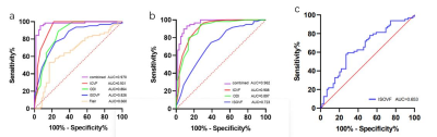 |
59 |
The Value of Neurite Orientation Dispersion and Density MR
Imaging in Diagnosing Temporal Lobe Epilepsy with Hippocampal
Sclerosis
Xiaonan Zhang1,
Guohua Zhao1,
Huiting Zhang2,
Eryuan Gao3,
and Jingliang Cheng1
1The First Affiliated Hospital of Zhengzhou University, Zhengzhou, China, 2MR Scientific Marketing, Siemens Healthineers Ltd., Wuhan, China, 3The First Affiliated Hospital of Zhengzhou University,, Zhengzhou, China Keywords: Quantitative Imaging, Brain The hippocampal microstructural alterations by using routine magnetic resonance imaging presents a challenge. This study aimed to evaluate the performance of the NODDI models in temporal lobe epilepsy (TLE) patients with hippocampal sclerosis (HS) by comparison with the routine Flair sequence. Our results found that all NODDI parameters had significant differences between ipsilateral HS and contralateral HS/HC, and had better diagnostic performance than Flair sequence. In addition, combined NODDI model had significant better diagnostic performance than all the single parameters. In conclusion, NODDI is superior to Flair image in diagnosing TLE with HS. |
|
4494.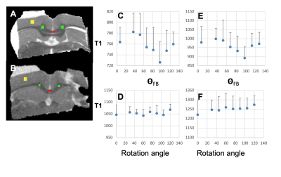 |
60 |
T1 Relaxation Anisotropy in White Matter is Directly Linked to
the Axon Fibre Orientation in the Magnetic Field
Risto Kauppinen1,
Jeromy Thotland2,
Henri P.P. Leskinen3,
Pramod K. Pisharady2,
Eppu Manninen3,
Mikko Kettunen3,
Christophe Lenglet2,
Olli H.J. Grohn3,
Michael Garwood2,
and Mikko J. Nissi3
1University of Bristol, Bristol, United Kingdom, 2University of Minnesota, Minneapolis, MN, United States, 3University of Eastern Finland, Kuopio, Finland Keywords: Relaxometry, Contrast Mechanisms Interrelationships between T1 relaxation time and axon fibre orientation in midsagittal corpus callosum (CC) were studied in human brain in vivo at 3T and 7T as well as in a rat brain ex vivo at 9.4T. It was observed that in the same CC tracts in vivo the change in T1 results from altered fibre orientation with respect to B0. In the rat brain, ex vivo rotation of a given midsagittal CC ROI produced angular T1 plots that match those observed in vivo. Thus, axon fibre orientation is directly linked to T1 relaxation anisotropy. |
|
 Back to Meeting Home
 Back to the Program-at-a-Glance
The International Society for Magnetic Resonance in Medicine is accredited by the Accreditation Council for Continuing Medical Education to provide continuing medical education for physicians.
 Back to Meeting Home
 Back to the Program-at-a-Glance
 View Presentation Video