Digital Poster
High- & Low-Field fMRI
ISMRM & ISMRT Annual Meeting & Exhibition • 03-08 June 2023 • Toronto, ON, Canada

| Computer # | |||
|---|---|---|---|
3663.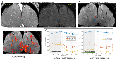 |
81 |
Measuring individual vein and artery BOLD responses to visual
stimuli in humans with multi-echo single-vessel functional MRI
at 7T
Divya Varadarajan1,2,
Paul Wighton1,2,
Jingyuan Chen1,2,
Sebastien Proulx1,2,
Robert Frost1,2,
Andre van der Kouwe1,2,
Avery Berman3,
and Jonathan R. Polimeni1,2
1Athinoula A. Martinos Center for Biomedical Imaging, Charlestown, MA, United States, 2Harvard Medical School, Boston, MA, United States, 3Carleton University, Ottawa, ON, Canada Keywords: fMRI, Blood vessels Here we apply single-vessel fMRI techniques to evaluate BOLD and non-BOLD contributions within individual arteries and veins. First we identified individual vessels from inflow effects in a small set of slices, and distinguished arteries and veins on the basis of their T2* decay. Then we applied a high-inplane-resolution multi-echo single-vessel fMRI approach to examine fMRI responses within these vessels. We find evidence for both BOLD and potentially non-BOLD responses within arteries, suggesting that in some cases BOLD may capture arterial responses BOLD which may be more neuronally specific (in space and time) than effects from downstream veins. |
|
3664.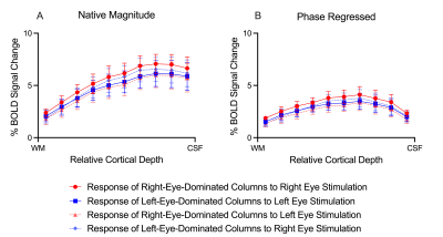 |
82 |
Ocular Dominance Columns Examined using BOLD fMRI with Phase
Regression at 7 T
Brett Liem1,2,
Atena Akbari1,2,
Joseph S Gati1,
Peter Zeman1,
and Ravi S Menon1,2
1Centre for Functional and Metabolic Mapping, Robarts Research Institute, London, ON, Canada, 2Medical Biophysics, The University of Western Ontario, London, ON, Canada Keywords: fMRI (task based), High-Field MRI, Phase Regression We investigated the signal spatial specificity of the GRE-BOLD contrast with and without phase regression in ocular dominance columns and layers of human V1. Phase regression is a post-processing technique that uses phase data to suppress the large vein signal, thus improving the GRE signal spatial specificity. Our results showed that the phase regressed laminar BOLD signal profile peaks towards the middle cortical depth, while that of GRE-BOLD was biased towards the cortical surface. Phase regression did not improve the contrast between columns, suggesting that the phase is not sensitive to intracortical veins running parallel to the cortical surface. |
|
3665.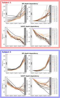 |
83 |
Dependence of the EPI and bSSFP resting-state fMRI signals on
the cortical orientation relative to B0: Initial observations at
9.4 Tesla
Dana Ramadan1,2,
Jonas Bause1,
Rüdiger Stirnberg3,
Philipp Ehses3,
and Klaus Scheffler1,4
1Department for High-field Magnetic Resonance, Max Planck Institute for Biological Cybernetics Tübingen, Tübingen, Germany, 2Graduate Training Centre of Neuroscience, University of Tübingen, Tübingen, Germany, 3German Center for Neurodegenerative Diseases (DZNE), Bonn, Germany, 4Department of Biomedical Magnetic Resonance, University of Tübingen, Tübingen, Germany Keywords: fMRI, High-Field MRI, Blood Vessels, Brain The spatial specificity of fMRI with GRE-EPI is significantly affected by large draining veins on the cortical surface. They cause susceptibility changes that are strongest when the veins are oriented perpendicular to B0. Since they follow the cortical curvature, their orientation to B0 can be approximated by the cortical orientation. Previous studies have shown a high dependence of the GRE-EPI signal on the cortical orientation, reflecting its disadvantage of being sensitive to these veins. In this preliminary two-subject study, this phenomenon is investigated with GRE-EPI and bSSFP in different cortical depths to experimentally explore the sensitivity of bSSFP to microvasculature. |
|
3666.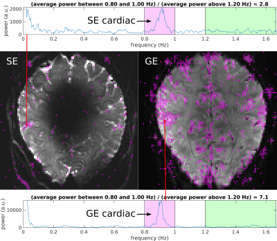 |
84 |
Application of SLR pulses to short-TR spin-echo fMRI at 7T: SNR
considerations and a direct demonstration of reduced cardiac
noise
Mukund Balasubramanian1,2,
Avery J. L. Berman3,4,
Robert V. Mulkern1,2,
Lawrence L. Wald1,5,
William A. Grissom6,
and Jonathan R. Polimeni1,5,7
1Harvard Medical School, Boston, MA, United States, 2Boston Children's Hospital, Boston, MA, United States, 3Carleton University, Ottawa, ON, Canada, 4University of Ottawa Institute of Mental Health Research, Ottawa, ON, Canada, 5Athinoula A. Martinos Center for Biomedical Imaging, Massachusetts General Hospital, Charlestown, MA, United States, 6Institute of Imaging Science, Vanderbilt University, Nashville, TN, United States, 7Harvard-MIT Division of Health Sciences and Technology, Massachusetts Institute of Technology, Cambridge, MA, United States Keywords: fMRI, RF Pulse Design & Fields, Ultra-High-Field MRI We show that SLR pulses provide much better slice profiles (and thus much higher SNR) than standard sinc pulses for short-TR spin-echo (SE) acquisitions, even when there is substantial B1+ inhomogeneity. As one application, we used SLR pulses and a TR of 300 ms in SE-EPI acquisitions at 7T, enabling the “direct” measurement of cardiac frequencies at ~1 Hz without aliasing. Far less cardiac fluctuation was seen in SE- versus GE-EPI data. While SE-fMRI is known to have reduced macrovascular weighting and thus improved spatial specificity relative to GE-fMRI, our results suggest that it may also have better temporal specificity. |
|
3667.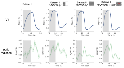 |
85 |
Mapping stimulus-driven hemodynamic changes in white matter
using 7T high-resolution fMRI
Jiawen Dakota Fan1,2,
Javier Gonzalez-Castillo3,
Peter A. Bandettini3,4,
Jonathan R. Polimeni2,5,6,
and Jingyuan E. Chen2,5
1Center for Data Science, New York University, New York City, NY, United States, 2Athinoula A. Martinos Center for Biomedical Imaging, Massachusetts General Hospital, Boston, MA, United States, 3Section on Functional Imaging Methods, Laboratory of Brain and Cognition, National Institute of Health, Bethesda, MD, United States, 4Functional MRI Core, National Institute of Health, Bethesda, MD, United States, 5Department of Radiology, Harvard Medical School, Boston, MA, United States, 6Harvard-MIT Division of Health Sciences and Technology, Massachusetts Institute of Technology, Cambridge, MA, United States Keywords: fMRI, White Matter In this study, by integrating 7T high-resolution imaging and massive data averaging, we show that white matter fMRI activations can be detected at a single-voxel level. Hemodynamic changes evoked by the flickering checkerboard stimuli were not homogenous within the optic radiation, and the averaged pattern exhibited a delayed time to peak longer than V1, consistent with previous literature. The current datasets also revealed stimulus-locked changes in certain white-matter tracts beyond the visual pathway. |
|
3668.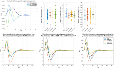 |
86 |
Characterizing across-trial variation in the hemodynamic
responses using ultra-fast fMRI data at 7T
Samuel Bianchi1,
Jakob Heinzle2,
Maria Engel1,
Stefan Frässle2,
Klaas Enno Stephan2,3,
and Klaas Paul Pruessmann1
1Institute for Biomedical Engineering, ETH Zurich and University of Zurich, Zurich, Switzerland, 2Translational Neuromodeling Unit (TNU), Institute for Biomedical Engineering, University of Zurich and ETH Zurich, Zurich, Switzerland, 3Max Planck Institute for Metabolism Research, Cologne, Germany Keywords: fMRI (task based), Modelling, Hemdoynamic response, HRF, Activation, BOLD Characterizing hemodynamic responses with great temporal precision is crucial for the interpretation of fMRI data with respect to the underlying neuronal activity. Recent literature highlights that analyses based on a canonical hemodynamic response function (HRF) may fail to capture relevant aspects of neuronal activity in certain conditions1. Ultra-fast fMRI enables precise temporally precise characterizations of hemodynamic responses and their trial-by-trial variation. Here, we analyzed fMRI timeseries sampled at which allowed us to model hemodynamic responses for individual trials during a visuo-motor task. Trial-by-trial variability of hemodynamic responses are analyzed by principal components analysis (PCA) and related to trial-specific conditions. |
|
3669.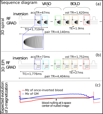 |
87 |
Combining the benefits of 3D acquisitions and spiral readouts
for VASO fMRI at UHF
Alejandro Monreal-Madrigal1,
Denizhan Kurban1,
Zoia Laraib1,2,
Renzo Huber1,
Dimo Ivanov1,
Nicolas Boulant3,
and Benedikt A Poser1
1Maastricht University, Maastricht, Netherlands, 2Polytechnic University of Turin, Turin, Italy, 3University-Paris-Saclay, CEA, CNRS, BAOBAB, NeuroSpin, Gif-sur-Yvette, France Keywords: fMRI, High-Field MRI, Pulse Sequence Design VASO fMRI can provide beneficial localization specificity and quantifiability compared to the commonly used BOLD contrast. Previous work has also shown the benefits of using spiral readouts compared to Cartesian EPI. In this work, we employ 3D stack-of-spirals readouts and compare it with the current state of the art 3D EPI readouts for VASO fMRI. The sequence implementation is done using Pulseq, images were reconstructed with MRIReco.jl; functional analysis with an openly available pipeline. We find that a VASO tSNR improvement of a factor of 2 over EPI is achieved using the proposed spiral implementation. |
|
3670. |
88 |
Pushing limits of spatial resolution in 3D EPI for fMRI on the
NexGen 7T scanner
Alexander JS Beckett1,2,
Samantha J Ma3,
An T Vu4,5,
and David A Feinberg1,2
1Helen Wills Neuroscience Institute, University of California, Berkeley, CA, United States, 2Advanced MRI Technologies, Sebastopol, CA, United States, 3Siemens Medical Solutions USA, Inc., Malvern, PA, United States, 4Radiology, University of California, San Francisco, CA, United States, 5San Francisco Veteran Affairs Health Care System, San Francisco, CA, United States Keywords: fMRI, Neuroscience Resolution in functional imaging can be increased by limiting the Field of View (FOV) for the image, increased acceleration and faster imaging using specialized gradient systems. Here we combine the high performance gradients of the NexGen 7T scanner with limited FOV imaging to collect ultra-high resolution fMRI in the occipital pole. Isotropic resolutions of 0.35mm are demonstrated using 3D EPI. |
|
3671.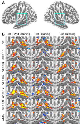 |
89 |
Entrainment between musical stimuli and cortical depth-dependent
fMRI signals
Hsin-Ju Lee1,2,
Hankyeol Lee3,
Kamil Uludag4,
and Fa-Hsuan Lin1,2
1Physical Sciences Platform, Sunnybrook Research institute, Toronto, ON, Canada, 2Department of Medical Biophysics, University of Toronto, Toronto, ON, Canada, 3Center for Neuroscience Imaging Research, Institute for Basic Science, Suwon, Korea, Republic of, 4Techna Institute & Koerner Scientist in MR Imaging, University Health Network, Toronto, ON, Canada Keywords: fMRI (task based), fMRI (task based) We use cortical-depth dependent fMRI to study the correlation between neural oscillations and fMRI signals across cortical depths in the auditory cortex during music listening. The correlation between fMRI signals and acoustic stimuli was the strongest at the primary auditory cortex in the intermediate cortical depths during the first listening, matching the hypothesis of feedforward signal supported by the connectivity. The correlation was suppressed in the repeated listening, superficial as well as deep depths, and at the secondary auditory cortex. |
|
3672. |
90 |
Direct imaging of Neuronal Activity (DIANA) in Humans
Shota Hodono1,
Reuben Rideaux2,
Timo van Kerkoerle3,
and Martijn A. Cloos1
1Centre for Advanced Imaging, University of Queensland, St Lucia, Australia, 2Queensland Brain Institute, University of Queensland, St Lucia, Australia, 3NeuroSpin, The French Alternative Energies and Atomic Energy Commission (CEA), Gif-Sur-Yvette, France Keywords: fMRI, Contrast Mechanisms, DIANA Here we describe our initial results attempting to observe neuronal activation in humans using the Direct Imaging of Neuronal Activity (DIANA) method. Both visual and auditory paradigms were explored. BOLD and anatomical ROI were tested and dedicated single slice control experiments were performed, but clear DIANA signals remained elusive. The translation of DIANA from animals to humans appears to be non-trivial. Nevertheless, considering the potential payoff continued effort towards this goal may be well worth it. To aid in this quest, parallel studies focused on DIANA’s ability to detect spiking and hyperpolarisation in controlled settings may be of great value. |
|
3673.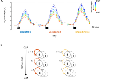 |
91 |
Using laminar fMRI and modeling to study auditory predictive
processing
Lonike Faes1,
Isma Zulfiqar1,
Luca Vizioli2,
Zidan Yu3,
Yuan-Hao Wu4,
Jiyun Shin4,
Ryszard Auksztulewicz5,
Lucia Melloni6,7,
Kamil Uludag8,
Essa Yacoub2,
and Federico De Martino1,2
1Deparment of Cognitive Neuroscience, Maastricht University, Maastricht, Netherlands, 2Center for Magnetic Resonance Research, Minneapolis, MN, United States, 3University of Hawaii, Honolulu, HI, United States, 4New York University Grossman School of Medicine, New York University, New York, NY, United States, 5Center for Cognitive Neuroscience Berlin, Free University Berlin, Berlin, Germany, 6Department of Neurology, New York University, New York, NY, United States, 7Department of Neurophysiology, Max-Planck Institute for Brain Research, Frankfurt am Main, Germany, 8Joint Department of Medical Imaging and Krembil Brain Institute, Techna Institute & Koerner Scientist in MR Imaging, Toronto, ON, Canada Keywords: fMRI (task based), High-Field MRI We use ultra-high field fMRI to explore the differential role of cortical layers in the information flow between top-down predictions and bottom up sensory evidence in a predictive coding (PC) framework. As the measured BOLD signal is affected by vascular draining, we use a computational model that combines neuronal dynamics with laminar vascular physiology to help us understand prediction signals at cortical depths and underlying neuronal responses. We show that a violation in prediction caused response modulation in superficial and deep layers in the primary auditory cortex, which is in line with the PC framework. |
|
3674. |
92 |
Increasing MT contrast in laminar fMRI using PUSH pTx pulses
Viktor Pfaffenrot1,
David Leitao2,3,
Annika Verheyen1,
Markus May1,4,
Raphael Tomi-Tricot5,
Shaihan Malik2,6,
and David Norris1,7
1Erwin L. Hahn Institute for Magnetic Resonance Imaging, University of Duisburg-Essen, Essen, Germany, 2Biomedical Engineering Department, School of Biomedical Engineering and Imaging Sciences, King's College London, London, United Kingdom, 3London Collaborative Ultra high field System (LoCUS), London, United Kingdom, 4High Field and Hybrid MR Imaging, University Duisburg-Essen, University Hospital Essen, Essen, Germany, 5MR Research Collaborations, Siemens Healthcare Limited, Frimley, United Kingdom, 6Centre for the Developing Brain, School of Biomedical Engineering and Imaging Sciences, King's College London, London, United Kingdom, 7Donders Institute for Brain, Cognition and Behaviour, Radboud University Nijmegen, Nijmegen, Netherlands Keywords: fMRI, Magnetization transfer, laminar fMRI The GRE-BOLD contrast used in laminar fMRI suffers from suboptimal specificity due to unwanted extravascular effects. Recently, efforts were made to increase specificity of GRE-BOLD by means of CBV-weighting using the magnetization transfer contrast. The specificity improvement depends on the amount of selective GM signal reduction. We investigate whether PUSH pTx pulses, designed to maximize MT contrast in a given area, can achieve a higher functional contrast. Our results suggest that when opting for high-power, off-resonant MT pulses, both PUSH and a CP2+ mode perform equally well in increasing the functional contrast at short TE compared to GRE-BOLD in V1. |
|
3675.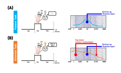 |
93 |
Separation of sensory and attention input to the human primary
somatosensory cortex by spin- and gradient-echo BOLD fMRI
SoHyun Han1,2,
Dongho Kim1,2,
and Seong-Gi Kim1,2
1Center for Neuroscience Imaging Research, Suwon, Korea, Republic of, 2Sungkyunkwan University, Suwon, Korea, Republic of Keywords: fMRI, Brain High spatial resolution laminar specific fMRI has a potential to separate between top-down and bottom-up signals. In this study, we investigated the effect of attention across cortical layers in the human S1 using SAGE-EPI sequence. fMRI experiments during vibrotactile stimulation with passive and attention tasks with 0.8mm isotropic resolution at 7T were performed. We demonstrated that the SE-BOLD can identify laminar profiles of bottom-up and top-down processes: the laminar profile of passive task showed the peak at layer 4, but that of attention task showed the peak at the superficial layers. |
|
3676.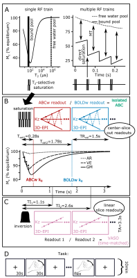 |
94 |
Isolating the arterial blood volume change to probe fMRI spatial
specificity
Nikos Priovoulos1,2,
Icaro Agenor Ferreira de Oliveira1,2,3,
Benedikt Poser4,
David G Norris5,6,
and Wietske van der Zwaag1,2
1Spinoza Center for Neuroimaging, Amsterdam, Netherlands, 2Computational Cognitive Neuroscience and Neuroimaging, Netherlands Institute for Neuroscience, Amsterdam, Netherlands, 3Techna Institute, University Health Network, Amsterdam, Netherlands, 4MR-Methods group, MBIC, Faculty of Psychology and Neuroscience, Maastricht University, Maastricht, Netherlands, 5Donders Institute for Brain, Cognition and Behaviour, Donders Centre for Cognitive Neuroimaging, Radboud University Nijmegen, Nijmegen, Netherlands, 6Erwin L. Hahn Institute for Magnetic Resonance Imaging, Essen, Germany Keywords: fMRI, fMRI, high field, cerebral blood volume BOLD fMRI is widely used in neuroscience, but has limited spatial specificity while alternative approaches based on tissue cerebral blood volume (CBV) change have limited sensitivity. Recently, arterial CBV change (Arterial Blood Contrast; tracked through magnetization-transfer) was suggested as an fMRI mechanism, but its localization and sensitivity remain unexplored, since efficiently isolating the arterial CBV is challenging. Here, we combine temporally-efficient saturation with center-slice-out readouts and BOLD-correction to isolate arterial CBV for high-resolution 7T human fMRI. Our novel sequence shows much-improved specificity compared to BOLD and similar sensitivity to VASO, suggesting Arterial Blood Contrast to be a promising fMRI approach. |
|
3677. |
95 |
Potential of diffusion fMRI for detecting white matter activity
in the human brain
Jasmine Khedidja Nguyen-Duc1,
Wiktor Olszowy2,
Jonathan Patino Lopez 3,
and Ileana Jelescu1
1Department of Radiology, CHUV, Lausanne, Switzerland, 2CIBM Center for Biomedical Imaging, Lausanne, Switzerland, 3Signal Processing Lab (LTS5), École Polytechnique Fédérale de Lausanne, Lausanne, Switzerland Keywords: fMRI (task based), Diffusion/other diffusion imaging techniques, simulations, diffusion fMRI Diffusion functional Magnetic Resonance Imaging (dFMRI) may be used to study the white matter activation in a more direct way than BOLD. The aim of this work is to extract the decrease in apparent diffusion coefficient (ADC) in the white matter observed in real data, and compare to that in a simulation of realistic axon swelling. Results suggest that the decrease is of low amplitude (<0.5%) but nonetheless detectable. |
|
3678.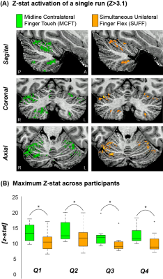 |
96 |
Proprioceptive engagement in the human cerebellum using 7T-fMRI
Emma Brouwer 1,2,
Julie Hashimoto 1,3,
Nikos Priovoulos1,2,
and Wietske van der Zwaag1,2
1Spinoza Centre for Neuroimaging, Amsterdam, Netherlands, 2Computational Cognitive Neuroscience and Neuroimaging, Netherlands Institute for Neuroscience, Amsterdam, Netherlands, 3Vrije Universiteit Amsterdam, Amsterdam, Netherlands Keywords: fMRI (task based), Brain, cerebellum The human cerebellum forms an important part of the sensory and motor networks. Specifically, cerebellar damage has been shown to result in difficulty to perform proprioceptive tasks. Hence, studying the functional cerebellar organisation can be of great neuroscientific and clinical interest. This requires high-resolution images due to the thin, highly-foliated cortex of the cerebellum. We investigated the difference between a simultaneous-unilateral-finger-flex (SUFF) and midline-contralateral-finger-touch (MCFT) using B1-shimmed fMRI at 7T. Movements with higher proprioceptive engagement (MCFT) resulted in stronger, more medially located activations on the cerebellar surface compared to movements which are less reliant on proprioception (SUFF). |
|
3679.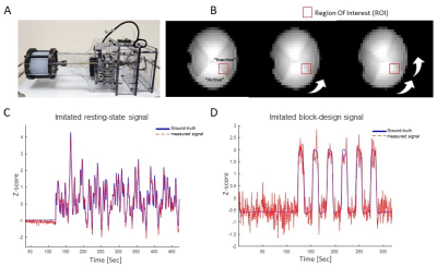 |
97 |
Multi-echo EPI for improving temporal resolution in task-based
fMRI at 7T- a dynamic phantom study
Guy Shlomo Baz1,2,
Edna Furman-Haran 2,3,
and Rita Shmidt1,2
1Department of brain sciences, Weizmann Institute, Rehovot, Israel, 2The Azrieli National Institute for Human Brain Imaging and Research, Weizmann Institute, Rehovot, Israel, 3Life Sciences Core Facilities, Weizmann Institute, Rehovot, Israel Keywords: fMRI (task based), High-Field MRI Ultra-high field MRI provides increased sensitivity, which we aim to utilize for improving the temporal resolution in functional studies. To investigate the achievable resolution at 7T MRI, a dynamic phantom that can generate an fMRI-like time-series was used. A dataset based on block-design with defined time shifts and a range of contrast-to-noise values was used to characterize the effective temporal resolution. Estimated temporal resolution was x1.7 times better for multi-echo compared to single-echo EPI, estimated as 146ms for a scan with TR of 600ms. This study offers a novel approach of optimizing protocols and new insights into fMRI temporal resolution. |
|
3680.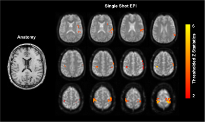 |
98 |
Investigating the feasibility of functional MRI using GRE EPI on
a high performance 0.5 T Scanner
Arjama Halder1,
Chad T. Harris2,
Curtis N. Wiens2,
Andrew T. Curtis2,
William B. Handler3,
Andrea Soddu3,
and Blaine A Chronik1,3
1Medical Biophysics, Western University, London, ON, Canada, 2Research and Development, Synaptive Medical, Toronto, ON, Canada, 3Physics and Astronomy, Western University, London, ON, Canada Keywords: fMRI, fMRI (task based) fMRI is typically not performed at field strengths < 1.5T due to low magnetic susceptibility contrast and inadequate gradient performance. Leveraging the high-performance gradient set of a head-only 0.5T MRI, the feasibility of motor task based fMRI was evaluated using a 4mm isotropic GRE-EPI acquisition. Activated regions within the PMC were consistent with expected behaviour. Furthermore, a significant change in signal during activation of 1.8+/-0.4% was measured within an ROI of activated voxels. These results suggest that motor task based BOLD fMRI is possible at 0.5T. |
|
 Back to Meeting Home
 Back to the Program-at-a-Glance
The International Society for Magnetic Resonance in Medicine is accredited by the Accreditation Council for Continuing Medical Education to provide continuing medical education for physicians.
 Back to Meeting Home
 Back to the Program-at-a-Glance
 View Presentation Video