Digital Poster
Contrast Agents
ISMRM & ISMRT Annual Meeting & Exhibition • 03-08 June 2023 • Toronto, ON, Canada

| Computer # | |||
|---|---|---|---|
4870.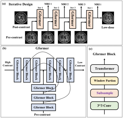 |
101 |
Arbitrary Level Contrast Dose Simulation in Brain MRI Using
Iterative Reconstruction with Transformer Models.
Dayang Wang1,
Srivathsa Pasumarthi1,
and Ryan Chamberlain1
1Subtle Medical Inc., Menlo Park, CA, United States Keywords: Simulations, Data Acquisition, MRI Dose Simulation In Gadolinium-based contrast agents (GBCAs) assisted MRI scans, dose simulation is a significant task that enables understanding of the perfusion process. In this work, we propose a new transformer-based iterative model to generate MRI images with arbitrary contrast dosages. By using datasets where only 0%, 10%, and 100% dose images are available, the proposed model can synthesize quantitatively and qualitatively accurate MRI images of varying dosages. The simulated images can be used for other downstream tasks like developing reduced-dose acquisitions and finding the minimum dose needed for a given pathology. |
|
 |
4871.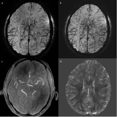 |
102 |
Imaging arterial and venous vessels using Iron Dextran enhanced
multi-echo susceptibility weighted imaging (SWI) MRI at 7T
Yinghao Li1,2,3,
Adrian Paez2,3,
Di Cao1,2,3,
Chunming Gu1,2,3,
Kaihua Zhang2,3,
Xinyuan Miao2,3,
Jay Pillai4,5,
Peter M van Zijl3,6,
Christopher Earley7,
Xu Li2,3,
and Jun Hua2,3
1Biomedical Engineering, Johns Hopkins University School of Medicine, Baltimore, MD, United States, 2F.M. Kirby Research Center for Functional Brain Imaging, Kennedy Krieger Institute, Baltimore, MD, United States, 3Neurosection, Division of MRI Research, Russell H. Morgan Department of Radiology and Radiological Science, Johns Hopkins University School of Medicine, Baltimore, MD, United States, 4Division of Neuroradiology, Russell H. Morgan Department of Radiology and Radiological Science, Johns Hopkins University School of Medicine, Baltimore, MD, United States, 5Department of Neurology, Johns Hopkins University School of Medicine, Baltimtore, MD, United States, 6F.M. Kirby Research Center for Functional Brain Imaging, Kennedy Krieger Institute, Baltimtore, MD, United States, 7Department of Neurology, Johns Hopkins University School of Medicine, Baltimore, MD, United States Keywords: Contrast Agent, Blood vessels Iron Dextran is a widely used FDA-approved ultra-small-superparamagnetic-iron-oxides (USPIO) to treat iron deficiency anemia in patients. Here, we evaluate the feasibility of using Iron Dextran as an MRI contrast agent for imaging arterial and venous blood vessels using multi-echo susceptibility weighted imaging (SWI) MRI at 7T. Phantom experiments were performed to measure relaxivity values (r1 and r2) for Iron Dextran in blood. Pre- and post-infusion MRI images were acquired in human subjects from which maps of arteries and veins were extracted. The post-contrast SWI images showed enhanced susceptibility difference between blood and the surrounding tissue in both arteries and veins. |
4872.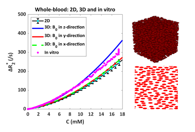 |
103 |
Dependency of R2 and R2* on Gd-DTPA Concentration: From
Whole-Blood to Realistic Brain Tumor Vasculature
Daniëlle van Dorth1,
Ahmad Alafandi2,
Krishnapriya Venugopal2,
Aurélien Delphin3,
Dirk H. J. Poot2,
Thomas Christen3,
Marion Smits2,4,5,
Jeroen de Bresser6,
Juan Hernandez Tamames2,
and Matthias J. P. van Osch1,4
1C. J. Gorter MRI Center, Department of Radiology, Leiden University Medical Center, Leiden, Netherlands, 2Radiology and Nuclear Medicine, Erasmus University Medical Center, Rotterdam, Netherlands, 3University Grenoble Alpes, Inserm, U1216, Grenoble Institut Neurosciences, Grenoble, France, 4Medical Delta, Delft, Netherlands, 5Brain Tumor Center, Erasmus MC Cancer Institute, Rotterdam, Netherlands, 6Radiology, Leiden University Medical Center, Leiden, Netherlands Keywords: Simulations, DSC & DCE Perfusion In quantitative DSC-MRI, the concentration-time course is obtained from the MR signal changes following a bolus injection of contrast agent. The generally assumed linear relationship between ΔR2* and contrast agent concentration is not always valid. In this study a realistic 3D simulation model was used to establish the characteristics of this relationship in whole-blood and brain tumor tissue. The results show an improved performance of the 3D simulation model compared to its 2D version. In addition, the relationship between ΔR2* and contrast agent concentration appeared to be dependent on the B0 direction and MR sequence. |
|
4873.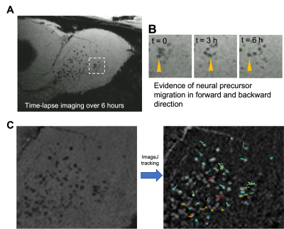 |
104 |
Ultra-high Moment Gold-coated Iron Particles Allows for
Longitudinal Time-lapse MRI of Individual Stem Cells in the
Brain
Nikorn Pothayee1,
Stephen Dodd1,
Li Liu1,
Gary Zabow2,
and Alan Koretsky1
1National Institutes of Health, Bethesda, MD, United States, 2National Institute of Standards and Technology, Boulder, CO, United States Keywords: Contrast Agent, Cell Tracking & Reporter Genes In this study, gold coated microfabricated gold-coated iron particles are used for in vitro and in vivo labeling of immune cell and neural precursors, respectively for MRI. These particles have a pure iron core and protective layer of gold and exhibit extremely high magnetic moment relative to more commonly used, chemically synthesized, iron-oxide based particles. Following in situ labeling of neural precursor cells, we show that the migration of a large pool of individual cells can be visualized in time-lapse and longitudinal MRI. |
|
4874.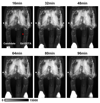 |
105 |
A Novel Iron-oxide-based T1 Contrast Agent outperforms Gd-DOTA
for Magnetic Resonance Lymphography
Yeon Ji Chae1,
Mi-hyun Kim2,3,
Chul-Woong Woo4,
Sang-Tae Kim4,
Do-Wan Lee5,
Hwon Heo1,
Monica Young Choi4,
Yoonseok Choi6,
Kyung Won Kim1,4,
and Dong-Cheol Woo1,4
1Department of Convergence Medicine, Asan Medical Center, University of Ulsan College of Medicine, Seoul, Korea, Republic of, 2Department of Radiation Science & Technology, Jeonbuk National University, Jeonbuk, Korea, Republic of, 3Trial Informatics. Inc, Seoul, Korea, Republic of, 4Convergence Medicine Research Center, Asan Institute for Life Sciences, Asan Medical Center, Seoul, Korea, Republic of, 5Department of Radiology, Asan Medical Center, University of Ulsan College of Medicine, Seoul, Korea, Republic of, 6Medical Research Institute, Gangneung Asan Hospital, Gangneung-si, Gangwon-do, Korea, Republic of Keywords: Contrast Agent, Contrast Agent, Magnetic resonance lymphography, Iron-oxide-based T1 contrast agent The novel iron-oxide-based T1 contrast agent may be an optimal contrast agent for MR lymphangiography, because it enhances the lymphatics only without venous contamination for more than 1 hour. It can overcome the limitations of gadolinium-based contrast agents such as venous contamination and rapid washout. |
|
4875.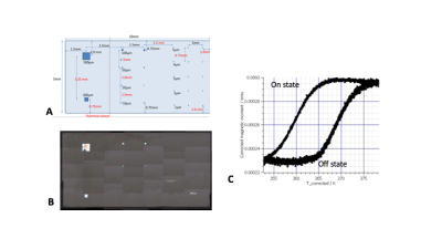 |
106 |
Towards cell-size and switchable magnetocaloric MRI contrast
agents using micofabricated thin-film FeRh.
Stephen Dodd1,
Natalia Gudino1,
O Zadorozhnii2,
M Staňo2,
J Hajduček2,
J. A. Arregi2,
Vojtech Uhlíř2,
H. Doug Morris3,
Mladen Barbic4,
and Alan Koretsky1
1Laboratory of Functional and Molecular Imaging, NINDS, National Institutes of Health, Bethesda, MD, United States, 2Ceitec Magnetism, Central European Institute of Technology, Brno, Czech Republic, 3Uniformed Services University of the Health Sciences, Bethesda, MD, United States, 4NYU Langone Health - Tech4Health Institute, New York, NY, United States Keywords: Contrast Agent, Cell Tracking & Reporter Genes, Magnetocaloric material We present progress towards smaller magnetocaloric samples, that drastically change magnetic moment and hence T2* contrast when switched with external magnetic field or temperature, with the eventual goal of achieving a size appropriate for cell tracking. To that end a FeRh sample was microfabricated with a thickness of 200 nm, and patterned with varying linear dimensions from 500 micron down to 1 micron square. We demonstrate that this sample may be switched with temperature and with external field through imaging at 4.7 and 11.7T and with the application of pulsed B0 insert coil. |
|
4876.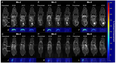 |
107 |
A new class of easily synthesized manganese contrast agents for
liver MRI
Sean William McRae1,
Michael Cleary2,
Francisco Martinez3,
Ying Xia3,
Peter Caravan2,
Eric Gale2,
John Ronald3,
and Timothy Scholl3
1Medical Biophysics, Western University, London, ON, Canada, 2Massachusetts General Hospital, Boston, MA, United States, 3Western University, London, ON, Canada Keywords: Contrast Agent, Molecular Imaging This work investigated the performance of a novel class of Mn(II) T1 contrast agents made from a commercially available chelator with various lipophilic targeting moieties added to promote uptake through the human derived OATP1B1 and OATP1B3 transporters. Agent performance was assessed in vitro using cells engineered to express the human OATPs, and in vivo by assessing liver uptake in mice where other OATP isoforms are present. In vitro measurements suggested targeting of agents to human OATPs where in vivo imaging revealed strong contrast enhancement in murine livers, with no evident toxicity at a 0.1 mmol/kg dose. |
|
4877.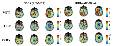 |
108 |
Resting perfusion measures in patients with steno-occlusive
disease; comparing two contrast agents Gadolinium and
hypoxia-induced dOHb
Ece Su Sayin1,
Vittorio Stumpo2,
Jacopo Bellomo2,
Julien Poublanc3,
Marco Piccirelli4,
James Duffin1,
Vepeson Wijeya 3,
Athina Pangalu4,
Andrea Bink4,
Bence Nemeth 4,
Zsolt Kulcsar4,
David John Mikulis3,
Joseph Arnold Fisher1,
Olivia Sobcyzk 3,
and Jorn Fierstra 2
1Physiology, University of Toronto, Toronto, ON, Canada, 2Department of Neurosurgery, University Hospital Zurich, Zurich, Switzerland, 3University Health Network, Toronto, ON, Canada, 4Department of Neuroradiology, University Hospital Zurich, Zurich, Switzerland Keywords: Contrast Agent, Perfusion Dynamic susceptibility contrast perfusion imaging is clinically acquired using Gadolinium based contrast agents (GBCA), however, there remain many drawbacks. We show that in patients with steno-occlusive disease perfusion imaging using hypoxia-induced dOHb have a high degree of similarity to those obtained using GBCA. |
|
4878.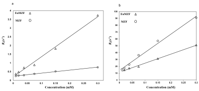 |
109 |
Relaxometry Measurements of Europium-doped Mn0.6Zn0.4Fe2O4 at
Increasing Magnetic Field
Hamidreza saeidi 1,2,
Morteza Mozaffari2,
Serhat Ilbey1,
Jochen Leupold1,
and Michael Bock1
1Division of Medical Physics, Department of Radiology, University Medical Center Freiburg, Freiburg, Germany, 2Faculty of Physics, University of Isfahan, Isfahan, Iran (Islamic Republic of) Keywords: Contrast Agent, Relaxometry, Relaxometery, longitudinal Relaxation Time Magnetic resonance imaging (MRI) contrast agents with both intrinsic high r1 and r2 relaxivities are desirable in medical diagnosis to achieve highly accurate and ambiguity-free MR images. Here, we report synthesis and MRI tests of Europium doped Mn-Zn ferrite (EuMZF) nanoparticles at different magnetic field strengths. EuMZF nanoparticles showed significant improvement in both positive (T1) and negative (T2) contrasts. The results showed that by increasing magnetic field, r1 relaxivity decreased while r2 increased so that the EuMZF is suitable as a dual-mode contrast at 1.5 and 3T but it is a T2 agent at 7 and 9.4T. |
|
4879.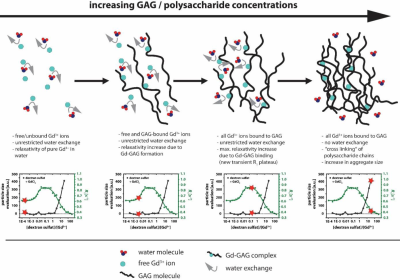 |
110 |
Molecular Weight of Sugar Structures impacts Shielding Effect of
Gd-Ions in Polysaccharides.
Patrick Werner1,2,
Matthias Taupitz2,
Sophia Ber3,
and Leif Schröder1
1German Cancer-research center (DKFZ), Heidelberg, Germany, 2Charite Berlin, Berlin, Germany, 3Universität Heidelberg, Heidelberg, Germany Keywords: Contrast Agent, Relaxometry, GBCA, gadolinium, long-term deposition, GAGs, dextran sulfate Detailed biochemical information on the long-term deposition of Gd3+ in the body after administration of GBCAs is still missing. Glycosaminoglycans should be considered as chelators of released Gd3+. We used dextran-sulfate to study the effect of polysaccharides with different molecular weights on the observable relaxivity. Chelation was observed through an increase in relaxation. Moreover, a molecular weight-specific shielding revealed by subsequent reduced R1 occurs at higher polysaccharide/Gd-ion ratios. This coincided with observations of Gd-induced aggregation. Similar shielding of re-chelated Gd3+can lead to an underestimation of deposited Gd-ions in the body and requires further research. |
|
4880. |
111 |
First Results of Novel Contrast Agent Based on Ultrasmall
Paramagnetic Nanoparticles for T1-Weighted MRI
Khallil Taverna Chaim1,
Mayara Klimuk Uchiyama1,2,
Robson Raphael Guimaraes2,
Koiti Araki2,
and Claudia da Costa Leite 1
1School of Medicine - Radiology, University of Sao Paulo, Sao Paulo, Brazil, 2Institute of Chemistry, University of Sao Paulo, Sao Paulo, Brazil Keywords: Contrast Agent, Contrast Agent, nanoparticles MRI is one of the best imaging techniques in clinical analysis because it is non-invasive and has high resolution. Contrast agents are used to further improve these images, as they increase the level of soft tissue detail, differentiating regions of interest. Currently, Gd-based contrast agents have important precautions. In this work we present a new nanoparticle based on iron oxide, with 4 nm and positive contrast (T1-weighted). Thus, effects similar to gadoteric acid, with reduced signal persistence and partial renal excretion, indicating great stability, this new paramagnetic nanoparticle has great potential to be applied as a contrast agent in MRI. |
|
4881. |
112 |
Robust in vivo MRI of OATP reporter gene expressing cells using
standard clinical dose of MRI contrast agent
Tapas Bhattacharyya1,
Jeremy M.-L. Hix1,
Christiane L. Mallett1,
and Erik M. Shapiro1
1Radiology and Institute for Quantitative Health Sciences and Engineering, Michigan State University, East Lansing, MI, United States Keywords: Molecular Imaging, Cell Tracking & Reporter Genes Hepatic OATPs are a promising molecular imaging reporter gene for MRI yet its translation for human studies is limited by the reported extremely high dose of contrast agent required (>40x clinical dose) for robust detection of engineered cells in vivo. Here we describe studies that culminated in the in vivo MRI detection of OATP-overexpressing cells using standard clinical doses. OATP-overexpression was accomplished by lentiviral transduction of tumor cells, followed by MRI screening to determine the best performing OATPs. In vivo MRI of OATP-overexpressing tumors was accomplished using a combination of both T1-weighted and dynamic contrast enhanced (DCE-) MRI. |
|
4882.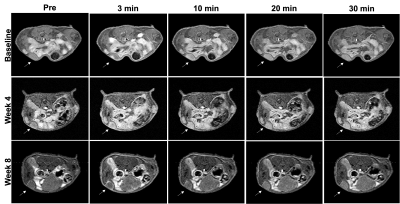 |
113 |
Monitoring triple negative breast cancer therapy against lncRNA
MANCR with MT218, a targeted MRMI contrast agent
Calin Nicolescu1,
Da Sun1,
Songqi Gao1,
and Zheng-Rong Lu1,2
1Department of Biomedical Engineering, Case Western Reserve University, Cleveland, OH, United States, 2Case Comprehensive Cancer Center, Case Western Reserve University, Cleveland, OH, United States Keywords: Molecular Imaging, Cancer, Breast Developing new molecular targeted therapies for triple negative breast cancer remains an obstacle. MANCR is a long noncoding RNA overexpressed in TNBC that sustains aggressive breast cancer growth. Silencing its expression with siRNA nanoparticles suppresses tumor growth, and treatment efficacy can be assessed by MRMI with a targeted contrast agent, MT218. Reduction in contrast-to-noise ratio following treatment confirmed effectiveness of siRNA therapy and allowed regular monitoring of tumor inhibition. RNA therapy shows promise as a new approach for treating aggressive breast cancer, and MRMI with the targeted contrast agent is a non-invasive imaging tool for TNBC detection and therapeutic surveillance. |
|
4883.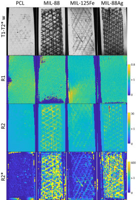 |
114 |
Relaxation Rate MR Contrast of Metal-Organic Framework or USPIO
Labelled Melt Electrowritten Polycaprolactone Scaffolds
Geoffrey J. Topping1,
Salma Mansi2,
Kilian M. A. Müller2,
Zahid Hussain3,
Sarah Dummert3,
Carolin Rickert4,
Sebastian P. Schwaminger5,
Younzhe Zou2,
Diana M. Rojas-González2,
Sonja Berensmeier5,
Oliver Lieleg4,
Roland Fischer3,
Petra Mela2,
and Franz Schilling1
1Department of Nuclear Medicine, School of Medicine, Klinikum rechts der Isar, Technical University of Munich, Munich, Germany, 2Medical Materials and Implants, Department of Mechanical Engineering, TUM School of Engineering and Design, & Munich Institute of Biomedical Engineering, Technical University of Munich, Garching, Germany, 3School of Natural Sciences and Catalysis Research Center, Technical University of Munich, Garching, Germany, 4Biomechanics, Department of Materials Engineering, TUM School of Engineering and Design, Technical University of Munich, Garching, Germany, 5Chair of Bioseparation Engineering, School of Engineering and Design, Technical University of Munich, Garching, Germany Keywords: Contrast Mechanisms, Preclinical, Tissue Engineering Polycaprolactone scaffolds were melt electrowritten with and without included metal-organic frameworks (MOF) or nanoparticles (USPIO). MR images and relaxation rate (R1, R2, R2*) maps were acquired of scaffolds in agar and blood. Pure polycaprolactone showed weak or no contrast. Addition of MOF or USPIOs produced strong T2* contrast. Tensile properties were preserved up to 0.2 w/w% USPIOs. MOFs produced strong w/w% dependent antibacterial effects when included in scaffolds. |
|
4884.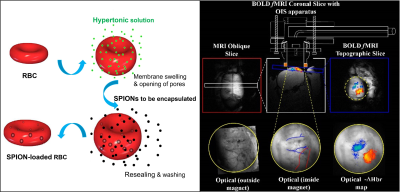 |
115 |
Engineering novel functional brain imaging contrast agents via
iron oxide encapsulation in human red blood cells
Elizabeth Jane Fear1,2,
Antonella Antonelli1,
Pasant Abdalla1,
Elisa Zamboni3,
Marie-Christine Labarthe-Last2,
Victoria Annis2,
Simon Benedict Duckett2,
Mauro Magnani1,
and Aneurin James Kennerley2,4
1Biomolecular Sciences, University of Urbino Carlo Bo, Urbino, Italy, 2Centre for Hyperpolarisation in Magnetic Resonance (CHyM), Chemistry, University of York, York, United Kingdom, 3Psychology, University of York, York, United Kingdom, 4Sport and Exercise Science, Institute of Science, Manchester Metropolitan University, Manchester, United Kingdom Keywords: Contrast Agent, Contrast Agent, red blood cells, Super Paramagnetic Iron Oxide Nanoparticles We have developed a next generation, safe, personalised iron-based contrast agent (CA) for empowering functional Magnetic Resonance Imaging (fMRI). Our proprietary method uses state-of-the-art red blood cell (RBC) encapsulation technology to engineer biocompatible super-paramagnetic CAs able to withstand rapid clearance from the bloodstream by macrophages (as part of the mononuclear phagocytic/reticuloendothelial system). The agent is validated in a multi-modal (concurrent fMRI and intrinsic optical imaging) preclinical rat model as a novel CA for the mapping of cerebral blood volume (CBV) in response to somatosensory stimulation, providing a non-BOLD alternative for accurate, high-resolution mapping of brain function. |
|
4885.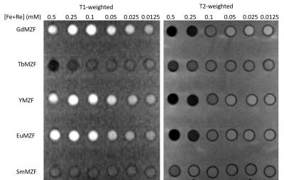 |
116 |
High Longitudinal and Transverse Relaxivities with Rare Earth
ions doped Mn0.6Zn0.4Fe2O4: a Potential for dual-mode Contrast
Agents
Hamidreza saeidi 1,2,
Morteza Mozaffari2,
Serhat Ilbey1,
and Michael Bock1
1Division of Medical Physics, Department of Radiology, University Medical Center Freiburg, Freiburg, Germany, 2Faculty of Physics, University of Isfahan, Isfahan, Iran (Islamic Republic of) Keywords: Contrast Mechanisms, Contrast Agent, Relaxometery, longitudinal Relaxation Time MRI contrast agents have gained extensive attention for providing contrast between normal and abnormal tissue. However, single-mode contrast agents do not always provide the required contrast enhancement for disease detection. One effective strategy for avoiding ambiguous images is to design MRI contrast agents with simultaneous T1 and T2 shortening effects. The purpose of this work is to synthesize rare earth ions doped Mn-Zn ferrite (ReMZF) nanoparticles and characterize them with a 1.5T MRI scanner in vitro. The ReMZF exhibited high r1 and r2 relaxivities but moderate r2/r1 which make them suitable to be used as dual-mode contrast agents. |
|
4886.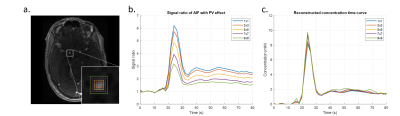 |
117 |
A dedicated arterial input function compensating for inflow and
partial voluming in dynamic contrast enhanced MRI
Chih-Hsien Tseng1,
Martijn Nagtegaal1,
Jaap Jaspers2,
Alejandra Mendez Romero2,
Piotr Wielopolski3,
Marion Smits3,4,
Matthias J.P. van Osch4,5,
and Frans Vos1,3,4
1Imaging Physics, Delft University of Technology, Delft, Netherlands, 2Radiation Oncology, Erasmus MC Cancer Institute, University Medical Center, Rotterdam, Netherlands, 3Radiation and Nuclear Medicine, Erasmus MC, University Medical Center, Rotterdam, Netherlands, 4Medical Delta, Delft, Netherlands, 5Radiology, C.J. Gorter MRI center, Leiden University Medical Center, Leiden, Netherlands Keywords: Contrast Mechanisms, DSC & DCE Perfusion Both inflow and partial volume effects (PVE) are sources of error when measuring the arterial input function (AIF) in DCE-MRI. We proposed a method that estimated the perceived pulse number of spins, and then corrected with these such that both effects were simultaneously compensated. Simulation data demonstrated that the reconstructed AIFs showed only marginal bias. In addition, the algorithm yielded highly correlated reconstructed curves over a wide range of PVEs in clinical data. Our findings show that the PVE can be compensated by considering it as part of the inflow correction as it exhibits similar confounding effects when measuring DCE-AIFs. |
|
4887.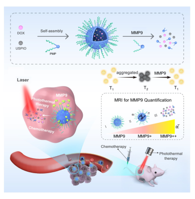 |
118 |
A MMP-Responsive Nanoplatform with Transformable Magnetic
Resonance Property for Quantitative Tumor Bioimaging and
Synergetic Therapy
Zhongling Wang1,
AN CHEN1,
Hongwei Lu2,
and Xiance Zhao3
1Shanghai General Hospital,School of Medicine ,Shanghai jiaotong University, Shanghai, China, 2Lthink Medical Institute, Guangzhou, China, 3Philips Healthcare, Shanghai, China Keywords: Molecular Imaging, Multimodal We developed a novel nanoplatform-based MMP9-responsive T2–T1 switching MRI contrast agent, which can be used not only for non-invasive visualization and quantitative analysis of MMP9 activity, but also as a carrier in photothermal sensitization chemotherapy. The nanoplatform acts as a T2 contrast agent in physiological conditions ,then transform into a T1 contrast agent by MMP9 stimulus. We have demonstrated that the changes R1 and R2/R1 values are MMP9 concentration dependent in tumors. The combination of PMPSD with laser irradiation was more effective than single chemo-/photothermal therapy, and up-regulation of MMP9 in the tumor could eanhance the nanoplatform’s therapeutic effect. |
|
4888. |
119 |
The magnetic resonance characteristics of SPIONs for imaging at
ultralow field (6.5 mT)
Sheng Shen1,
David E. J. Waddington2,
Marie Zhang3,
and Matthew S. Rosen1,4,5
1MGH/A. A. Martinos Center for Biomedical Imaging, Boston, MA, United States, 2The University of Sydney, Sydney, Australia, 3Imagion Biosystems, Ltd, San Diego, CA, United States, 4Department of Physics, Harvard University, Cambridge, MA, United States, 5Harvard Medical School, Boston, MA, United States Keywords: Contrast Agent, Modelling Characterization of the parameters of superparamagnetic iron oxide nanoparticles (SPIONs) can guide synthesis of optimized particles for use in magnetic resonance imaging (MRI). With a goal of application of SPIONs in ultra-low field (ULF) MRI, we investigated the properties of SPIONs, and conducted imaging on a 6.5 mT MRI scanner. Specifically, we measured the NMR relaxivity and susceptibility of different SPIONs sample, both dominated by the core size of SPIONs. We also implemented susceptibility imaging using an bSSFP sequence to obtain positive contrast images of SPION phantoms, and present here a method to optimize the ULF susceptibility MRI images. |
|
 Back to Meeting Home
 Back to the Program-at-a-Glance
The International Society for Magnetic Resonance in Medicine is accredited by the Accreditation Council for Continuing Medical Education to provide continuing medical education for physicians.
 Back to Meeting Home
 Back to the Program-at-a-Glance
 View Presentation Video