Digital Poster
Elastography Throughout the Brain & Body
ISMRM & ISMRT Annual Meeting & Exhibition • 03-08 June 2023 • Toronto, ON, Canada

Digital Poster
Elastography Throughout the Brain & Body
| Computer # | |||
|---|---|---|---|
5143.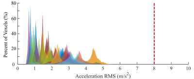 |
41 |
Vibration Exposure in Brain MR Elastography
Safoura Sadegh Pour Aji Bishe1,2,
Kay Pepin1,2,
Jeremiah Heilman1,2,
Xiang Shan1,
Ziying Yin1,
and Richard Ehman1
1Radiology, Mayo Clinic, Rochester, MN, United States, 2Resoundant, Inc., Rochester, MN, United States Keywords: Safety, Brain, Elastography Magnetic resonance elastography (MRE) requires application of mechanical vibrations to assess the stiffness of tissue noninvasively and quantitatively. This study assessed the levels of vibration exposure in brain tissue during a typical brain MRE exams performed for clinical research. Voxel-level vibration exposure levels throughout the brain were measured in 45 subjects in this IRB approved study. Histograms of voxel-level vibration in all cases demonstrated maximum exposure levels that were well below the very conservative EU 2002 Directive 2002/44/EC of the European Parliament guideline for total body vibration. |
|
5144.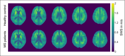 |
42 |
The impact of multiple sclerosis on cortical brain stiffness.
Helge Herthum1,
Rafaela V. Silva2,
Anna S. Morr3,
Mehrgan Shahryari3,
Matthias Anders3,
Yasmine Safraou3,
Stefan Hetzer1,
Jürgen Braun3,
Michael Scheel4,
Friedemann Paul4,
Carmen Infante-Duarte2,
and Ingolf Sack3
1Berlin Center for Advanced Neuroimaging, Charité Universitätsmedizin Berlin, Berlin, Germany, 2Experimental and Clinical Research Center, Charité - Universitätsmedizin Berlin, Corporate Member of Freie Universität Berlin and Humboldt-Universität zu Berlin, Berlin, Germany, 3Department of Radiology, Charité - Universitätsmedizin Berlin, Corporate Member of Freie Universität Berlin and Humboldt-Universität zu Berlin, Berlin, Germany, 4Department of Neuroradiology, Charité - Universitätsmedizin Berlin, Corporate Member of Freie Universität Berlin and Humboldt-Universität zu Berlin, Berlin, Germany Keywords: Elastography, Brain, Cortical stiffness Multiple sclerosis (MS) is a chronic neuroinflammatory disease that affects both white matter and cortical areas. While magnetic resonance imaging (MRI) can image pathological changes in the white matter, it is limited in quantifying cortical tissue damage in MS. Therefore, cerebral 3D-MR elastography based on multifrequency wave excitation and tomoelastography postprocessing was developed to measure cortical stiffness. We found that the cerebral cortex in MS patients is markedly softer than global brain matter and deep gray matter indicating the use of cerebral tomoelastography as a potential new imaging marker for monitoring MS disability. |
|
5145.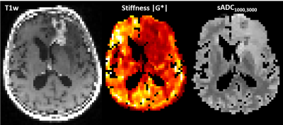 |
43 |
Testing the potential of DWI-based virtual elastography in
patients with brain cancer
Siri Fløgstad Svensson1,2,
Oliver Geier1,
Elies Fuster-Garcia1,3,
Gunnhild Ager-Wick1,
Robin Anthony Birkeland Bugge1,
Anne-Hilde Farstad4,
Karoline Skogen5,
and Kyrre Eeg Emblem1
1Department for Physics and Computational Radiology, Oslo University Hospital, Oslo, Norway, 2Department for Physics, University of Oslo, Oslo, Norway, 3Universitat Politècnica de València, BDSLab, Instituto Universitario de Tecnologías de la Información y Comunicaciones, València, Spain, 4Department for Radiography, Oslo University Hospital, Oslo, Norway, 5Department for Radiology, Oslo University Hospital, Oslo, Norway Keywords: Elastography, Elastography A previous study in the liver suggested that DWI can be used for virtual elastography. Here, we investigate the potential correlations between DWI-derived parameters and MR elastography (MRE) stiffness measurements in sixteen patients with brain cancer. The highest cross-correlation was obtained between stiffness and the shifted ADC map with b-values 1000 and 3000 s/mm² (median cross correlation 0.91). However, no correlation between mean values of MRE and sADC1000,3000 was found in normal-appearing white and gray matter, nor in tumor regions. This could be due to a too simple diffusion model and the anisotropic tissue structure of the brain. |
|
5146.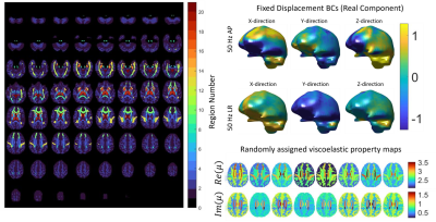 |
44 |
Realistic simulated MR Elastography data generated from in vivo
brain images
Dhrubo Jyoti1,
Diego Caban-Rivera2,
Mary Kramer2,
Alexa Diano2,
Curtis Johnson2,
Elijah Van Houten3,
Keith Paulsen1,
and Matthew Mcgarry1
1Dartmouth College, Hanover, NH, United States, 2University of Delaware, Newark, DE, United States, 3University of Sherbrooke, Sherbrooke, QC, Canada Keywords: Elastography, In Silico Simulations can play an important role in validating the accuracy of MR elastography (MRE) inversion algorithms. A realistic brain MRE simulation is created through a finite element model from in vivo MRE data, with 20 randomly generated simulated brains created by assigning properties of anatomical segmentations of brain structures within a range given by a published MRE atlas. Multi-frequency and multi-direction synthetic MRE data was generated via boundary conditions from in vivo measured motions to allow a range of inversion algorithms to be tested. These data will be supplied to the MRE study group for an inversion reconstruction challenge. |
|
5147.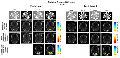 |
45 |
Characterization of Functional MR Elastography Responses to
Variations in Visual Stimulus Frequency and Contrast
Harish Palnitkar1,
Matthew C Murphy1,
Yi Sui1,
Kevin J Glaser1,
Armando Manduca1,
John Huston 3rd1,
Richard L Ehman1,
and Arvin Arani1
1Department of Radiology, Mayo Clinic, Rochester, MN, United States Keywords: Elastography, Elastography, Functional MR Elastography The response of BOLD fMRI to changes in both visual contrast frequency and contrast intensity have been well documented. Recently, an increase in the stiffness of activated regions of the visual cortex due to a controlled variation of block duration has been demonstrated with fMRE elastograms. The current study aims to investigate the relationship between BOLD fMRI and fMRE due to a controlled variation of both contrast intensity and contrast frequency of a visual stimulation. This study demonstrates that the fMRE-measured stiffness change varies linearly with the underlying neural activity, which can be modulated by stimulus parameterization. |
|
5148.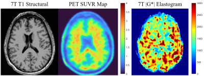 |
46 |
A Biomechanical Analysis Framework for Co-Correlation of 7T MR
Elastography Measures and Amyloid Beta Deposition
Emily Triolo1,
Mackenzie Langan2,3,
Oleksandr Khegai2,
Akbar Alipour2,
Carolina Ferreira-Atuesta3,
Aymeric Pionteck1,
Jonathan Sutkowski3,
Trey Hedden3,
Priti Balchandani2,
and Mehmet Kurt1,2
1Mechanical Engineering, University of Washington, Seattle, WA, United States, 2Biomedical Engineering and Imaging Institute, Ichan School of Medicine at Mount Sinai, New York City, NY, United States, 3Graduate School of Biomedical Sciences, Ichan School of Medicine at Mount Sinai, New York City, NY, United States Keywords: Elastography, Brain, 7T, Alzheimer's Disease, PET Previous studies have implied the future use of MRE metrics to track disease progression and as a tool for AD diagnosis, potentially replacing or augmenting methods that use ionizing radiation. In this pilot study, we have therefore developed a novel framework for performing ultrahigh field (7T) MRE at high resolution on subjects who have previously undergone PET scans and performing joint analysis of biomechanical and pathologic markers on these subjects. We have successfully created a framework for measurement of high-resolution brain mechanical properties for co-correlation with traditional PET measures to be used on AD patients in the future. |
|
5149.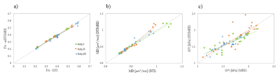 |
47 |
Development of in vivo human brain multifrequency DTIMRE
(mDTIMRE)
Shujun Lin1,
Bradley Sutton2,
Richard Magin1,
Aaron Anderson2,
and Dieter Klatt1
1Biomedical Engineering, University of Illinois Chicago, Chicago, IL, United States, 2Beckman Institute, University of Illinois Urbana-Champaign, Urbana, IL, United States Keywords: Elastography, Elastography Simultaneous acquisition of diffusion tensor imaging and multifrequency magnetic resonance elastography was examined in this preliminary study on in vivo human brain. The mDTIMRE was achieved by ensuring the same diffusion encoding using experiment parameters with different mechanical frequencies, while fulfilling the timing condition of simultaneous acquisition. By applying a multifrequency dual elasto-visco inversion approach, the effective image resolution of mechanical property maps is improved and enables quantitatively assessment of tissue properties in brain sub-regions. The experiment results show a good correlation between mDTIMRE and conventional measurements. |
|
5150.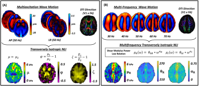 |
48 |
Transversely Isotropic MR Elastography with a Power Law
Multi-Frequency Reconstruction
Diego A. Caban-Rivera1,
Elijah E. W. Van Houten2,
Matthew D. J. McGarry3,
L. Tyler Williams1,
Phil V. Bayly4,
Keith D. Paulsen3,
and Curtis L. Johnson1
1Biomedical Engineering, University of Delaware, Newark, DE, United States, 2Département de Génie Mécanique, Université de Sherbrooke, Sherbrooke, QC, Canada, 3Thayer School of Engineering, Dartmouth College, Hanover, NH, United States, 4McKelvey School of Engineering, Washington University in St. Louis, St. Louis, MO, United States Keywords: Elastography, Brain This study combined multiexcitation MRE (ME-MRE), and multifrequency MRE (MF-MRE) with a transversely isotropic nonlinear inversion (TINLI) and axonal fiber directions from diffusion tensor imaging to investigate anisotropic and frequency-dependent material properties simultaneously. These preliminary results show that multifrequency-TINLI MRE can be readily applied to in vivo human data at distinct frequencies. |
|
5151.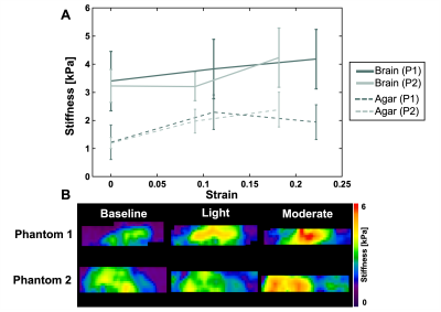 |
49 |
Response of Brain Tissue Mechanical Properties to Applied
Pre-Strain in Magnetic Resonance Elastography
Olivia M Bailey1,
Alexa M Diano1,
Ali H Lateef1,
Elise A Corbin1,
and Curtis L Johnson1
1Biomedical Engineering, University of Delaware, Newark, DE, United States Keywords: Elastography, Traumatic brain injury This study uses magnetic resonance elastography (MRE) with incremental compression to measure the apparent viscoelastic properties of bovine brain tissue as pre-strain is applied. Results from our tissue-agar phantom showed an increase in stiffness, and a decrease in damping ratio for the bovine brain. The agar showed an increase in stiffness and no consistent change in damping ratio. This research serves as the foundation for future studies measuring the nonlinear properties of human cadaveric brain tissue. Output from this work will be instrumental in advancing current traumatic brain injury (TBI) models. |
|
5152.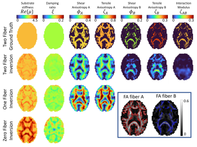 |
50 |
Anisotropic MR elastography with a DTI-informed crossing fiber
mechanical model
Matthew Mcgarry1,
Damian Sowinski1,
Diego Caban-Rivera2,
Elijah Van Houten3,
Curtis Johnson2,
Keith Paulsen1,
and Philip Bayly4
1Dartmouth College, Hanover, NH, United States, 2University of Delaware, Newark, DE, United States, 3University of Sherbrooke, Sherbrooke, QC, Canada, 4Washington University in St Louis, St Louis, MO, United States Keywords: Elastography, White Matter Anisotropic MRE has shown some promise in estimating mechanical properties of fiber-reinforced biological tissues. However, these methods are restricted to modeling a single fiber and ignores complexity that occurs such as regions of brain white matter with crossing fibers. Here we implement an inversion algorithm capable of modeling material with two fiber directions obtained from diffusion MRI in order to reduce model data mismatch and provide fiber-specific properties, which may show promise in correlating with brain health and function. Performance of this algorithm is demonstrated in simulation and in vivo brain data and compared with one-fiber and zero-fiber (isotropic) inversions. |
|
5153. |
51 |
Improved Brain MRE Performance on a Compact, Lightweight 3T
Scanner with High-Performance Gradients
Yi Sui1,
Matthew C. Murphy1,
Ziying Yin1,
Kevin J. Glaser1,
Richard L. Ehman1,
Matt A. Bernstein1,
John Huston III1,
and Yunhong Shu1
1Department of Radiology, Mayo Clinic, Rochester, MN, United States Keywords: Elastography, Brain, MRE, Compact 3T We conducted a direct comparison of Brain MRE performance between a compact, lightweight 3T scanner (C3T) and a whole-body scanner (WB3T). We demonstrate in phantom and volunteers that the C3T scanner provides superior image quality and scanning efficiency for commonly performed brain MRE pulse sequences, especially for higher-resolution scanning. |
|
5154.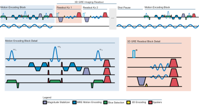 |
52 |
EMPIRE: Elastographic Magnetization Prepared Imaging with Rapid
Encoding
Alex M Cerjanic1,2,
Alexa M Diano2,
Matthew DJ McGarry3,
and Curtis L Johnson2
1Department of Medicine, Massachusetts General Hospital, Boston, MA, United States, 2Department of Biomedical Engineering, University of Delaware, Newark, DE, United States, 3Dartmouth College, Hanover, NH, United States Keywords: Elastography, Data Acquisition This study demonstrates the use of magnetization preparation for MR elastography in combination with a 3D GRE stack of spirals pulse sequence. By decoupling the motion encoding from the readouts, rapid encoding strategies, such as 3D GRE, can be used. MRE images, displacement, and inverted elastograms are obtained on in vivo human brain. 3D GRE readouts with EMPIRE motion encoding can acquire a complete MRE dataset 33% faster than 2D SE-EPI with similar parallel imaging acceleration factors. |
|
5155.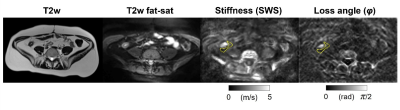 |
53 |
Mesenteric Adipose Tissue in Crohn’s Disease assessed by in vivo
MR Elastography
Rolf Reiter1,
Florian Nino Loch2,
Carsten Kamphues3,
Mehrgan Shahryari1,
Stephan Rodrigo Marticorena Garcia1,
Britta Siegmund4,
Carl Weidinger4,
Anja Kühl5,
Bernd Hamm1,
Jürgen Braun6,
Ingolf Sack1,
Patrick Asbach1,
and Laura Jensen1
1Radiology, Charité - Universitätsmedizin Berlin, Berlin, Germany, 2Surgery, Charité - Universitätsmedizin Berlin, Berlin, Germany, 3Surgery, Parkklinik Weißensee, Berlin, Germany, 4Gastroenterology, Infectious Diseases and Rheumatology, Charité - Universitätsmedizin Berlin, Berlin, Germany, 5iPATH.Berlin-Immunopathology for Experimental Models, Charité - Universitätsmedizin Berlin, Berlin, Germany, 6Medical Informatics, Charité - Universitätsmedizin Berlin, Berlin, Germany Keywords: Elastography, Body, inflammatory bowel disease Despite increasing evidence that the functional involvement and structural changes of mesenteric adipose tissue influence the course of Crohn's disease, its viscoelastic properties remain elusive. We demonstrate the feasibility of in vivo MR elastography of mesenteric adipose tissue and present preliminary reference values for Crohn's disease patients and healthy controls in this explorative study. Our preliminary results show an excellent diagnostic performance in detecting Crohn's disease by assessing the viscoelastic properties of mesenteric adipose tissue using histopathology of surgical specimens as reference. Our results motivate further studies for the biophysical characterization of mesenteric adipose tissue in inflammatory bowel disease. |
|
5156.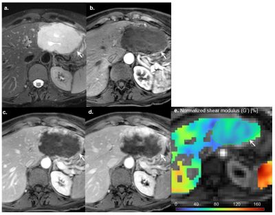 |
54 |
Intrinsic MR elastography for classification of focal liver
lesions using normalized viscoelastic parameters: a pilot study
Amirhosein Baradaran Najar1,2,
Elige Karam2,3,
Guillaume Gilbert4,
Maxime Barat2,3,
Emmanuel Montagnon3,
Hélène Castel5,
Jeanne-Marie Giard5,
Bich Nguyen6,
Guy Cloutier2,7,8,
An Tang2,7,
and Elijah Van Houten1,9
1Département du génie mécanique, Université de Sherbrooke, Sherbrooke, QC, Canada, 2Centre de recherche du Centre hospitalier de l'Université de Montréal (CRCHUM), Montréal, QC, Canada, 3Department of Radiology, Radiation Oncology and Nuclear Medicine, Université de Montréal, Montréal, QC, Canada, 4Philips Healthcare, Mississauga, ON, Canada, 5Centre Hospitalier de l’Université de Montréal (CHUM), Montréal, QC, Canada, 6Service of Pathology, Centre Hospitalier de l’Université de Montréal (CHUM), Montréal, QC, Canada, 7Institute of Biomedical Engineering, Université de Montréal, Montréal, QC, Canada, 8Laboratory of Biorheology and Medical Ultrasonics(LBUM), Centre de recherche du Centre hospitalier de l’Université de Montréal (CRCHUM), Montréal, QC, Canada, 9Centre de recherche du Centre hospitalier de l'Université de Sherbrooke (CRCHUS), Sherbrooke, QC, Canada Keywords: Elastography, Elastography, Intrinsic magnetic resonance elastography, Cancer Intrinsic activation MR elastography (iMRE) method has been applied to a cohort of patients undergoing liver MRI for assessment of focal liver lesions. We used segmentation, masking, and normalization of viscoelastic parameters to investigate the capability of this method to differentiate benign and malignant liver lesions. The normalized storage modulus (G') computed by nonlinear inversion-iMRE showed significant differences between benign and malignant lesions. |
|
5157.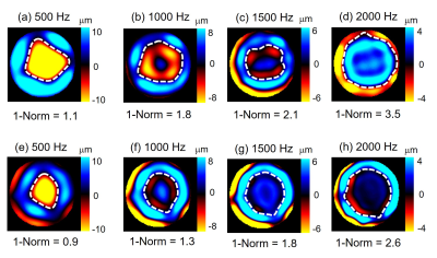 |
55 |
Quantifying inhomogeneity of kidney, liver and muscle before and
after the freeze-thaw cycle using 1-Norm waveform analysis and
MR elastography
Rolf Reiter1,2,
Harish Palnitkar3,
Shreyan Majumdar4,
Joseph Crutison4,
Shujun Lin4,
Thomas J. Royston4,
and Dieter Klatt4
1Radiology, Charité - Universitätsmedizin Berlin, Berlin, Germany, 2BIH Biomedical Innovation Academy, BIH Charité Digital Clinician Scientist Program, Berlin Institute of Health at Charité – Universitätsmedizin Berlin, Berlin, Germany, 3Radiology, Mayo Clinic, Rochester, MN, United States, 4Richard and Loan Hill Department of Biomedical Engineering, University of Illinois Chicago, Chicago, IL, United States Keywords: Elastography, Tissue Characterization The recently developed 1-Norm technique can be used to quantify the extend of scattering of mechanical waves generated by inhomogeneities. This explorative study shows that 1-Norm can detect mechanical inhomogeneity of ex vivo kidney, liver and muscle without the need to use ill-posed wave inversion techniques. The 1-Norm technique has potential to serve as an MRE-based diagnostic biomarker independent of stiffness for the characterization of pathological conditions associated with changes in tissue mechanical inhomogeneity. |
|
5158.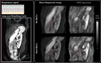 |
56 |
Sagittal free-breathing MR elastography of the pancreas with
motion-correction at 3T
Anne-Sophie van Schelt1,2,
Nienke Petronella Maria Wassenaar1,
Jurgen H. Runge1,
Jaap Stoker1,2,3,
Aart J Nederveen1,
and Eric M Schrauben1
1Radiology and Nuclear Medicine, Amsterdam UMC, location AMC, Amsterdam, Netherlands, 2Imaging and Biomarkers, Cancer Center Amsterdam, Amsterdam, Netherlands, 3Endocrinology, Metabolism, Amsterdam Gastroenterology, Amsterdam, Netherlands Keywords: Elastography, Pancreas Breathing motion can be detrimental to quantitative accuracy in pancreatic MR elastography (MRE). We investigated motion-correction strategies on sagittal multi-frequency free-breathing SE-EPI MRE acquisitions to mitigate breathing-motion and increase accuracy. Breathing-states were determined through a respiratory-belt and multiple number of bins were tested. Three intensity-based registration methods (with and without non-rigid post-processing) and non-rigid registration were ranked on MRE quality measures. The best method showed improved data quality, inversion precision and repeatability compared to no correction, but resulted in apparently increased shear wave speed (SWS), which warrants further investigation. |
|
5159.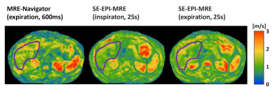 |
57 |
MRE navigator for rapid estimation of liver stiffness.
Matthias Anders1,
Carsten Warmuth1,
Josef Pfeuffer2,
Heiko Tzschätzsch1,
Helge Herthum1,
Katja Degenhardt3,
Oliver Wieben4,
Sebastian Schmitter3,
Jeanette Schulz-Menger1,5,6,
Jürgen Braun1,
and Ingolf Sack1
1Charité - Universitätsmedizin Berlin, Berlin, Germany, 2MR Application Development, Siemens Healthcare, Erlangen, Germany, 3Physikalisch-Technische Bundesanstalt(PTB), Braunschweig and Berlin, Berlin, Germany, 4University of Wisconsin, Madison, WI, United States, 5Experimental and Clinical Research Center (ECRC), DZHK partner site Berlin, Berlin, Germany, 6HELIOS Klinikum Berlin Buch, Berlin, Germany Keywords: Elastography, Data Acquisition MR elastography (MRE) can noninvasively detect liver fibrosis based on elevated stiffness values. However, a complete MRE scan is time-consuming and typically extends over multiple breath-holds. Therefore, a multi-shot gradient echo sequence with spiral readout was developed to provide full two-dimensional elastograms of the liver in less than one second. The new sequence can be used as an MRE navigator to provide immediate feedback on wave penetration and data consistency for parameter optimization prior to running full multi-dimensional MRE. Moreover, the method can be used to rapidly track potential changes in liver stiffness such as those induced by respiration. |
|
5160.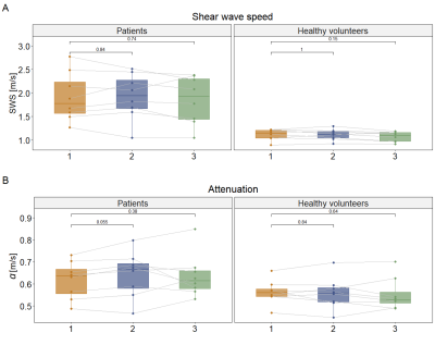 |
58 |
Repeatability of multifrequency MR elastography in pancreatic
ductal adenocarcinoma patients and healthy volunteers
Nienke P.M. Wassenaar1,2,
Anne-Sophie van Schelt1,2,
Eric M. Schrauben1,
Hanneke W.M. van Laarhoven2,3,
Jaap Stoker1,2,
Aart J. Nederveen1,
and Jurgen H. Runge1
1Radiology and Nuclear Medicine, Amsterdam University Medical Centers, Amsterdam, Netherlands, 2Imaging and Biomarkers, Cancer Center Amsterdam, Amsterdam, Netherlands, 3Medical Oncology, Amsterdam University Medical Centers, Amsterdam, Netherlands Keywords: Elastography, Pancreas MR elastography (MRE) could be useful as an imaging biomarker in pancreatic cancer. To this end, pancreatic MRE parameters should be repeatable and be able to differentiate between healthy and tumour tissue. In this study, 10 patients with pancreatic ductal adenocarcinoma and 8 sex- and age-matched healthy volunteers underwent three MRE scans in a test-retest set-up. Results showed that MRE parameters were repeatable in patients and healthy volunteers. Furthermore, a significant increase in shear wave speed was found in pancreatic tumours compared to healthy pancreatic tissue. |
|
5161. |
59 |
Imaging of Propagating Broadband Transient Waves with
Multi-scale MR Elastography Motion Encoding: A Validation Study
Yuan Le1,
Jun Chen1,
Phillip J. Rossman1,
Armando Manduca1,
Kevin J. Glaser1,
Bradley D. Bolster Jr.2,
Stephan Kannengiesser3,
and Richard L. Ehman1
1Radiology, Mayo Clinic, Rochester, MN, United States, 2US MR R&D Collaboration, Siemens Medical Solutions Inc., Salt Lake City, UT, United States, 3MR Application Predevelopment, Siemens Healthcare GmbH, Erlangen, Germany Keywords: Elastography, Elastography, Transient wave, multi-scale encoding We demonstrate imaging of the propagation of transient waves using multi-scale motion encoding gradient waveforms. Displacement values are calculated using the inverse Haar transforms. We validated the results by comparison with wave images obtained using standard MRE acquisition and processing. The approach provides the ability to image broadband motion more efficiently and accurately compared with previous methods and promises to be a useful approach for biomechanical studies of traumatic brain injury. |
|
5162.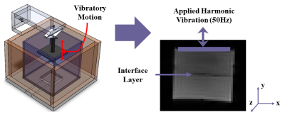 |
60 |
Utilizing MR Elastography to Visualize Higher Harmonic
Components of Wave Propagation in Phantoms with Interfaces
Emily Triolo1,
Melih Eriten2,
Curtis Johnson3,
and Mehmet Kurt1,4
1Mechanical Engineering, University of Washington, Seattle, WA, United States, 2Mechanical Engineering, University of Wisconsin–Madison, Madison, WI, United States, 3Biomedical Engineering, University of Delaware, Newark, DE, United States, 4Biomedical Engineering and Imaging Institute, Ichan School of Medicine at Mount Sinai, New York City, NY, United States Keywords: Elastography, Phantoms, Wave Propagation, Biointerface While MRE can be used to calculate mechanical properties of tissue, bio-interfaces can cause distortions in the shear-wave propagation patterns which are difficult to account for with current MRE techniques. Therefore, we utilized three specialized MRE sequences to visualize the first three harmonics of wave propagation in phantoms with simulated bio-interfaces. We were able to measure the doubling and tripling of wave number in the phantoms excited at the fundamental vibration frequency using this optimized MRE protocol. We characterized differences in wave propagation at higher harmonics along phantom interfaces, and observed substantial differences in wave characteristics based on adhesion level. |
|
 Back to Meeting Home
 Back to the Program-at-a-Glance
The International Society for Magnetic Resonance in Medicine is accredited by the Accreditation Council for Continuing Medical Education to provide continuing medical education for physicians.
 Back to Meeting Home
 Back to the Program-at-a-Glance
 View Presentation Video