Digital Poster
fMRI: Acquisition, Analysis, Applications
ISMRM & ISMRT Annual Meeting & Exhibition • 03-08 June 2023 • Toronto, ON, Canada

Digital Poster
fMRI: Acquisition, Analysis, Applications
| Computer # | |||
|---|---|---|---|
3838.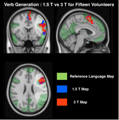 |
81 |
FMRI-based language mapping at 1.5 and 3T: the influence of
methodological choices.
Khaliesah Bolhassan1,2,
Rachael Franklin3,
Enrico De Vita4,
Jozef Jarosz2,
and Marco Borri2
1School of Biomedical Engineering & Imaging Sciences, King's College London, London, United Kingdom, 2Neuroradiology, King's College Hospital, London, United Kingdom, 3MRI Physics, Guy’s and St Thomas NHS Foundation Trust, London, United Kingdom, 4Radiology Department, Great Ormond Street Hospital for Children, London, United Kingdom Keywords: fMRI (task based), Data Analysis This work aims to assess the influence of methodological aspects in fMRI language mapping at 1.5T and 3T. Probability maps from healthy volunteer cohorts were compared to a published reference using a Dice Index vs probability threshold plot. T-value normalisation to the peak within the classic Broca/Wernicke’s language area as well as the inclusion of additional language tasks increased overlap to the reference. The extent of reproducible activation was larger at 3T. |
|
3839. |
82 |
Dynamic analysis of resting-state brain fMRI signal from a novel
perspective: Avalanche
Junlin Guo1,
Nazirah Mohd Khairi1,
Lyuan Xu1,2,
and Don Mitchell Wilkes1
1Electrical and Computer Engineering, Vanderbilt University, Nashville, TN, United States, 2Vanderbilt University Institute of Imaging Science, Vanderbilt University Medical Center, Nashville, TN, United States Keywords: fMRI (resting state), Brain, Spatiotemporal The human brain, at rest, is complex and many functional studies focus on the brain patterns around criticality. This work addresses a novel perspective, the avalanche, in resting-state fMRI. In this work, we design two data-driven signal modeling approaches that dynamically measure and visualize the signal entropy from both spatial and temporal aspects. The first approach applies a clustering-based scheme with the Markov chain. The second method utilizes the autoregressive model with a sliding window. The results show a consistent, less complex pattern at the avalanche state, from which the interpretation of the brain can be clearer than at criticality. |
|
3840.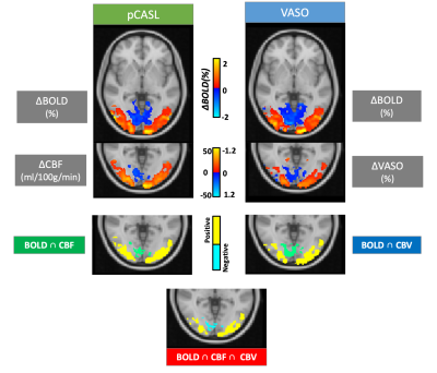 |
83 |
Investigation of cerebral blood flow and volume changes during
positive and negative BOLD responses at 3T
Ratnamanjuri Devi1,
Torsten Schlumm1,
Toralf Mildner1,
and Harald E Möller1
1Max Planck Institute for Human Cognitive and Brain Sciences, Leipzig, Germany Keywords: fMRI (task based), Arterial spin labelling, Vascular Space Occupancy CBF and CBV changes were measured sequentially, in regions of positive and negative BOLD responses, using a multi-echo center-out readout, known to reduce BOLD contaminations in pCASL and VASO measurements due to its short echo times. The CBV-CBF coupling was found to differ between positive and negative BOLD regions. The temporal relation of the CBF and VASO timecourses to their corresponding T2* timecourses, on the other hand, was found to be almost identical in the two ROIs. |
|
3841.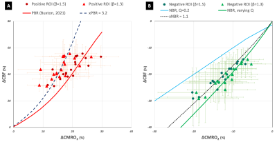 |
84 |
Evaluation of neuronal feed-forward control of cerebral blood
flow in negative BOLD response
Ratnamanjuri Devi1,
Jöran Lepsien1,
Kathrin Lorenz1,
Torsten Schlumm1,
Toralf Mildner1,
and Harald E Möller1
1Max Planck Institute for Human Cognitive and Brain Sciences, Leipzig, Germany Keywords: fMRI (task based), Arterial spin labelling The application of a simplistic model of neuronal control of changes in cerebral blood flow and oxygen metabolism to experimental data in regions of the positive and negative BOLD response, suggested differences in neuronal contributions and inhibitory control of changes in cerebral blood flow between the two regions. |
|
3842.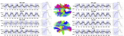 |
85 |
Whole-brain, gray and white matter time-locked functional signal
changes with simple tasks and model-free analysis
Kurt G Schilling1,
Muwei Li1,
Francois Rheault2,
Yurui Gao1,
Leon Y Cai3,
Yu Zhao1,
Zhongliang Zu1,
Zhaohua Ding1,
Adam W Anderson3,
Bennett A Landman3,
and John C Gore1
1Vanderbilt University Medical Center, Nashville, TN, United States, 2Sherbrooke University, Sherbrooke, QC, Canada, 3Vanderbilt University, Nashville, TN, United States Keywords: fMRI, White Matter Recent studies have revealed that blood oxygenation-level dependent (BOLD) signal changes correlate with task timings throughout a majority of the cortex, challenging the idea of sparse and localized brain functions, and highlighting the pervasiveness of potential false negative fMRI findings. However, these studies have focused on gray matter only. We extend the analysis to white matter and find widespread BOLD signal changes in both white and gray matter, challenging the idea of sparse functional localization, but also the prevailing wisdom of treating white matter BOLD signals as artefacts to be removed. |
|
3843.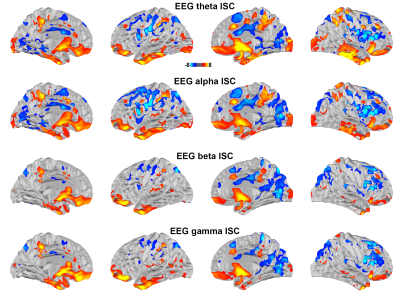 |
86 |
Neurovascular coupling of the human brain in complex
naturalistic stimuli viewing
Hsin-Ju Lee1,2,
Emily Rutledge1,3,
Jiahui Veron Cheng1,4,
Lauri Nummenmaa5,
Severi Santavirta5,
and Fa-Hsuan Lin1,2
1Physical Sciences Platform, Sunnybrook Research institute, Toronto, ON, Canada, 2Department of Medical Biophysics, University of Toronto, Toronto, ON, Canada, 3University of Waterloo, Waterloo, ON, Canada, 4University of Toronto, Toronto, ON, Canada, 5Turku PET Centre, University of Turku, Turku, Finland Keywords: fMRI (task based), fMRI (task based), EEG-fMRI Simultaneously acquired EEG and fMRI data allow for elucidating the neurovascular coupling. Here, we use temporally sparse fast fMRI and continuous EEG to study the correlations between hemodynamic signals and neural oscillations across the cerebral cortex during movie watching. |
|
3844.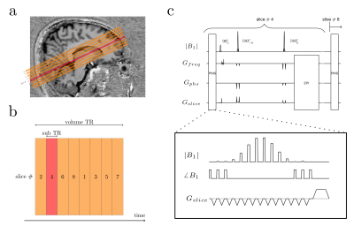 |
87 |
Using PINS pulses to saturate inflow effects from slice gaps in
functional MRI
Shota Hodono1,
Chia-yin Wu1,2,3,
Carl Dixon1,
Donald Maillet1,
Jin Jin4,
Jonathan R Polimeni5,6,
and Martijn A Cloos1
1Centre for Advanced Imaging, The University of Queensland, St Lucia, Australia, 2ARC Training Centre for Innovation in Biomedical Imaging Technology, The University of Queensland, St. Lucia, Australia, 3chool of Information Technology and Electrical Engineering, The University of Queensland, St. Lucia, Australia, 4Siemens Healthcare Pty Ltd, Brisbane, Australia, 5Athinoula A. Martinos Center for Biomedical Imaging, Massachusetts General Hospital, Charlestown, MA, United States, 6Harvard-MIT Division of Health Sciences and Technology, Massachusetts Institute of Technology, Cambridge, MA, United States Keywords: fMRI, Velocity & Flow In this work, we demonstrate the use of PINS pulses to mitigate unwanted inflow effects in 2D multi-slice sequences. A PINS pulse was played prior to slice-selective excitation to saturate the magnetization within all slice gaps. Bloch simulation and flow-phantom experiments show inflow effects can be removed completely. Functional scans using twice-refocused spin echo suggest that inflow effects may have noticeable contributions to SE-BOLD fMRI response. This PINS implementation allows us to further understand fMRI signal dynamics by modulating the inflow effect. |
|
3845.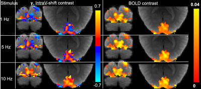 |
88 |
Intravascular Shifted fMRI Contrast for Visual Stimulations at 3
Tesla Using DANTE-Prepared Dual-Echo EPI
Linqing Li1,
Yuhui Chai2,
Andy John Derbyshire1,
and Peter Bandettini3
1Functional MRI Facility, National Institute of Mental Health, Bethesda, MD, United States, 2Beckman Institute for Advanced Science and Technology, University of Illinois at Urbana-Champaign, Urbana, IL, United States, 3Section on Functional Imaging Methods, National Institute of Mental Health, Bethesda, MD, United States Keywords: fMRI, fMRI (task based), Intravascular fMRI contrast, extravascular fMRI contrast By using dual-echo DANTE-EPI for functional visual stimulation studies, the contrast of fractions of intravascular signal to total signal change can be generated. We show an intravascular shifted (IVS) contrast can provide simultaneous visualization of intra-extra vascular contrast under different frequencies of visual stimulations at 3T. |
|
3846.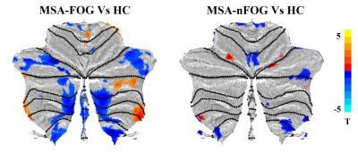 |
89 |
Cerebellar gray matter volume and cerebellum-cerebral
connectivity associated with freezing of gait in patients with
multiple system atrophy
Huaguang Yang1,
Weiyin Vivian Liu2,
Liang Li1,
Zhi Wen1,
Lanhua Hu1,
XiaoGuang Luo3,
and Yunfei Zha1
1Renmin Hospital of Wuhan University, Wuhan, China, 2MR Research, GE Healthcare, Wuhan, China, 3Department of Neurology, The First Affiliated Hospital of South University of Science and Technology, The Second Clinical Medical College of Jinan University, Shenzhen People's Hospital, ShenZhen, China Keywords: fMRI (resting state), Parkinson's Disease Cerebellum is responsible for posture and gait control. 65.93% of patients with multiple system atrophy experienced FOG. Patients with the damaged cerebellar locomotor region had gait-freezing-like symptoms, suggesting that the cerebellum may be involved in the occurrence and development of FOG symptoms. This study suggested that the cerebellum volume atrophy may be involved in FOG development in MSA patients and subsequently induce functional abnormality in the cerebellum-cerebral circuit. This study provided neuroimaging evidence for clinical understanding of cerebellum role in MSA patients with FOG injury. |
|
3847.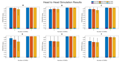 |
90 |
Quantifying Organizational Changes of Functional Connectivity
Linked to Hypertension in Resting-State Functional MRI
William D. Reeves1,
Ishfaque Ahmed1,
Brooke S. Jackson2,
Wenwu Sun1,
Michelle L. Brown3,
Celestine F. Williams3,
Catherine L. Davis3,
Jennifer E. McDowell2,
Nathan E. Yanasak4,
Shaoyong Su3,
and Qun Zhao1
1Department of Physics, University of Georgia, Athens, GA, United States, 2Department of Psychology, University of Georgia, Athens, GA, United States, 3Georgia Prevention Institute, Medical College of Georgia, Augusta, GA, United States, 4Department of Radiology and Imaging, Medical College of Georgia, Augusta, GA, United States Keywords: fMRI (resting state), Hypertension Using a graph theory approach, functional connectivity differences in a 52 (32 hypertensive, 20 normotensive) subject cohort were tracked using resting-state fMRI. A null generation modification to a difference degree test (DDT) is profiled that resulted in increased true positivity rates in simulations while maintaining nominal false positivity rates. Applying the modified DDT to the hypertension cohort resulted in the discovery of 7 brain regions that exhibit significant groupwise differential expression along with 33 unique differentially expressed connections to other areas in the brain. The results presented agree with previous studies and represent a promising application of the modified DDT. |
|
3848.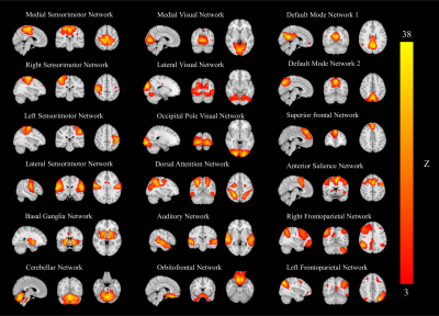 |
91 |
Altered resting state networks associated with REM sleep
behavioral disorder in Parkinson’s disease
Destaw Bayabil Mekbib1,
Miao Cai2,
Weiying Dai3,
Xiaoli Liu4,
and Li Zhao1
1Key Laboratory for Biomedical Engineering of Ministry of Education, Zhejiang University, Hangzhou, China, 2Neurology, Zhejiang Hospital, Hangzhou, China, 3Computer Science, Binghamton Univeristy, State University of New York, Binghamton, NY, United States, 4Zhejiang Hospital, Hangzhou, China Keywords: fMRI (resting state), Parkinson's Disease Resting-state functional MRI has played a fundamental role in the study of Parkinson’s disease (PD) with rapid eye movement sleep behavior disorder (RBD). In this work, we investigated changes in functional connectivity within and between resting-state networks and their relationship to global fluctuations in 13 PD with RBD, 28 PD without RBD, 6 RBD without PD, and 20 healthy controls. Based on an in-house reliable data selection strategy, unique effects on the lateral visual network in RBD patients and on the sensorimotor network in PD patients were found. These may provide novel clues to understanding the pathology and improve diagnosis. |
|
3849.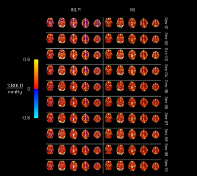 |
92 |
Bayesian and lagged general linear modelling strategies in
breath-hold induced cerebrovascular reactivity mapping with
muti-echo BOLD fMRI
Genevieve Hayes1,
Joana Pinto1,
Stefano Moia2,
Martin Craig3,
Michael Chappell3,
Cesar Caballero-Gaudes2,
and Daniel P Bulte1
1Institute of Biomedical Engineering, University of Oxford, Oxford, United Kingdom, 2Basque Centre on Cognition, Brain and Language, Donostia-San Sebastian, Spain, 3Sir Peter Mansfield Imaging Centre, University of Nottingham, Nottingham, United Kingdom Keywords: fMRI, Blood vessels, Cerebrovascular reactivity Cerebrovascular reactivity (CVR), the ability of blood vessels to dilate and constrict in response to a vasoactive stimulus, is an important indicator of cerebrovascular health and can be estimated using BOLD functional MRI. In this work, we evaluated a novel variational Bayesian method for mapping CVR dynamics which yielded similar CVR results and reproducibility to a time-shifted, “lagged” general linear model approach, and uncovered the need for more research into negative CVR. This novel approach is more time efficient and has the potential to improve CVR mapping by incorporating non-linear modelling and physiologically meaningful information (in the form of priors). |
|
3850.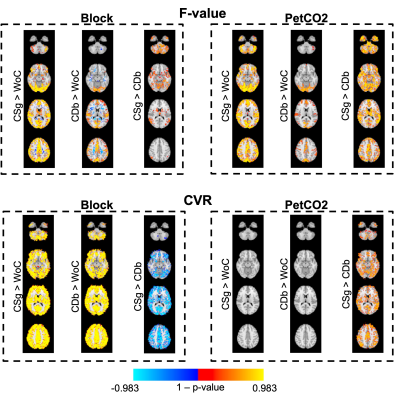 |
93 |
Cerebrovascular reactivity mapping using breath-hold BOLD-fMRI:
comparison of signal models when using voxelwise lag
optimization
Catarina Domingos1,
Inês Esteves1,
Ana R. Fouto1,
Amparo Ruiz-Tagle1,
Gina Caetano1,
and Patrícia Figueiredo1
1Institute for Systems and Robotics - Lisboa and Department of Bioengineering, Instituto Superior Técnico, Universidade de Lisboa, Lisbon, Portugal Keywords: fMRI, fMRI We investigate the best modeling approach for mapping cerebrovascular reactivity (CVR) using voxelwise lag optimization. We considered two types of regressors (Block, PetCO2), three convolution models (no convolution, single gamma, double gamma), and a variable haemodynamic delay. We found that the CVR values obtained when using the PetCO2 signal without convolution, or convolved with a single gamma, are better than those obtained with the canonical HRF (double gamma, time-to-peak = 6s), while convolution with a canonical HRF remains the best option when using a Block design. |
|
3851.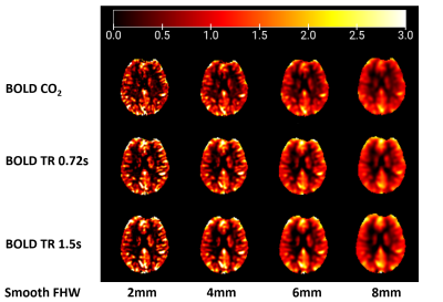 |
94 |
Cerebrovascular reactivity mapping using intermittent breath
modulation: temporal resolution dependence and comparisons with
CO2 inhalation
Beini Hu1,
Lori Donaldson1,
Parimal Joshi1,
Lincoln Kartchner1,
Sara Akar1,
and Peiying Liu1
1Department of Diagnostic Radiology and Nuclear Medicine, University of Maryland Baltimore, Baltimore, MD, United States Keywords: fMRI, Neuro Cerebrovascular reactivity (CVR) is typically measured using a carbon dioxide (CO2) stimulus combined with BOLD fMRI. However, this requires considerable subject cooperation. Although resting-state BOLD fMRI has shown its potential to generate CVR maps, the CVR results could be unreliable due to little fluctuation in some subjects’ spontaneous breathing. A new method utilizing intermittent breath modulation requires no gas-inhalation and presents higher sensitivity than resting-state CVR mapping. In this study, we investigated the effect of temporal resolution on CVR mapping obtained from breath modulation BOLD data. Our results showed that good CVR quality can be achieved with TR of 0.72s. |
|
3852.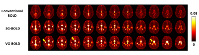 |
95 |
Toward vessel-suppressed cerebrovascular reactivity (CVR)
mapping by using crusher gradients
Yimei Cao1,
Hongli Fan1,
Cuimei Xu1,
and Hanzhang Lu1
1Department of Radiology, Johns Hopkins University, Baltimore, MD, United States Keywords: fMRI, fMRI Cerebrovascular reactivity (CVR) is an indicator of cerebrovascular reserve and provides important information about vascular health in a range of brain conditions and diseases. However, current CVR maps often have artifactual bright spots in arterial/venous areas due to the complexity of the BOLD signal mechanism. In this work, we proposed a novel CVR acquisition scheme by adding single-direction or varying-direction crusher gradients to the BOLD sequence. The results demonstrated that these modified sequences could successfully suppress large-vessel artifacts in CVR map. |
|
3853.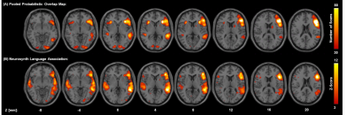 |
96 |
Clinical Functional MRI-derived Probabilistic Template of
Primary Language Areas of Patients with Brain Tumors
Jian Ming Teo1,2,
Jina Lee3,
Ping Hou1,
Vinodh A Kumar3,
Kyle R Noll4,
Sujit S Prabhu5,
and Ho-Ling Liu1
1Department of Imaging Physics, The University of Texas MD Anderson Cancer Center, Houston, TX, United States, 2The University of Texas MD Anderson Cancer Center UTHealth Graduate School of Biomedical Sciences, Houston, TX, United States, 3Department of Neuroradiology, The University of Texas MD Anderson Cancer Center, Houston, TX, United States, 4Department of Neuro-Oncology, The University of Texas MD Anderson Cancer Center, Houston, TX, United States, 5Department of Neurosurgery, The University of Texas MD Anderson Cancer Center, Houston, TX, United States Keywords: fMRI, Data Processing This study aims to develop a functional template of primary language areas based on 306 presurgical language fMRI scans from 102 patients with brain tumors. The template was constructed from voxelwise probabilistic distribution of fMRI results of three common clinical language paradigms. An independent dataset of 38 patients was used to test the template from this study and compare with a template derived from the meta-analysis of 1101 published studies. The results showed that our template agreed better with the test dataset, with significantly higher dice coefficients in both anterior and posterior primary language areas, comparing with the literature-based template. |
|
3854.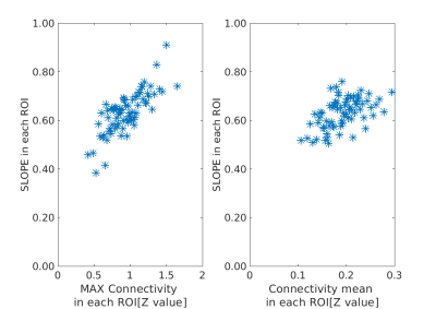 |
97 |
Slope of the spectrum of low-frequency BOLD signal relation to
the connectivity strength
Kazuhiro Nakamura1 and
Toshibumi Kinoshita1
1Research Institute for Brain and Blood Vessels Akita, Akita, Japan Keywords: fMRI (resting state), fMRI (resting state), spectrum analysis For the valuation of BOLD fluctuations, we have investigated the slope of the spectrum (SLOPE) to exclude the respiratory and heartrate fluctuation. Functional connectivity (ROI to ROI) was evaluated by Conn v19.b software. The SLOPE was evaluated in the frequency range of 0.01 – 0.1Hz. Maximum connectivity in each ROI correlated well with the value of SLOPE, but the mean connectivity did not. Therefore, it will be possible to evaluate the strength of connectivity of rs-fMRI by evaluating SLOPE. |
|
 Back to Meeting Home
 Back to the Program-at-a-Glance
The International Society for Magnetic Resonance in Medicine is accredited by the Accreditation Council for Continuing Medical Education to provide continuing medical education for physicians.
 Back to Meeting Home
 Back to the Program-at-a-Glance
 View Presentation Video