Digital Poster
CEST I
ISMRM & ISMRT Annual Meeting & Exhibition • 03-08 June 2023 • Toronto, ON, Canada

| Computer # | |||
|---|---|---|---|
2979.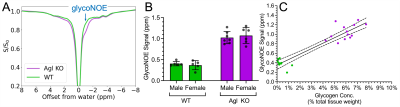 |
101 |
Characterization of glycogen storage disease type III using
glycoNOE MRI
Qing Zeng1,2,
Chongxue Bie1,2,
Peter C.M. van Zijl1,2,
Yuguo Li1,2,
Valentina D'souza1,2,
Michael Machado3,
Kirsten Achilles Poon3,
Andrew Grimm3,
and Nirbhay N. Yadav1,2
1The Russell H. Morgan Department of Radiology, The Johns Hopkins University School of Medicine, Baltimore, MD, United States, 2F.M. Kirby Research Center for Functional Brain Imaging, Kennedy Krieger Institute, Baltimore, MD, United States, 3Ultragenyx Pharmaceutical Inc., Novato, CA, United States Keywords: CEST & MT, CEST & MT, liver Glycogen storage disease type III (GSD III) is characterized by abnormally high glycogen accumulation in liver, muscle, and heart. Treatments such as diet modification and drugs are currently being developed, but there is a lack of suitable methods for assessing disease load and possible treatment efficacy. Recently, it was shown that glycoNOE MRI can image glycogen levels in vivo and here we apply this technique to distinguish GSD III from controls in a mouse model. The results show that glycoNOE can clearly distinguish GSD III and that glycoNOE contrast exhibits a linear correlation with ex vivo quantification of glycogen levels. |
|
2980.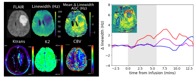 |
102 |
Dynamic Glucose Enhanced MRI of Brain Tumors using Direct Water
Saturation
Peter van Zijl1,
Nirbhay Yadav2,
Sajad Mohammed Ali3,
Anina Seidemo3,
David Olayinka Kamson4,
Lindsay Blair5,
John Laterra6,
and Linda Knutsson7
1Radiology, F.M. Kirby Research Center, Johns Hopkins University, Kennedy Krieger Institute, Baltimore, MD, United States, 2Radiology/F.M. Kirby Research Center, Johns Hopkins University, Kennedy Krieger Institute, Baltimore, MD, United States, 3Department of Medical Radiation Physics, Lund University, Lund, Sweden, 4Department of Oncology, Johns Hopkins University, Baltimore, MD, United States, 5Neurology, Johns Hopkins University, Baltimore, MD, United States, 6Neurology, Johns Hopkins University, Kennedy Krieger Institute, Baltimore, MD, United States, 7F.M. Kirby Research Center, Radiology, Medical Radiation Physics, Kennedy Krieger Institute, Johns Hopkins University, Lund University, Baltimore, MD, United States Keywords: CEST & MT, Cancer, Glucose The transverse relaxation time (T2) of water is affected by the presence of exchangeable protons that are chemically shifted with respect to the water protons. We use direct saturation (DS) MRI to dynamically measure this effect during infusion of D-glucose to assess its uptake in brain tumors. The change in T2 becomes apparent as a line broadening of the DS spectrum, which is acquired using a whole-brain water saturation shift reference (WASSR) acquisition. First results using 0.5g/kg D-glucose show linewidth changes on the order of a few Hertz in glioma patients, allowing separation of tumor tissue from healthy brain tissue. |
|
2981.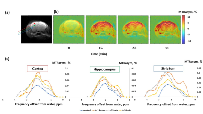 |
103 |
Towards Metabolic Brain Imaging with Glucosamine CEST MRI: In
Vivo Characterization and First Insights
Michal Rivlin1,
Or Perlman2,3,
and Gil Navon1
1School of Chemistry, Tel-Aviv University, Tel Aviv, Israel, 2Department of Biomedical Engineering, Tel Aviv University, Tel Aviv, Israel, 3Sagol School of Neuroscience, Tel-Aviv University, Tel Aviv, Israel Keywords: CEST & MT, Brain, Glucosamine, MRI, CEST, Metabolism, Brain Disorders The uptake of glucosamine (GlcN), a non-toxic food supplement, can be monitored by CEST MRI. While previously demonstrated in breast cancer, here we show that GlcN metabolism can be detected in the brain. Following GlcN administration in mice, the MTRasym signals were significantly elevated in the cortex, hippocampus, and striatum. A Lorentzian multi-pool fitting pointed to a significant increase in the hydroxyl, amide, and rNOE signals. An in vitro BSA study confirmed the interactions between brain compounds and GlcN shown in vivo. This study suggests that GlcN CEST has the potential to serve as a metabolic biomarker in brain disorders. |
|
2982.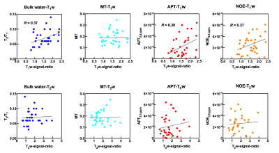 |
104 |
Evaluation of Biological Metabolic Activity within an
Atherosclerotic Plaque using Chemical Exchange Saturation
Transfer Imaging
Yuki Kanazawa1,
Tosiaki Miyati2,
Masafumi Harada1,
Mitsuharu Miyoshi3,
Yuki Matsumoto1,
Hiroaki Hayashi2,
Yasuhisa Kanematsu1,
and Yasushi Takagi1
1Tokushima University, Tokushima, Japan, 2Kanazawa University, Kanazawa, Japan, 3Global MR Applications and Workflow, GE Healthcare Japan, Hino, Japan Keywords: CEST & MT, Atherosclerosis We demonstrated biological metabolic activity within an atherosclerotic plaque of the carotid artery using CEST imaging. 35 patients with carotid stenosis, of which all were pathologically diagnosed with carotid endarterectomy, were evaluated. The following estimation parameters in CEST image were evaluated; bulk water, magnetization transfer, amide proton transfer (APT), and nuclear Overhauser effect (NOE). As a result, there were weak positive correlations between T1w-signal-ratio and the above-mentioned parameters: bulk water (R = 0.37), APT (R = 0.39), and NOE (R = 0.37). Multi-parametric analysis of CEST imaging can obtain detailed information concerning the activity in an atherosclerotic plaque. |
|
2983.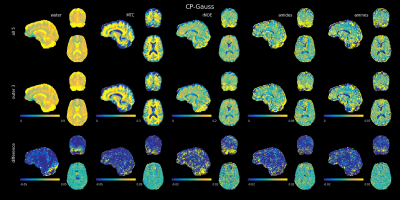 |
105 |
PUSHUP-CEST: Calibration free homogeneous saturation for
ulta-high field CEST imaging
Yannik Völzke1,
Rüdiger Stirnberg1,
Eberhard Pracht1,
Daniel Löwen1,
Vincent Gras2,
Nicolas Boulant2,
Moritz Zaiss3,
and Tony Stöcker1,4
1German Center for Neurodegenerative Diseases (DZNE), Bonn, Germany, 2Université Paris-Saclay, Commissariat à l’Energie Atomique, CNRS, NeuroSpin, Baobab,, Gif sur Yvette, France, 3Institute of Neuroradiology, University Hospital Erlangen, Friedrich-Alexander- Universität Erlangen-Nürnberg (FAU), Erlangen, Germany, 4Department of Physics and Astronomy, University of Bonn, Bonn, Germany Keywords: CEST & MT, CEST & MT CEST imaging benefits from the increased spectral resolution at ultra-high field. Due to the inhomogeneous RF field, a B1+-correction is necessary, which requires repeated measurements. The number of required repetitions can be reduced to two by using a ptx-based saturation scheme, like MIMOSA. We present an alternative saturation scheme that is based on PUSH saturation and universal pulses, which we name PUSHUP. Using this approach, we could reduce the inhomogeneity of the CEST saturation as effectively as MIMOSA while being more SAR efficient. |
|
2984.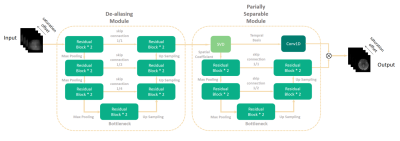 |
106 |
Highly Accelerated Chemical Exchange Saturation Transfer Imaging
with Partially Separable Network
Chuyu Liu1,
Zhensen Chen2,
and Xiaolei Song1
1Center for Biomedical Imaging Research, Department of Biomedical Engineering, Tsinghua University, Beijing, China, 2Institute of Science and Technology for Brain-Inspired Intelligence, Fudan University, Shanghai, China Keywords: CEST & MT, Machine Learning/Artificial Intelligence Herein, we developed a partially separable network (PSN) for CEST acceleration. Our contributions are: 1) We found that the reconstruction error of CEST mainly exists in the spatial subspace. 2) A deep learning network based on partially separable model was developed to optimize CEST images in spatial subspace. Retrospective results suggested that our method enabled a highly accelerated CEST imaging (14X for healthy adults and 11X for brain tumor patients) with contrast maps and Z-spectrum consistent with gold standard, which could have great clinical utility. |
|
2985.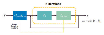 |
107 |
Joint Model-Based Optimization of Sampling Pattern and
Reconstruction for Human Brian CEST MRI at 3T
Chuyu Liu1,
Zhongsen Li1,
and Xiaolei Song1
1Center for Biomedical Imaging Research, Department of Biomedical Engineering, Tsinghua University, Beijing, China Keywords: CEST & MT, Machine Learning/Artificial Intelligence Here we introduced a model-based deep learning approach which enabled joint optimization of both sampling and reconstruction for CEST MRI. The main purpose is to investigate an efficient undersampling pattern for CEST acceleration. Retrospective results (4X) showed that the proposed workflow is capable of leveraging redundancy information from optimized sampling pattern, further reconstructing gold-standard-consistent contrast maps. |
|
2986.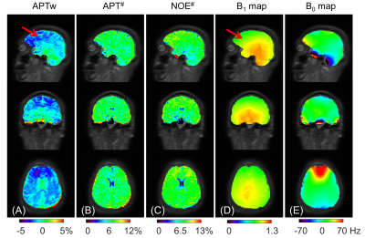 |
108 |
Prospective acceleration of whole-brain chemical exchange
saturation transfer by Joint K-space and Image-space Parallel
Imaging (KIPI)
Tao Zu1,
Zhechuan Dai1,
Xingwang Yong1,
Tongling Jiang1,
and Yi Zhang1
1Key Laboratory for Biomedical Engineering of Ministry of Education, Department of Biomedical Engineering, College of Biomedical Engineering & Instrument Science, Zhejiang University, Hangzhou, China Keywords: CEST & MT, Parallel Imaging The clinical use of chemical exchange saturation transfer (CEST) imaging is limited by its relatively long scan time due to the measurements of multiple frames at the same location, especially for 3D whole-brain imaging. In this study, the recently proposed reconstruction method by joint K-space and Image-space Parallel Imaging (KIPI) is utilized for prospectively accelerating 3D CEST imaging. Prospective KIPI allows an acceleration factor of up to 8-fold for acquiring source images, reducing the scan time to 5.5 min for the whole-brain quantitative CEST imaging, with 17 saturation frames and 2.9 mm isotropic resolution. |
|
2987.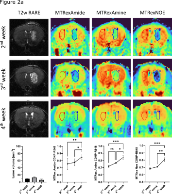 |
109 |
Metabolic Imaging of malignant gliomas during immunotherapeutic
intervention using chemical exchange saturation transfer (CEST)
MRI at 9.4T
Kianush Karimian-Jazi1,2,
Volker Sturm1,
Katharina Schregel1,2,
Jessica Hunger1,3,
Verena Turco3,4,
Noah Enbergs1,
Berin Boztepe1,3,
Manuel Fischer1,
Yannik Streibel1,
Nikolaus von Knebel-Doeberitz5,
Andreas Korzowski6,
Steffen Görke6,
Florian Kroh6,
Mark E. Ladd6,
Heinz-Peter Schlemmer5,
Daniel Paech5,7,
Christopher B. Rodell8,
Michael Platten3,4,
Wolfgang Wick2,9,
Sabine Heiland1,
Martin Bendszus1,
and Michael O. Breckwoldt1,3
1Department of Neuroradiology, Heidelberg University Hospital, Heidelberg, Germany, 2Clinical Cooperation Unit Neurooncology, German Cancer Consortium (DTK) within the German Cancer Research Center (DKFZ), Heidelberg, Germany, 3Clinical Cooperation Unit Neuroimmunology and Brain Tumor Immunology, German Cancer Consortium (DTK) within the German Cancer Research Center (DKFZ), Heidelberg, Germany, 4Department of Neurology, University Medical Center Mannheim, Heidelberg University, Mannheim, Germany, 5Department of Radiology, German Cancer Research Center (DKFZ), Heidelberg, Germany, 6Department of Medical Physics in Radiology, German Cancer Research Center (DKFZ), Heidelberg, Germany, 7Clinic for Neuroradiology, University Hospital Bonn, Bonn, Germany, 8School of Biomedical Engineering, Science and Health Systems, Drexel University, Philadelphia, PA, United States, 9Department of Neurology, Heidelberg University Hospital, Heidelberg, Germany Keywords: CEST & MT, High-Field MRI, Glioma CDNP-R848, an experimental immunotherapeutic TLR7/8 agonist, showed high treatment efficacy with a significant tumor volume reduction and led to a re-normalization of the metabolic properties (MTRex Amide, MTRex Amine and MTRex NOE) of the tumor in relation to the healthy brain, with distinct differences to vehicle treatment. The clinical relevance of CEST imaging appears to be localizing active tumor areas and a better understanding of tumor heterogeneity. In the future, we aim to better characterize the origin of the CEST contrast by correlated histological and spatial metabolic analysis. |
|
2988.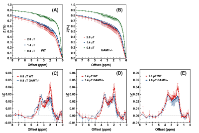 |
110 |
The exchange rate of creatine CEST in mouse brain
Ziqin Zhang1,2,
Kexin Wang1,2,
Sooyeon Park2,3,
Anna Li2,
Yuguo Li2,4,
Robert Weiss4,5,
and Jiadi Xu2,4
1Department of Biomedical Engineering, Johns Hopkins University, Baltimore, MD, United States, 2F.M. Kirby Research Center for Functional Brain Imaging, Kennedy Krieger Research Institute, Baltimore, MD, United States, 3Department of Neuroscience, Johns Hopkins University, Baltimore, MD, United States, 4Russell H. Morgan Department of Radiology and Radiological Science, Johns Hopkins University School of Medicine, Baltimore, MD, United States, 5Division of Cardiology, Department of Medicine, Johns Hopkins University School of Medicine, Baltimore, MD, United States Keywords: CEST & MT, CEST & MT, amide CEST, amine CEST, guanidinium CEST, creatine CEST, exchange rate, concentration, polynomial Lorentzian line-shape fitting (PLOF), high spectral resolution (HSR) CEST, two-step Bloch-McConnell (BM) fitting. We aim to use three different approaches to estimate the exchange rate of creatine (Cr) CEST, i.e., the pure CrCEST line-shape, B1-dependent CrCEST, and the pH response with different B1 values. The pure CrCEST signal extracted using wild type and GAMT-/- mice with low Cr and PCr concentrations; the pH in the brain cells altered by hypercapnia to demonstrate the pH sensitivity of GuanCEST; a two-step Bloch-McConnell fitting implemented to quantify the exchange rates. in vivo CrCEST exchange rate was found slow (~260-350 s-1). CrCEST is the major contribution to the opposite pH-dependence of GuanCEST signal under different B1. |
|
2989.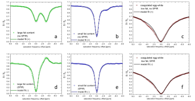 |
111 |
Z-spectral fitting for fat-correction of APT/CEST-MRI including
fat-suppression pulses to improve body applications
Jochen Keupp1,
Ivan E. Dimitrov2,3,
Holger Eggers1,
and Elena Vinogradov3,4
1Philips Research, Hamburg, Germany, 2Philips Healthcare, Gainesville, FL, United States, 3Department of Radiology, University of Texas Southwestern Medical Center, Dallas, TX, United States, 4Advanced Imaging Research Center, University of Texas Southwestern Medical Center, Dallas, TX, United States Keywords: CEST & MT, Fat, Z-spectral fitting, Fat suppression pulses Fat signal correction remains a challenge in APT/CEST-MRI for body-oncology. We show that signal background models that include direct water saturation, MT-effect, and modified fat spectra can be fitted to Z-spectra acquired with/without fat-suppression. APT/CEST signals are then extracted as the residual from the background fit (APT#). The model for fat-suppressed spectra uses a mirrored fat contribution. After B0-correction, 5 model parameters are sufficient. This allows for the acquisition of clinically feasible protocols using less than 20 Z-spectral offsets. Fat-suppression potentially increases the precision of APT# imaging by lowering the initial fat contribution. |
|
2990.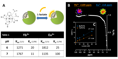 |
112 |
Imaging Extracellular Lactate with Lanthanide-PCTA-based
PARACEST Agents
Remy Chiaffarelli1,2,
Paul Jurek3,
Pedro Cruz4,
Max Zimmermann1,2,
Carlos Geraldes4,
Garry Kiefer3,
and André Ferreira Martins1,2
1Werner Siemens Imaging Center, Department of Preclinical Imaging and Radiopharmacy, University Hospital Tuebingen, Tuebingen, Germany, 2Cluster of Excellence iFIT (EXC 2180) “Image-Guided and Functionally Instructed Tumor Therapies”, University of Tuebingen, Tuebingen, Germany, 3Macrocyclics, Inc., Plano, TX, United States, 4Coimbra Chemistry Center - Institute of Molecular Sciences (CQC-IMS), University of Coimbra, Coimbra, Portugal Keywords: CEST & MT, CEST & MT, PARACEST, Metabolism Lactate accumulation in the tumor microenvironment is a hallmark of cancer aggressiveness. Here, we report a method to image extracellular lactate using shiftCEST MRI and Yb- and EuPCTA Shift Reagents (SRs). In the presence of lactate, the SRs shift the lactate OH-CEST signal away from the water signal by ~14 ppm (EuPCTA) and ~95 ppm (YbPCTA), allowing for quantitative detection of extracellular lactate produced by cancer cells. In vivo studies confirmed the detection and fast renal elimination of the lactate*YbPCTA into the bladder by shiftCEST MRI. Overall, these results provide new insights into developing innovative non-invasive metabolic MRI. |
|
2991.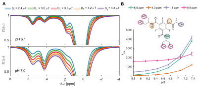 |
113 |
In silico optimization of iopamidol CEST MRI for renal pH
mapping at 3 Tesla
Julia Stabinska1,2,
Moritz Zaiss3,4,
Adnan Bibic1,
Farzad Sedaghat2,
Cristina L Sadowsky5,6,
Peter CM van Zijl1,2,
and Michael T McMahon1,2
1F.M. Kirby Research Center for Functional Brain Imaging, Kennedy Krieger Institute, Baltimore, MD, United States, 2The Russell H. Morgan Department of Radiology and Radiological Science, The Johns Hopkins University School of Medicine, Baltimore, MD, United States, 3High-field Magnetic Resonance Center, Max Planck Institute for Biological Cybernetics, Tuebingen, Germany, 4Institute of Neuroradiology, Friedrich-Alexander University Erlangen-Nürnberg (FAU), Erlangen, Germany, 5International Center for Spinal Cord Injury, Kennedy Krieger Institute, Baltimore, MD, United States, 6Physical Medicine and Rehabilitation, The Johns Hopkins University School of Medicine, Baltimore, MD, United States Keywords: CEST & MT, CEST & MT In vivo optimization of CEST iopamidol contrast in human subjects is complicated and requires multiple examinations and injections of the agent. To address this challenge, we propose application of a numerical approach that utilizes exchange rates determined under physiological conditions at 17.6T to perform kidney-like multi-pool Bloch-McConnell simulations for in silico optimization of saturation parameters for 3T applications. Our results suggest that the iopamidol-based CEST MRI is sensitive to pH in the range between 6 and 7.2 with the optimal results when short CEST saturation pulses (3x100 ms), low B1 strength (B1~0.8 µT) and short recovery time (Trec~T1w) are applied. |
|
2992. |
114 |
Learned spatiotemporal correlation priors for CEST image
denoising using incorporated global-spectral convolution neural
network
Huan Chen1,
Liangjie Lin2,
Lin Chen1,
and Zhong Chen1
1Department of Electronic Science, Xiamen University, Xiamen, China, 2Clinical & Technical Support, Philips Healthcare, Beijing, China Keywords: CEST & MT, Machine Learning/Artificial Intelligence Chemical exchange saturation transfer (CEST) MRI is a versatile technique that exploits the saturation transfer between exchangeable protons and water for non-invasive detection of diluted metabolites. Although theoretically promising, the practical application of CEST MRI is still challenged by low CEST contrast and low signal-to-noise ratio (SNR) of acquired images. Here, we proposed a deep learning-based method, dubbed denoising CEST network (DCEST-Net), to fully exploit the spatiotemporal correlation prior embedded in the CEST images and restore noise-free images from their noisy observations. Results suggested that DCEST-Net can achieve better performance compared to the state-of-the-art denoising methods. |
|
2993.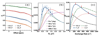 |
115 |
The exchange rates of protein guanidinium protons in the mouse
brain
Kexin Wang1,2,
Ran Sui1,2,
Lin Chen3,4,
Yuguo Li2,3,
and Jiadi Xu2,3
1Biomedical Engineering, Johns Hopkins University, Baltimore, MD, United States, 2F.M. Kirby Research Center for Functional Brain Imaging, Kennedy Krieger Research Institute, Baltimore, MD, United States, 3Russell H. Morgan Department of Radiology and Radiological Science, Johns Hopkins University, Baltimore, MD, United States, 4Department of Electronic Science, Fujian Provincial Key Laboratory of Plasma and Magnetic Resonance, Xiamen University, Xiamen, China Keywords: CEST & MT, Alzheimer's Disease, GuanCEST We develop a novel two-step multi-B1 Bloch-McConnell fitting approach for calculating the exchange rate of CEST protons in vivo, and apply it to guanidinium protons, the exchange rate of which is 70.1 ± 5.5 s-1 with a concentration of 40.4 ± 5.2 mM in mouse brain at an optimized B1 of 0.8 µT. Guanidinoacetate N-methyltransferase deficiency (GAMT-/-) mice that have low creatine and phosphocreatine concentrations in brain are studied for protein guanidinium, i.e., arginineCEST (ArgCEST). The low exchange rate of ArgCEST suggests that the inverse pH dependence in GuanCEST with low B1 is dominated by CrCEST compared to ArgCEST. |
|
2994.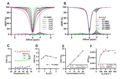 |
116 |
CEST effect of dimethyl sulfoxide (DMSO) at negative offset
frequency
Haoyun Su1,2,
Jianpan Huang1,
and Kannie W.Y. Chan1,2,3,4,5
1Department of Biomedical Engineering, City University of Hong Kong, Kowloon, Hong Kong, 2Hong Kong Centre for Cerebro-Cardiovascular Health Engineering (COCHE), New Territories, Hong Kong, 3Russell H. Morgan Department of Radiology and Radiological Science, Johns Hopkins University School of Medicine, Baltimore, MD, United States, 4City University of Hong Kong Shenzhen Research Institute, Shenzhen, China, 5Tung Biomedical Sciences Centre, City University of Hong Kong, Kowloon, Hong Kong Keywords: CEST & MT, Molecular Imaging CEST MRI can detect mM range of endogenous and exogenous molecules, and bound small molecules such as glycogen and lactate via rNOE. Here we reported for the first time that DMSO and its structural analogs in aqueous solution had distinctive CEST peaks at the negative offsets of Z-spectrum. CEST effect of DMSO was dependent on saturation power and concentration, and less sensitive to tested temperature. Alcohols, acetone, acetonitrile, acetic acid and N,N-dimethylformamide also showed observable CEST peaks at the negative offsets. Interaction between DMSO and water molecule, such as hydrogen bonding, could contribute to the observed CEST effect. |
|
2995.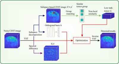 |
117 |
Subspace denoising for CEST MRI with non-local low-rank
constraint and spectral-smoothness regularization
Xinran Chen1,
Jian Wu1,
Liangjie Lin2,
Zhiliang Wei3,
Lin Chen1,
and Zhong Chen1
1Department of electronic science, Xiamen University, Xiamen, China, 2Clinical & Technical Support, Philips Healthcare, Beijing, China, 3Russell H. Morgan Department of Radiology and Radiological Science, Johns Hopkins University School of Medicine, Baltimore, MD, United States Keywords: CEST & MT, Data Processing, Denoising Chemical exchange saturation transfer (CEST) is a powerful technique that enables non-invasive detection of endogenous metabolites in living tissues. Since the observed water signal is decreased due to the transfer of saturated spins, CEST imaging inherently suffers from low SNR, hence degrading accuracy and reproducibility. Inspired by the spatial-spectral correlation of CEST images, here we propose a Subapace denoising method with Non-Local Low-Rank constraint and Spectral-Smoothness regularization (SNLRSS) to diminish the noise, which improves the accuracy of subsequent quantitative analyses of CEST images. |
|
2996.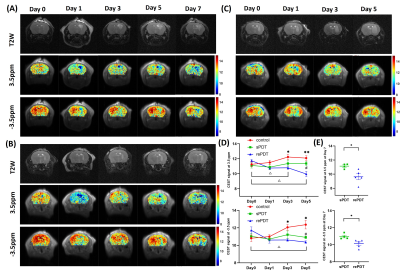 |
118 |
Imaging treatment efficacy of repeated photodynamic therapy in
glioblastoma using CEST MRI
Vivian W.M. Leung1,
Joseph H.C. Lai1,
Zilin Chen1,
Jianpan Huang1,
Se Weon Park1,2,
Yang Liu1,2,
Charles Hu3,
and Kannie W. Y. Chan1,2,4,5,6
1Department of Biomedical Engineering, City University of Hong Kong, Hong Kong, China, 2Hong Kong Centre for Cerebro-Cardiovascular Health Engineering (COCHE), Hong Kong, China, 3Incando Therapeutics Pte Ltd, Singapore, Singapore, 4Russell H. Morgan Department of Radiology and Radiological Science, The Johns Hopkins University School of Medicine, Baltimore, MD, United States, 5City University of Hong Kong Shenzhen Research Institute, Shenzhen, China, 6Tung Biomedical Sciences Centre, City University of Hong Kong, Hong Kong, China Keywords: CEST & MT, Tumor Glioblastoma (GBM) is hard to treat and has poor prognosis. Photodynamic therapy (PDT) is a promising treatment for GBM. Here, we detect the treatment efficacies of different PDT schemes (repeated (re-) and single (s-) PDT) on a rodent model of GBM using CEST MRI to monitor the molecular changes associated with tumor physiology and necrosis. Significant decreases in APT and rNOE signals were detected in the rePDT group as well as decreased proliferative activities in histology when compared with sPDT. This indicates that both APT and rNOE can be a reliable approach to assess PDT treatment efficacy against GBM. |
|
2997.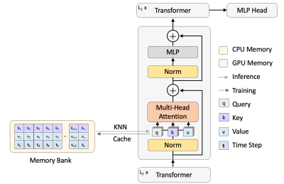 |
119 |
Memorizing Transformer for Small-scale Multi-parametric MRI
Brain Tumor Diagnosis
Yiqing Shen1,2,
Nhat Le1,2,
Jingpu Wu1,3,
Pengfei Guo1,2,
Jinyuan Zhou1,
Mathias Unberath2,
and Shanshan Jiang1
1Department of Radiology, School of Medicine, Johns Hopkins University, Baltimore, MD, United States, 2Department of Computer Science, Whiting School of Engineering, Johns Hopkins University, Baltimore, MD, United States, 3Department of Applied Mathematics and Statistics, Whiting School of Engineering, Johns Hopkins University, Baltimore, MD, United States Keywords: CEST & MT, Machine Learning/Artificial Intelligence Deep learning approaches have been widely applied to the MRI field. Among them, transformers have received increasing popularity due to their capability in handling multi modalities. Yet, transformers are hungry for large-scale data, which is expensive to collect. Here, we develop a novel memorizing transformer for small-scale multi-parametric MRI analysis. Empirically, we evaluate the proposed method on a dataset curated from 147 brain post-treatment malignant glioma cases for classifying treatment effect and tumor recurrence. The proposed memorizing transformer boosted a 5.15% improvement in the test area under the receiver-operating-characteristic curve (AUC) to the baseline transformer approaches. |
|
2998.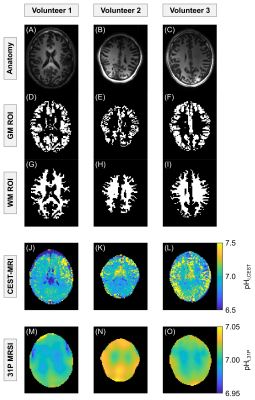 |
120 |
Quantitative volumetric mapping of intracellular pH in the human
brain at 7 T using endogenous CEST-MRI: A proof of principle
study
Philip S Boyd1,
Florian Kroh1,2,
Johannes Breitling1,
Vanessa L Franke1,2,
Mark E Ladd1,2,3,
Peter Bachert1,2,
Steffen Goerke1,
and Andreas Korzowski1
1Division of Medical Physics in Radiology, German Cancer Research Center (DKFZ), Heidelberg, Germany, 2Faculty of Physics and Astronomy, University of Heidelberg, Heidelberg, Germany, 3Faculty of Medicine, University of Heidelberg, Heidelberg, Germany Keywords: CEST & MT, Molecular Imaging, intracellular pH, guanidyl protons, 7 Tesla In this study, our method for quantitative pHi mapping using endogenous CEST-MRI was successfully transferred to examinations of the human brain at B0=7T. Applicability in vivo was demonstrated in n=3 healthy volunteers, showing an overall median pHi,CEST of 7.00 and 6.96 for gray and white matter, respectively. In order to proof the plausibility of the presented approach, the obtained pHi,CEST maps were additionally validated directly in vivo via 31P MRSI at 7T, showing an overall median pHi,31P of 7.02 and 7.01 for gray and white matter, respectively. Consequently, reliable CEST-based pHi mapping is now also possible in the human brain. |
|
 Back to Meeting Home
 Back to the Program-at-a-Glance
The International Society for Magnetic Resonance in Medicine is accredited by the Accreditation Council for Continuing Medical Education to provide continuing medical education for physicians.
 Back to Meeting Home
 Back to the Program-at-a-Glance
 View Presentation Video