Digital Poster
Spectroscopy
ISMRM & ISMRT Annual Meeting & Exhibition • 03-08 June 2023 • Toronto, ON, Canada

| Computer # | |||
|---|---|---|---|
3681.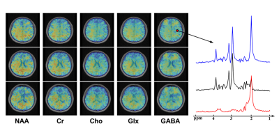 |
101 |
High-Resolution J-Edited MRSI Using Multi-Slab Acquisition and
Learned Subspaces
Rong Guo1,2,
Yibo Zhao2,3,
Yudu Li2,4,
Chao Ma5,
Wen Jin2,3,
Yao Li6,
Georges El Fakhri5,
Brad Sutton2,3,4,7,8,
and Zhi-Pei Liang2,3,4
1Siemens Medical Solutions USA, Inc., Urbana, IL, United States, 2Beckman Institute for Advanced Science and Technology, University of Illinois at Urbana-Champaign, Urbana, IL, United States, 3Department of Electrical and Computer Engineering, University of Illinois at Urbana-Champaign, Urbana, IL, United States, 4National Center for Supercomputing Applications, University of Illinois at Urbana-Champaign, Urbana, IL, United States, 5Gordon Center for Medical Imaging, Department of Radiology, Massachusetts General Hospital, Boston, MA, United States, 6School of Biomedical Engineering, Shanghai Jiao Tong University, Shanghai, China, 7Department of Bioengineering, University of Illinois at Urbana-Champaign, Urbana, IL, United States, 8Carle Illinois College of Medicine, University of Illinois at Urbana-Champaign, Urbana, IL, United States Keywords: Spectroscopy, Spectroscopy High-resolution mapping of GABA in the brain has long been desired by the neuroscience community. But it is still challenging due to several long-standing technical obstacles including low SNR, long scan time, and spectral overlapping. This work proposes an MRSI method integrating multi-slab EPSI acquisition, MEGA spectral editing, and subspace modeling to overcome these difficulties, successfully achieving 3D GABA and metabolite mapping (at 3.0×3.0×4.0 mm3 nominal resolution) in a 12-minute scan time. |
|
3682.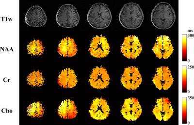 |
102 |
Whole-Brain Multi-Parametric Molecular Imaging Using Accelerated
J-Resolved Subspace 1H-MRSI
Zepeng Wang1,2,
Yahang Li1,2,
and Fan Lam1,2
1Bioengineering, University of Illinois at Urbana-Champaign, Champaign, IL, United States, 2Beckman Institute for Advanced Science and Technology, Urbana, IL, United States Keywords: Spectroscopy, Quantitative Imaging J-resolved MRSI is a powerful molecular imaging tool for measuring brain metabolites, neurotransmitters and other important biophysical parameters. The inherent SNR challenge of MRSI and prolonged scan time for multi-TE data limit the imaging resolution. This work presents a brand-new capability of whole-brain multiparametric, quantitative MRSI, by integrating a fast-scanning J-resolved MRSI sequence with SNR-efficient multi-band excitation, task-specific experiment designs, subspace imaging and optimized parameter estimation. Experimental studies and initial validation were performed to demonstrate this capability for high-resolution metabolite, neurotransmitter and metabolite T2 mapping from a single scan. |
|
3683.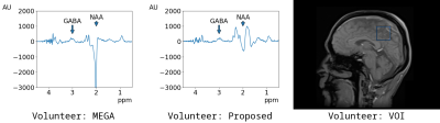 |
103 |
Selective Extraction of J-coupled Signals Using MEGA-PRESS with
Conditional Saturation Pulses
Hidenori Takeshima1,
Shuki Maruyama2,
and Masao Yui3
1Imaging Modality Group, Advanced Technology Research Department, Research and Development Center, Canon Medical Systems Corporation, Kanagawa, Japan, 2Imaging Modality Group, Advanced Technology Research Department, Research and Development Center, Canon Medical Systems Corporation, Tochigi, Japan, 3Research and Development Center, Canon Medical Systems Corporation, Tochigi, Japan Keywords: Pulse Sequence Design, Spectroscopy In this abstract, a spectral editing method using MEGA-PRESS with conditional frequency-selective saturation pulses is proposed. The proposed method used additional frequency-selective saturation blocks before the excitation block. The saturation pulses were activated only when the MEGA pulses were inactive. When saturation pulses were activated, the sequence acted as the conventional MEGA-PRESS whose editing pulses were inactive except that the passband of the saturation pulse was not excited. Spectrum reconstruction for the proposed method was same as that for the conventional MEGA-PRESS. Experimental results showed that the proposed method selectively extracted J-coupled signals with all frequencies. |
|
3684.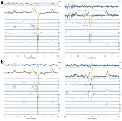 |
104 |
Test-retest reliability of PRESS- and sLASER-localized
multi-metabolite spectral editing
Mark Mikkelsen1,
Ralph Noeske2,
and Dikoma Shungu1
1Department of Radiology, Weill Cornell Medicine, New York, NY, United States, 2GE Healthcare, Berlin, Germany Keywords: Spectroscopy, Spectroscopy, Reproducibility Multi-metabolite spectral-edited MRS enables the efficient detection of multiple low-concentration metabolites. Two such approaches, HERMES and HERCULES, directly edit two or more metabolites in a single scan. However, the reproducibility of these techniques has yet to be fully established. This study investigated PRESS- and sLASER-localized HERMES and HERCULES test-retest reliability, with the additional aim of demonstrating that sLASER localization is the better of the two sequences for obtaining reproducible measurements. Overall, the test-retest reliability of sLASER was found to be higher than that of PRESS. |
|
3685.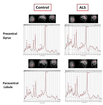 |
105 |
Short-TE semi-LASER 1H MRS of the primary motor cortex in ALS
and controls at 7 Tesla
Zeinab Eftekhari1,2,3,
Thomas B Shaw1,4,5,
Jin Jin2,6,
Kieran O'Brien6,
Gary Cowin1,
Dinesh K Deelchand7,
Robert Henderson5,8,
Frederik Steyn5,8,
Sicong Tu9,
Wolfgang Bogner10,
and Markus Barth1,2,4
1Centre for Advanced Imaging, The University of Queensland, Brisbane, Australia, 2ARC Training Centre for Innovation in Biomedical Imaging Technology, The University of Queensland, Brisbane, Australia, 3Queensland Brain Institute, The University of Queensland, Brisbane, Australia, 4School of Information Technology and Electrical Engineering, The University of Queensland, Brisbane, Australia, 5Neurology Department, Royal Brisbane and Women’s Hospital, Brisbane, Australia, 6Siemens Healthcare Pty Ltd, Brisbane, Australia, 7Centre for Magnetic Resonance Research, Department of Radiology, University of Minnesota, Minneapolis, MN, United States, 8School of Medicine, The University of Queensland, Brisbane, Australia, 9Brain and Mind Centre, The University of Sydney, Sydney, Australia, 10High-field MR Centre, Department of Biomedical Imaging and Image-guided Therapy, Medical University of Vienna, Vienna, Austria Keywords: Spectroscopy, Spectroscopy, High-Field MRI, Magnetic Resonance Spectroscopy Magnetic Resonance Spectroscopy (MRS) can offer a unique, non-invasive tool for measuring the neurochemicals in the brain in vivo. This technique may be useful for studying the ratio of metabolites in both health and disease, including in Amyotrophic Lateral Sclerosis (ALS), where differing neuronal populations of the motor cortex are impacted. This study aimed to develop a 7T MRS protocol for ALS patients and measured metabolite ratios in the upper and lower limb regions of the motor cortex at 7T in controls and in ALS. This has the potential to improve progression monitoring and diagnostic certainty in early-stage disease. |
|
3686.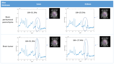 |
106 |
Optimizing MRSI semi-LASER for mouse brain tumor studies:
evaluation of increased spatial resolution and k-space sampling
strategies
Zoona Javed1,2,
Gary V Martinez3,
Marta Mulero-Acevedo1,4,5,
Ana Paula Candiota1,4,5,
Carles Arús 1,4,5,
Miquel Cabañas Egaña2,4,
and Silvia Lope-Piedrafita2,4
1Departament de Bioquímica i Biologia Molecular, Universitat Autònoma de Barcelona, Cerdanyola del Valles, Spain, 2Servei de Ressonància Magnètica Nuclear, Universitat Autonoma de Barcelona, Cerdanyola del Valles, Spain, 3Department of Imaging Physics,, The University of Texas M.D. Anderson Cancer Center, Houston, TX, United States, 4Centro de Investigación Biomédica en Red en Bioingeniería, Biomateriales y Nanomedicina (CIBER-BBN), Cerdanyola del Valles, Spain, 5Institut de Biotecnologia i de Biomedicina (IBB), Universitat Autònoma de Barcelona, Cerdanyola del Valles, Spain Keywords: Spectroscopy, Cancer, Brain Tumor MRSI-based nosological images have been previously applied to monitor therapy response in a murine model of GL261 glioblastoma using a commercially available MRSI-PRESS sequence. Due to the heterogeneous nature of glioblastomas, increasing spatial resolution could provide additional insight into changes related to therapy response. The in-house implementation of the MRSI-semi-LASER sequence, using weighted acquisition, has allowed us a 20% slice thickness reduction without losing relevant spectral quality. Moreover, different k-space sampling strategies have been evaluated showing significant SNR increase with elliptical sampling, which may allow further increase in the spatial resolution or reduction in the total experimental time. |
|
3687.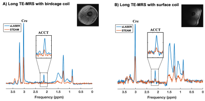 |
107 |
Comparison of STEAM and sLASER to quantify acetylcarnitine at
rest using long TE 1H-MRS in human skeletal muscle with a
surface or birdcage coil at 7T
Pandichelvam Veeraiah1,2,
Rick Voncken1,
Kim Brouwers1,3,
Julian Mevenkamp3,
Job van den Hurk1,4,
Joachim E Wildberger3,
and Vera B Schrauwen-Hinderling3,5,6
1Scannexus (Ultra-High Field Imaging Center), Maastricht, Netherlands, 2Faculty of Health Medicine and Life sciences (FHML), Maastricht University, Maastricht, Netherlands, 3Department of Radiology and Nuclear Medicine, Maastricht University Medical Center (MUMC), Maastricht, Netherlands, 4Department of Cognitive Neuroscience, Faculty of Psychology and Neuroscience, Maastricht University, Maastricht, Netherlands, 5Nutrition & Movement Sciences, Maastricht University, Maastricht, Netherlands, 6German Diabetes Center, Dusseldorf, Germany Keywords: Spectroscopy, Muscle, Ultra-high field MRS, proton MR spectroscopy, Muscle Long echo time 1H-MRS has been used to determine in vivo acetylcarnitine (ACCT) concentrations in the skeletal muscle. At ultra-high field (UHF), STEAM-based 1H-MRS was used for this purpose, in combination with a knee birdcage coil. However, STEAM suffers from an inherent 50% signal loss and a knee coil often does not fit around the upper leg in obese volunteers. Here, we demonstrated that sLASER, in combination with a surface coil, provides high signal-to-noise ratio (SNR) and can be used as an alternative method at 7T to detect physiologically low ACCT concentrations at rest in both lean and obese volunteers. |
|
3688.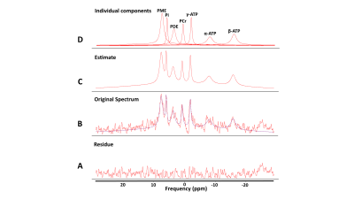 |
108 |
Localized 31P Magnetic Resonance Spectroscopy of Normal Human
Kidneys at 3T
Limin Zhou1,
Keith Hulsey1,
Durga Udayakumar1,2,
Andrea J. Wiethoff3,
and Ananth J. Madhuranthakam1,2
1Department of Radiology, University of Texas Southwestern Medical Center, DALLAS, TX, United States, 2Advanced Imaging Research Center, University of Texas Southwestern Medical Center, DALLAS, TX, United States, 3Novartis Institutes for BioMedical Research, Cambridge, MA, United States Keywords: Spectroscopy, Kidney, Phosphorous (31P) MR spectroscopy (MRS), healthy volunteers Phosphorous (31P) MR spectroscopy (MRS) can measure high energy phosphate metabolism non-invasively in vivo, which can provide metabolic insights into kidney pathophysiology. However, obtaining spectra from healthy volunteers for reference is challenging. In this study, we investigate the feasibility of localized 31P MRS in kidneys of healthy volunteers with a 3T clinical MR scanner. 31P spectra were successfully obtained in 10 healthy volunteers with metabolite ratio calculated to provide information about the energy metabolism. |
|
3689.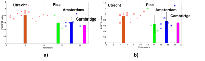 |
109 |
Feasibility of a multi-site and cross-platform liver 31P MRSI
processing pipeline for three 7T clinical MRI vendors
Jabrane Karkouri1,
Lieke van den Wildenberg2,
Bobby Runderkamp3,
Jeanine Prompers2,
Paolo Cecchi4,
Michela Tosetti4,5,
Dennis Klomp2,
and Christopher T. Rodgers1
1University of Cambridge, Cambridge, United Kingdom, 2Center for Image Sciences, University Medical Center Utrecht, Utrecht, Netherlands, 3Department of Radiology and Nuclear Medicine, Amsterdam UMC, Amsterdam, Netherlands, 4IMAGO7 Research Foundation, Pisa, Italy, 5Laboratory of Medical Physics and Magnetic Resonance, IRCCS Stella Maris, Pisa, Italy Keywords: Spectroscopy, Non-Proton We present preliminary baseline results from three-vendor 7 T MRSI for applications on liver 31P MRS. Liver scans were performed on 21 healthy volunteers, at 4 sites and 3 different 7T MR scanner platforms. Data were analysed using a new Matlab pipeline combining the best methods from our labs. This pipeline is operator independent and we report here the liver metabolites as ratios of PME/PDE and Pi/ATP concentrations. The cross-site average PME/PDE was 0.63 ± 0.12, and the cross-site average Pi/ATP was 0.76 ± 0.11. This compares favourably with literature reports [Purvis et. al., 2017]. |
|
3690.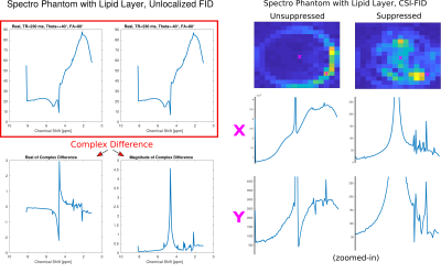 |
110 |
Removal of lipid signals and other short T2 components in
GRE-MRI and FID-MRSI using quadratic radio frequency phase
increments
Lukas Hingerl1,
Bernhard Strasser1,
Gilbert Hangel1,2,
Stanislav Motyka1,
Fabian Niess1,
Eva Niess1,
Alexandra Lipka1,
Dario Goranovic1,
Philipp Lazen1,
Stephan Gruber1,
Siegfried Trattnig1,3,
and Wolfgang Bogner1
1Department of Biomedical Imaging and Image-guided Therapy, Radiology and Nuclear Medicine, Medical University of Vienna, HFMR Centre, Vienna, Austria, 2Department of Neurosurgery, Medical University of Vienna, Vienna, Austria, 3Institute for Clinical Molecular MRI in Musculoskeletal System, Karl Landsteiner Society, Vienna, Austria Keywords: Spectroscopy, Spectroscopy, Lipid Fat Suppression Removal We present an elegant and easy to implement method for MRI and CSI steady-state sequences to remove or suppress lipids or other components with short transverse relaxation times by neither introducing additional pulses nor hardware and by just altering the excitation pulse phase. |
|
3691.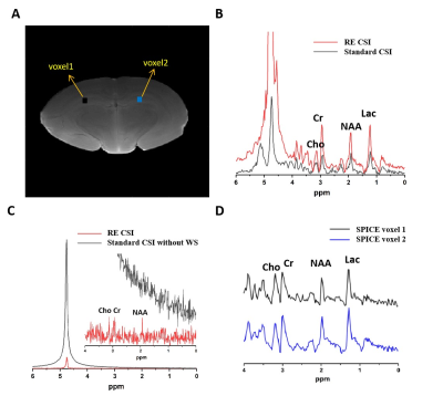 |
111 |
High-Resolution 1H-MRSI of the Brain at 9.4T Integrating
Relaxation Enhancement and Subspace Imaging
Yizun Wang1,2,
Stanislav S. Rubakhin1,2,3,
and Fan Lam1,2,4
1Department of Bioengineering, University of Illinois at Urbana-Champaign, Urbana, IL, United States, 2Beckman Institute for Advanced Science and Technology, University of Illinois at Urbana-Champaign, Urbana, IL, United States, 3Department of Chemistry, University of Illinois at Urbana-Champaign, Urbana, IL, United States, 4Department of Electrical and Computer Engineering, University of Illinois at Urbana-Champaign, Urbana, IL, United States Keywords: Spectroscopy, Data Acquisition, MRSI Data Processing Ultrahigh-field systems offer sensitivity and specificity advantages for metabolite mapping using MRSI. We present here a method that integrates relaxation enhancement acquisition, a unique strategy leveraging higher fields, and subspace imaging, for high-resolution 1H-MRSI at a 9.4 T system. Both in vitro and in vivo experiments have been performed to demonstrate the feasibility of the proposed method. We are able to achieve significantly enhanced SNR compared to no relaxation enhancement and produce high-quality spatially-resolved spectra and high-resolution metabolite images using subspace imaging with 0.6×0.6×2 mm3 nominal resolution in 18 minutes or 1×1×2 mm3 in 10 minutes. |
|
3692.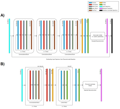 |
112 |
Tailoring a convolutional neural network towards spectral
quantification and reconstruction of simulated 7T brain FID-MRSI
data
Dario Goranovic1,
Stanislav Motyka1,
Bernhard Strasser1,
Paul Weiser2,
Georg Langs2,
and Wolfgang Bogner1
1High Field MR Center - Department of Biomedical Imaging and Image-Guided Therapy, Medical University of Vienna, Vienna, Austria, 2Computational Imaging Research Lab - Department of Biomedical Imaging and Image-guided Therapy, Medical University of Vienna, Vienna, Austria Keywords: Spectroscopy, Brain The abstract investigates the possibilities of using convolutional neural networks for enhanced and robust spectral quantification of simulated 7T FID-MRSI brain spectra. The proposed network architecture predicts wavelet parameters for baseline correction, as well as spectral parameters and metabolite amplitudes for spectral reconstruction. The potential reduction in quantification times could thus mitigate some disadvantages of current MRSI processing techniques. |
|
3693.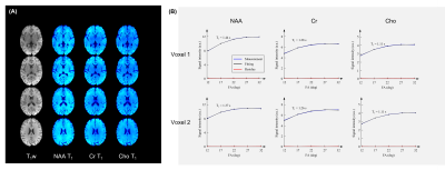 |
113 |
3D High-Resolution T1 Mapping of Brain Metabolites
Yibo Zhao1,2,
Rong Guo1,3,
Yudu Li1,4,
Wen Jin1,2,
Yao Li5,
Jie Luo5,
and Zhi-Pei Liang1,2
1Beckman Institute for Advanced Science and Technology, University of Illinois at Urbana-Champaign, Urbana, IL, United States, 2Department of Electrical and Computer Engineering, University of Illinois at Urbana-Champaign, Urbana, IL, United States, 3Siemens Medical Solutions USA, Inc., Urbana, IL, United States, 4National Center for Supercomputing Applications, University of Illinois at Urbana-Champaign, Urbana, IL, United States, 5School of Biomedical Engineering, Shanghai Jiao Tong University, Shanghai, China Keywords: Spectroscopy, Relaxometry In MRSI studies, T1 values of metabolites are desirable for correcting relaxation and B1 inhomogeneity effects, and for evaluating microenvironmental changes in pathological conditions. Current metabolite T1 mapping has been limited to single-voxel or single-slice experiments due to SNR and imaging time constraints. In this work, we demonstrated the feasibility of 3D high-resolution T1 mapping of brain metabolites using a novel data acquisition and processing method featuring physics-based low-rank tensor modelling and FID acquisitions. The proposed method has been validated using phantom and in vivo data, producing encouraging results. |
|
3694.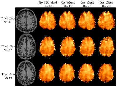 |
114 |
Compressed Sensing for MRSI with Concentric Ring Trajectories at
7 T
Bernhard Strasser1,
Ovidiu C Andronesi2,
Lukas Hingerl1,
Stanislav Motyka1,
Siegfried Trattnig1,
Wolfgang Bogner1,
and Antoine Klauser3
1Department of Biomedical Imaging and Image-guided Therapy, Medical University of Vienna, Vienna, Austria, 2Martinos Center for Biomedical Imaging, Massachusetts General Hospital, Charlestown, MA, United States, 3Department of Radiology and Medical Informatics, University of Geneva, Geneva, Switzerland Keywords: Spectroscopy, New Trajectories & Spatial Encoding Methods We show the feasibility of compressed sensing for MRSI using concentric ring trajectories. Four volunteers (three for 2D- and one for 3D-MRSI) were measured in about 9 minutes (2D-MRSI), and 14 minutes (3D-MRSI) using an FID-based MRSI sequence at 7 T. An iterative compressed sensing reconstruction including coil sensitivities and a total general variation spatial regularization was performed with retrospective undersampling factors of 1.5, 2.0 and 2.9. RMSE values were below 22 % and SSIM values above 0.8 for medium accelerations below 2.9 in the 2D case. The metabolic maps and spectra are similar to the gold standard without acceleration. |
|
3695.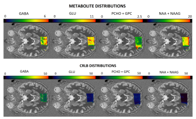 |
115 |
Implementation of Zoom MRSI at 7T for High-resolution GABA and
Glutamate Mapping
Onur Ozyurt1,
Diana Rotaru2,
Nicholas Farley3,
Zoe Kourtzi2,
Guy Williams1,
and Uzay Emrah Emir3,4
1Wolfson Brain Imaging Center, Department of Clinical Neurosciences, University of Cambridge, Cambridge, United Kingdom, 2Department of Psychology, University of Cambridge, Cambridge, United Kingdom, 3School of Health Sciences, Purdue University, West Lafayette, IN, United States, 4Weldon School of Biomedical Engineering, Purdue University, West Lafayette, IN, United States Keywords: Spectroscopy, Brain, GABA, Glutamate, MRSI High-resolution neurochemical mapping using 2D zoom or reduced field of view (rFOV) magnetic resonance spectroscopy (MRSI) enables the high-resolution metabolic assessment of brain regions that is difficult to probe with standard MRSI sequences. To our knowledge this is the first study to have demonstrated the application of zoom MRSI at 7T for GABA and glutamate mapping. |
|
3696.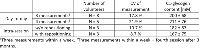 |
116 |
Reproducibility of non-localized 13C-MRS using a quadrature
surface coil for assessing hepatic carbohydrate metabolism in
humans
Marc Jonuscheit1,2,
Benedict Korzekwa1,2,
Yuliya Kupriyanova1,2,
Julian Mevenkamp3,
Michael Roden1,2,4,
and Vera B. Schrauwen-Hinderling1,2,3
1Institute for Clinical Diabetology, German Diabetes Center, Leibniz Institute for Diabetes Research at Heinrich Heine University, Düsseldorf, Germany, 2German Center for Diabetes Research (DZD e.V.), München-Neuherberg, Germany, 3Department of Radiology and Nuclear Medicine, Maastricht University Medical Center, Maastricht, Netherlands, 4Department of Endocrinology and Diabetology, Medical Faculty, Heinrich Heine University, Düsseldorf, Germany Keywords: Spectroscopy, Spectroscopy, 13C-MRS, Quadrature Coil, Absolute Quantification We determined day-to-day variation and intra-session reproducibility of 13C-MRS based hepatic glycogen quantification by phantom replacement, using a custom-created rigid quadrature surface coil and determining the C1-glycogen signal in eight healthy volunteers. The quadrature coil allows measurements at high distances, which can be important for applications in overweight and obese people. Coefficients of variation were less than 22% for day-to-day variation and less than 9% for intra-session reproducibility (with repositioning of volunteer). In conclusion, our 13C-MRS protocol yields robust absolute concentrations of the hepatic glycogen and the day-to-day variation assessed here can be the basis for sample size calculations. |
|
3697.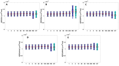 |
117 |
Effects of denoising on diffusion-weighted MRS data
Guglielmo Genovese1 and
Małgorzata Marjańska1
1Center for Magnetic Resonance Research, University of Minnesota, Minneapolis, MN, United States Keywords: Spectroscopy, Data Processing, Diffusion-Weighted MRS, High Field (3T), Denoising, Low-Rank Model Diffusion-weighted (DW) MRS is a useful tool for detecting microstructural changes linked to neurological diseases. However, DW-MRS suffers from low signal-to-noise ratio, which makes human application extremely difficult. To overcome this issue, a denoising algorithm based on low-rank approximation was recently utilized. Here, effects of the application of low-rank based denoising algorithm to DW-MRS data are described based on synthetic data. Severely artifactually low group SDs for the apparent diffusion coefficients (ADCs) of all metabolites and biased mean ADC values were observed after denoising. These findings suggest that low-rank approximations are detrimental to a reliable quantification of ADCs of metabolites. |
|
3698.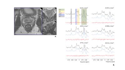 |
118 |
Diffusion-weighted MR spectroscopy of the prostate
Angeliki Stamatelatou1,
Rudy Rizzo2,3,
Kadir Simsek4,
Jack J.A. van Asten1,
Arend Heerschap1,
Tom WJ Scheenen1,
and Roland Kreis2,3
1Department of Medical Imaging, Radboud University Medical Center, Nijmegen, Netherlands, 2Magnetic Resonance Methodology, Institute of Diagnostic and Interventional Neuroradiology, University of Bern, Bern, Switzerland, 3Translational Imaging Center, sitem-insel, Bern, Switzerland, 4Cardiff University Brain Research Imaging Centre (CUBRIC), School of Psychology, Cardiff University, Cardiff, United Kingdom Keywords: Spectroscopy, Diffusion/other diffusion imaging techniques Diffusion-weighted Magnetic Resonance Spectroscopy (DW-MRS) is ideally suited to explore complex microstructure with metabolites selectively distributed in different subspaces. So far this technique was applied in brain and muscle only. In this work, we explored DW-MRS for the first time in the prostate, an organ, with potentially more motion problems. Thus, dedicated acquisition and post-processing techniques were used including the measurement of water next to that of metabolites for corrections. ADC values of citrate, total choline, total creatine and spermine were estimated and evaluated according to the compartmental structure of the prostate indicating hindered metabolite diffusion in the luminal space. |
|
3699.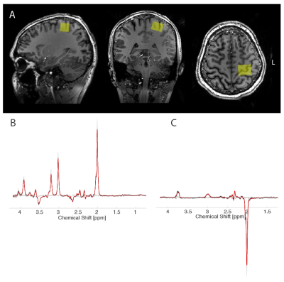 |
119 |
Comparison of short-interval afferent inhibition measures with
tonic GABA and Glutamate levels
Michal Považan1,
Mads Just Madsen1,
and Leo Tomasevic1
1Danish Research Centre for Magnetic Resonance, Hvidovre, Denmark Keywords: Spectroscopy, Neuroscience The neurochemical substrate of the measures observed with transcranial magnetic stimulation remains unclear. MR spectroscopy may provide valuable insights into understanding the synaptic GABAergic and glutamatergic activity, however studies comparing these methods haven’t provided an adequate explanation. We aimed to compare GABA and glutamate concentrations with short-interval afferent inhibition measured with TMS. We found positive correlation between the amount of inhibition and tonic GABA levels, however this finding needs to be carefully scrutinized. |
|
3700. |
120 |
Ex-vivo MR spectroscopy in paraformaldehyde-fixed mouse brain
Alireza Abaei1,
Dinesh K Deelchand 2,
Stefano Antonucci3,
Jelena Scekic-Zahirovic3,
Florian olde Heuvel3,
Francesco Roselli3,
and Volker Rasche4
1Ulm University, Ulm, Germany, 2Center for Magnetic Resonance Research, University of Minnesota, Minneapolis, MN, United States, 3German Center for Neurodegenerative Diseases (DZNE), Ulm, Germany, 4Core Facility Small Animal Imaging (CF-SANI), University of Ulm, Ulm, Germany Keywords: Spectroscopy, Spectroscopy, in vivo ex vivo MRS The goal of this study is to demonstrate the feasibility and reliability of metabolite characterization by MR spectroscopy ex vivo in order to maximize the information obtained from a single brain specimen, without the limitations such as scanning time, degraded MRI quality due to intervening tissue and motion artifacts. We demonstrate that ex-vivo spectroscopy obtained after rapid PBS/PFA perfusion is strongly correlated with the MR spectrum previously acquired in the same animal but in vivo but distinct abnormalities due to hypoxia and incomplete fixation do appear; the quality of the spectrum quickly degrades upon longer fixation times. |
|
 Back to Meeting Home
 Back to the Program-at-a-Glance
The International Society for Magnetic Resonance in Medicine is accredited by the Accreditation Council for Continuing Medical Education to provide continuing medical education for physicians.
 Back to Meeting Home
 Back to the Program-at-a-Glance
 View Presentation Video