Digital Poster
CEST & MT
ISMRM & ISMRT Annual Meeting & Exhibition • 03-08 June 2023 • Toronto, ON, Canada

| Computer # | |||
|---|---|---|---|
3272.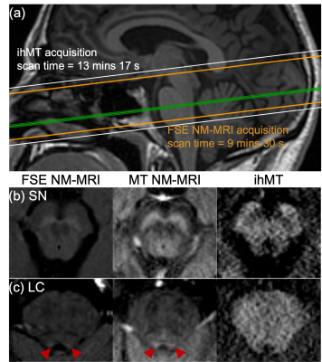 |
41 |
Extending inhomogeneous MT to assess both neuromelanin and
myelin in brainstem structures
Gopal Varma1,
Jane Fergusson2,
Asma Hassani2,
Aaron K Grant1,
Aron S Buchman3,
David C Alsop1,
and Veronique VanderHorst2
1Radiology, Division of MR Research, Beth Israel Deaconess Medical Center, Harvard Medical School, Boston, MA, United States, 2Department of Neurology, Beth Israel Deaconess Medical Center, Harvard Medical School, Boston, MA, United States, 3Department of Neurological Sciences, Rush University Medical Center, Rush Alzheimer's Disease Center, Chicago, IL, United States Keywords: Magnetization transfer, Neuro, Brainstem Application of neuromelanin MRI (NM-MRI) in the brainstem has been utilized in studies of neurological disorders such as Parkinson's disease. Acquisition of two types of MT preparation for inhomogeneous MT, MT applied at a single offset and then dual off-resonance frequencies, presents an advancement to NM-MRI by providing an acquisition with a myelin sensitive signal for complementary information. We demonstrate this in ex-vivo brainstem samples at 9.4 T, which allows comparison with other MRI microstructural techniques, as well as in-vivo at 3 T, where MT images provide a contrast comparable to NM-MRI by fast-spin-echo. |
|
3273.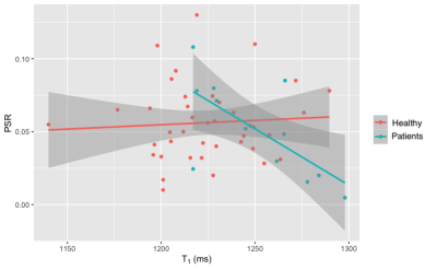 |
42 |
Quantitative Magnetisation Transfer Imaging using bSSFP to
assess diffuse myocardial fibrosis.
Luke Pleva1,
John Farrant1,2,
Christopher Miller1,2,
and Josephine Naish1,2
1The University of Manchester, Manchester, United Kingdom, 2MCMR, Manchester University NHS Foundation Trust, Manchester, United Kingdom Keywords: Magnetization transfer, Cardiovascular Patients with chronic kidney disease often suffer from cardiac complications where increased diffuse myocardial fibrosis is present. Gadolinium is contraindicated in these patients, so cannot benefit from techniques such as ECV. We present a bSSFP based quantitative magnetisation transfer technique to assess diffuse myocardial fibrosis through the calculation of a pool size ratio and compare against ECV. 6 bSSFP images, T1, and T2 maps were acquired where a two pool model was fitted. No significant correlation between PSR and T1 in healthy patients was found. Significant correlations were found between PSR and T1 and PSR and ECV in clinical patients. |
|
3274.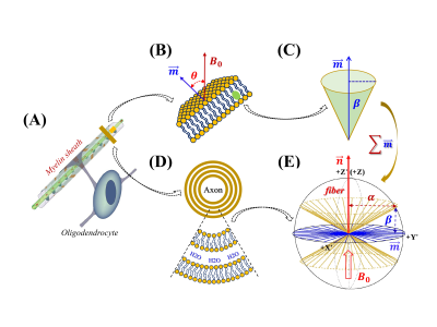 |
43 |
Characterization of semisolid proton $$$R_2$$$ orientation
dependence in WM
Yuxi Pang1
1University of Michigan, Ann Arbor, MI, United States Keywords: Relaxometry, White Matter Quantitative magnetization transfer (qMT) imaging can indirectly probe $$$R_2$$$ orientation dependence in WM for semisolid methylene protons from lipid bilayers. Prior experimental and simulation studies indicate that the long lipid chain not only rotates rapidly around itself, but it also wobbles in a cone. The existing modeling methods, however, did not take the latter motional mode into account, potentially leading to a biased measure of myelin-specific anisotropic $$$R_2$$$ relaxation. This work thus proposes a new model encompassing both motional modes for better characterizing an anisotropic $$$R_2$$$ profile of semisolid protons in WM. |
|
3275.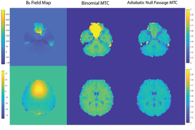 |
44 |
Adiabatic Null Passage for on resonance magnetization transfer
preparation
Shahrokh Abbasi-Rad1,2,3,4 and
David Norris1,2
1Donders Institute for Brain, Cognition and Behaviour, Radboud University, Nijmegen, Netherlands, 2Erwin L. Hahn Institute for Magnetic Resonance Imaging, University Duisburg-Essen, Essen, Germany, 3Department of Radiology, Harvard Medical School, Boston, MA, United States, 4Athinoula A. Martinos Center for Biomedical Imaging, Massachusetts General Hospital, Charlsetown, MA, United States Keywords: Magnetization transfer, RF Pulse Design & Fields, Adiabatic Null Passage On-resonance bound pool saturation is the most efficient way of generating MTC. However, it suffers from excessive direct free water saturation due to RF pulse instabilities, and potential T2-weighting. We proposed a time-reversed adiabatic pulse (adiabatic null passage) for on-resonance MT preparation, by reversing the time-domain phase modulation function of a symmetric adiabatic pulse at its mid-point. We compared the MTC performance of ANP with binomial pulses using their MTR images. MTR values were reported through lines crossing CSF, showing high values at GM/WM and zero at CSF voxels. ANP improved the excessive saturation at regions with high B0 inhomogeneity. |
|
3276.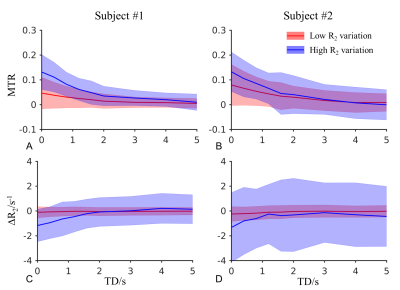 |
45 |
Imaging magnetization exchange between cerebrospinal fluid and
brain parenchyma in humans at 3 T
Jiaen Liu1
1Advanced Imaging Research Center, UT Southwestern Medical Center, Dallas, TX, United States Keywords: Magnetization transfer, Neurofluids Imaging mass exchange between cerebrospinal fluid (CSF) and brain parenchyma tissue is promising for enhancing our understanding about the role of CSF in clearance of metabolic waste and contributes to clinical diagnosis of neurological disorders and degeneration. In this study, we evaluated the feasibility of achieving this goal based on the magnetization transfer effect and CSF-selective spin echo contrast at 3 T in human subjects. The results suggested that techniques with low sensitivity to flow and partial volume effect are required for robust clinical application. |
|
3277.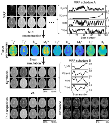 |
46 |
Saturation Transfer MR Fingerprinting (ST-MRF): free bulk water,
semisolid macromolecule, and amide proton parameter
quantification
Munendra Singh1,
Peter van Zijl1,
Shanshan Jiang1,
Jinyuan Zhou1,
and Hye-Young Heo1
1Russell H. Morgan Department of Radiology and Radiological Science, Johns Hopkins University, Baltimore, MD, United States Keywords: CEST & MT, CEST & MT Conventional saturation transfer MRI approaches, such as magnetization transfer contrast (MTC) and chemical exchange saturation transfer (CEST) MRI, acquire qualitative contrast weighted images, without specific information on the quantitative parameters affecting contrast, namely proton exchange rate and concentration. In addition, the contrast weighted images are highly dependent on scan parameters and data acquisition strategies. Here, we developed a fast, quantitative saturation transfer (ST) imaging technique based on MR fingerprinting principles to simultaneously estimate free bulk water, semisolid macromolecule, and amide proton-related parameters. The approach was evaluated with Bloch simulation and synthetic MRI analysis using in vivo human brains. |
|
3278.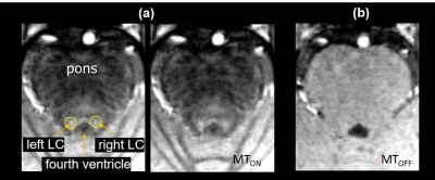 |
47 |
Asymmetric magnetization transfer ratio of locus coeruleus with
sleep disorder: A 7T MRI study
Wenwen Yu1,
Ying-Hua Chu2,
Changyong Xie3,
Shuanghong Chen3,
Jianping Zhang3,
Zidong Yang1,
He Wang1,4,
and Yuchuan Qiao1,4
1Institute of Science and Technology for Brain-inspired Intelligence,Fudan university, Shanghai, China, 2MR Collaboration, Siemens Healthineers Ltd., Shanghai, China, 3Center of Naval Special Medicine, Naval Medical University, Shanghai, China, 4Key Laboratory of Computational Neuroscience and Brain-Inspired Intelligence (Fudan University), Ministry of Education, Shanghai, China Keywords: CEST & MT, Magnetization transfer, locus coeruleus, magnetization transfer ratio, neuromelanin sensitive MRI Neuromelanin (NM), mainly found in locus coeruleus (LC), is a well-known biomarker for neurodegenerative diseases such as Parkinson's disease (PD) and can be detected with neuromelanin-sensitive MRI (NM-MRI). However, whether NM content is associated with sleep disorders is largely unclear. We acquired 3D turbo flash MTC images from 20 normal and four dyssomnia adult males in a 7T scanner to examine the relationship between the sleep quality scores and the NM content in LC. Our preliminary results reveal that the magnetization transfer ratio (MTR), an indicator of NM concentration, of bilateral LC is significantly asymmetric in the dyssomnia group, and the mean value of MTR is lower in the left LC and higher in the right LC in dyssomnia group than normal group. |
|
3279.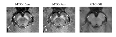 |
48 |
Simultaneous Neuromelanin Sensitive Imaging and Quantitative
Susceptibility Mapping by 3D Multi-echo GRE sequence with
Optimized MTC Pulse
Mengying Chen1,
Yupeng Wu1,
Qifan Pang1,
Haodong Zhong1,
Gaiying Li1,
Yang Song2,
Yi Wang3,
and Jianqi Li1
1Shanghai Key Laboratory of Magnetic Resonance, East China Normal University, Shanghai, China, 2MR Scientific Marketing, Siemens Healthineers, Shanghai, China, 3Department of Radiology, Weill Medical College of Cornell University, New York, NY, United States Keywords: CEST & MT, Magnetization transfer, Neuromelanin Although 3D magnetization transfer contrast (MTC) GRE sequence can image neuromelanin and magnetic susceptibility simultaneously, MTC saturation pulse takes too long time, and whether MTC affects accuracy of the susceptibility is unclear. Six subjects were scanned using 3D multi-echo GRE sequences with different durations of MTC pulse. 3D GRE sequence with 5ms of MTC pulse provided same saturation effects in highlighting neuromelanin as 10ms of MTC pulse, and yielded susceptibility values in the deep gray matter nuclei similar to sequence without MTC pulse. Short MTC pulse provides a practical means to simultaneously image the neuromelanin and magnetic susceptibility. |
|
3280. |
49 |
Model-based technique for rapid magnetization transfer corrected
T1 mapping
Zhitao Li1,
Johe Pauly2,
and Shreyas Vasanawala1
1Department of Radiology, Stanford University, Stanford, CA, United States, 2Electrical Engineering, Stanford University, Stanford, CA, United States Keywords: CEST & MT, Brain, T1 A model-based technique for rapid magnetization transfer corrected T1 mapping is proposed, the T1 maps generated from the proposed technique is free of the under-estimation caused by MT effect. The technique is validated in both simulation and in-vivo experiments. |
|
3281.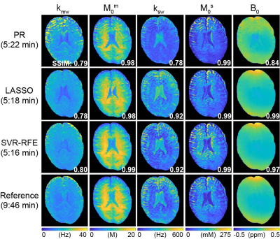 |
50 |
Sequence optimization for saturation transfer MR fingerprinting
Munendra Singh1 and
Hye-Young Heo1
1Russell H. Morgan Department of Radiology and Radiological Science, Johns Hopkins University, Baltimore, MD, United States Keywords: CEST & MT, CEST & MT An optimal saturation transfer MR fingerprinting (ST-MRF) acquisition schedule is critical for efficient and accurate tissue parameter mapping. To optimize RF saturation-encoded MRF acquisitions to a minimal number of saturation scan parameters for magnetization transfer contrast (MTC) and chemical exchange saturation transfer (CEST) parameter determination, we developed an optimization framework using a support vector regression-recursive feature elimination (SVR-RFE) method. Bloch simulations and in vivo studies showed that the proposed optimization method outperformed the quantification accuracy compared to existing methods. The SVR-RFE-based optimization method allowed us to reduce scan time by ~50% without sacrificing reconstruction accuracy. |
|
| 3282. | 51 |
MT and CEST MRI of in vitro gastric protein digestion
Morwarid Mayar1,2,
Mart de Vries1,
Paul Smeets2,3,
John van Duynhoven1,
and Camilla Terenzi1
1Laboratory of Biophysics, Wageningen University & Research, Wageningen, Netherlands, 2Human Nutrition and Health, Wageningen University & Research, Wageningen, Netherlands, 3Image Sciences Institute, University Medical Centre Utrecht, Utrecht, Netherlands Keywords: CEST & MT, Body, in vitro Gastric digestion of dietary protein is commonly studied using in vitro digestion models, which need to be verified with in vivo data. Here, we used MT and CEST MRI to monitor in vitro gastric digestion of milk proteins. We show that MT and CEST measurements can be used to monitor the breakdown of the initially-formed semi-solid protein coagulum and hydrolysis of soluble proteins. We also demonstrate that RF-based ratiometric CEST analysis can be used for pH mapping in both acid- and base-catalyzed regions. Our results open the way to quantification of in vivo protein digestion with the use of MRI. |
|
3283.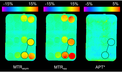 |
52 |
The Influences on Amide Proton Transfer (APT) Signal Metrics at
3T: A Simulation and In-Vitro Study
Aisling Fothergill1,
David Higgins2,
Owen Thomas3,
David Coope4,
Ibrahim Djoukhadar3,
and Laura Parkes1
1Geoffrey Jefferson Brain Research Centre, School of Health Sciences, Faculty of Biology, Medicine and Health, The University of Manchester, Manchester, United Kingdom, 2Philips, Farnborough, United Kingdom, 3Department of Radiology, Geoffrey Jefferson Brain Research Centre, Salford Royal Hospital, Northern Care Alliance NHS Foundation Trust, Manchester Academic Health Science Centre, Manchester, United Kingdom, 4Department of Academic Neurological Surgery, Geoffrey Jefferson Brain Research Centre, Salford Royal Hospital, Northern Care Alliance NHS Foundation Trust, Manchester, United Kingdom Keywords: CEST & MT, Quantitative Imaging, APT Amide Proton Transfer MRI shows clinical benefit in glioma imaging. Clinically useful APTw metrics should be sensitive to changes in amide concentration, a potential marker of tumour proliferation, and insensitive to T1 changes. To investigate sensitivity, simulations and in-vitro studies were performed over a range of amide concentrations and T1 values. In-vitro data included two varied concentration series, with and without T1-maintenance, and a constant concentration T1-vaired series. In-vitro results showed APT* had poor sensitivity, MTRrex was most sensitive to concentration and T1, AREX may overcorrect for T1, and MTRasym appears least sensitive to T1 while remaining sensitive to concentration. |
|
3284.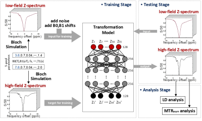 |
53 |
A CEST Z-spectral learning network from 3T to higher B0 field: a
simulation-based preliminary study
Mengdi Yan1,
Chongxue Bie1,
Yibin Chen1,
Xiaowei He1,
and Xiaolei Song2
1Northwest University, Xi'an, Shanxi, China, 2Tsinghua University, Beijing, China Keywords: CEST & MT, Data Analysis Z-spectrum acquired under ultra-high field (> 3T) features stronger and better isolated CEST peaks than those under 3T. But from imaging aspect, 3T scanners perform better and are clinically accessible. Herein, we built a deep neural network (DNN) for predicting Z-spectrum under higher B0 from the corresponding measurement at 3T. The network was trained by 10 million Z-spectra calculated from Bloch-equation models. Simulations with various B0 shifts and noise suggested that 3T Z-spectra could be rapidly and accurately transformed to those under 7T or 9.4T. This network may help improve signal extraction and interpretation of CEST data acquired at 3T. |
|
3285.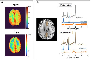 |
54 |
Optimization and validation of Creatine- and Glutamate-CEST
weighted imaging in the human brain at 7T
Bárbara Schmitz-Abecassis1,2,
Chloé Najac1,
Jaimy Plugge1,
Matthias J.P. van Osch1,2,
and Ece Ercan1,3
1Department of Radiology, Leiden University Medical Center, Leiden, Netherlands, 2Medical Delta Cancer Diagnostics 3.0, South-Holland, Netherlands, 3Philips Healthcare, Best, Netherlands Keywords: CEST & MT, High-Field MRI Amine chemical exchange saturation transfer (CEST) is highly sensitive for imaging metabolites in vivo. However, it lacks specificity. We aimed to develop and validate optimal CEST protocols to image creatine and glutamate in the human brain at 7T. Simulations were used to define optimal acquisition parameters, followed by in vitro validation. The optimal B1rms and total saturation (tsat) parameters were determined for creatine (B1rms = 2.5μT & tsat = 1500ms) and glutamate (B1rms = 3.5μT & tsat = 1000ms). These protocols were then used in healthy volunteers to investigate the correlation of glutamate- and creatine-weighted CEST with magnetic resonance spectroscopy results. |
|
3286.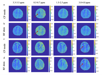 |
55 |
Demonstration of B1 mapping based RF shimming for CEST imaging
at 7T
Eleni Demetriou1,2,
Ivan E Dimitrov1,3,
Peter van Zijl4,5,
Hans Hoogduin6,
Elena Vinogradov7,
Bei Zhang8,
and Anke Henning8
1Advanced imaging research center, University of Texas Southwestern Medical Center, Dallas, TX, United States, 2Brain repair and Rehabilitation, UCL, London, United Kingdom, 3Philips healthcare, Florida, FL, United States, 4F.M. Kirby Research Center for Functional Brain Imaging, Kennedy Krieger Institute, Baltimore, United Kingdom, 5Department of Radiology, Johns Hopkins University, Baltimore, MD, United States, 6Department of Radiology, University Medical Center Utrecht, Utrecht, The Netherlands, Ultrecht, Netherlands, 7Department of Radiology, University of Texas Southwestern Medical Center, Dallas, TX, United States, 8Advanced Imaging Research Center, University of Texas Southwestern Medical Center, Dallas, TX, United States Keywords: CEST & MT, Brain, B1 inhomogeneities In this work, we sought to evaluate CEST in combination with RF shimming that is based on the modulation of the amplitudes and phases of the RF pulses transmitted by an eight channel 7T MR system. |
|
3287.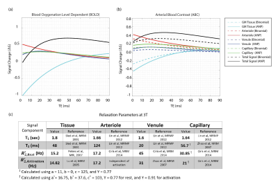 |
56 |
Bloch-McConnell Simulation for Arterial Blood Contrast at 3T and
7T: Adiabatic Null Passage vs Binomial Magnetization Transfer
Preparation
Shahrokh Abbasi-Rad1,2,3,4 and
David Norris1,2
1Donders Institute for Brain, Cognition and Behaviour, Radboud University, Nijmegen, Netherlands, 2Erwin L. Hahn Institute for Magnetic Resonance Imaging, University Duisburg-Essen, Essen, Germany, 3Department of Radiology, Harvard Medical School, Boston, MA, United States, 4Athinoula A. Martinos Center for Biomedical Imaging, Massachusetts General Hospital, Charlestown, MA, United States Keywords: fMRI, Magnetization transfer, Arterial Blood Contrast Arterial blood contrast (ABC) uses on-resonance binomial pulse for magnetization transfer preparation to saturate the tissue signal and highlights the contribution of arterioles. Previously, using Bloch-McConnel simulations, we showed that Binomial MT contaminates ABC with T2 contrast. We suggested the use of adiabatic null passage to decrease the T2 effect. We performed Bloch-McConnel simulations at 3T and 7T comparing the performance of binomial and ANP pulses. ANP increased the arterial contribution from 32% to 42% at 3T and from 18% to 30% at 7T. This study concludes that ANP reduces the T2-contamination imposed by binomial MT block and improves ABC. |
|
3288.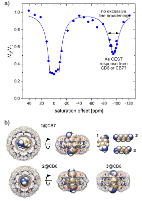 |
57 |
HyperCEST Potential of Members of the Cucurbit[n]uril Family:
Revised Signal Assignment and Suitable Host Identification with
Accelerated MRI
Leif Schröder1,
Hen Amit Morik1,
and Patrick Schuenke2
1Translational Molecular Imaging, Deutsches Krebsforschungszentrum (DKFZ), Heidelberg, Germany, 2Biomedical Magnetic Resonance, Physikalisch-Technische Bundesanstalt (PTB), Berlin, Germany Keywords: Contrast Agent, CEST & MT The full potential of the ultra-sensitive approach of HyperCEST MRI crucially depends on host structures with efficient release of hyperpolarized 129Xe. Cucurbit[n]urils (CB6 and CB7) have been proposed as promising candidates but a quantitative analysis of CEST signatures yet remained an open question. Here, we use MRI-derived z-spectra of multiple samples which provide strong evidence that CB7 is unsuitable as CEST agent and that previously observed signals are solely assigned to CB6 which is also side product in CB7 synthesis. Image acquisition with >20-fold acceleration for the 4-dimensional datasets is possible without compromising a detailed quantitative analysis. |
|
3289.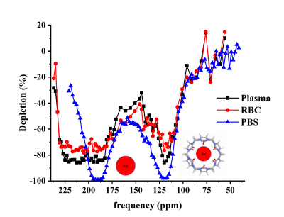 |
58 |
Maximization of cucurbit[6]uril hyperpolarized chemical exchange
saturation transfer (HyperCEST) in bovine blood at 3.0 T
Vira Grynko1,2,
Viktoriia Batarchuk2,3,
Yurii Shepelytskyi2,3,
Hannah Aalto4,
Joseph Deschamps4,
Iulian Constantin Russet5,
and Mitchell Albert2,3,6
1Chemistry and Materials Science Program, Lakehead University, Thunder Bay, ON, Canada, 2Thunder Bay Regional Health Research Institute, Thunder Bay, ON, Canada, 3Chemistry, Lakehead University, Thunder Bay, ON, Canada, 4Applied Life Science Program, Lakehead University, Thunder Bay, ON, Canada, 5Xemed LCC, Durham, NH, United States, 6Northern Ontario School of Medicine, Thunder Bay, ON, Canada Keywords: Hyperpolarized MR (Gas), Contrast Agent Cucurbit[6]uril is a well-studied contrast agent for hyperpolarized 129Xe (HP 129Xe) MRI. Although the in vivobiodistribution of CB6 was measured with hyperpolarized chemical exchange saturation transfer (HyperCEST), there were no previous studies reported for maximization of the HyperCEST effect by optimization of depolarization pre-pulse trains. In the present work, we maximized the CB6 HyperCEST effect in bovine blood, and found for the first time, a HyperCEST depletion of the RBC resonance. In addition, the minimum detectable concentration was found to be four times lower than previously reported. |
|
3290.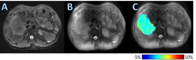 |
59 |
Amide Proton Transfer-Weighted MRI in Preoperative Assessment of
Microvascular Invasion of Hepatocellular Carcinoma: A
Preliminary Study
Jingcheng Huang1,
Chengshi Hou1,
Qingqing Wen2,
Weiqiang Dou2,
and Xianfu Luo1
1Clinical Medical School of Yangzhou University, Northern Jiangsu People’s Hospital, Yangzhou, China, Yangzhou City, China, 2GE Healthcare, MR Research China, Beijing, P.R. China, Beijing City, China Keywords: Quantitative Imaging, CEST & MT, APTw Conventional MRI methods are difficult to reveal histologic and molecular characteristic of HCC. In this study, we aimed to e explore the feasibility of amide proton transfer weighted (APTw) imaging for predicting microvascular invasion (MVI) of hepatocellular carcinoma. Significant difference in APTw values were observed for MVI positive and negative lesions. With these findings, APTw imaging can be considered a potential technique for noninvasive preoperational assessment of MVI in hepatocellular carcinoma. |
|
 Back to Meeting Home
 Back to the Program-at-a-Glance
The International Society for Magnetic Resonance in Medicine is accredited by the Accreditation Council for Continuing Medical Education to provide continuing medical education for physicians.
 Back to Meeting Home
 Back to the Program-at-a-Glance
 View Presentation Video