Digital Poster
CEST II
ISMRM & ISMRT Annual Meeting & Exhibition • 03-08 June 2023 • Toronto, ON, Canada

| Computer # | |||
|---|---|---|---|
3156.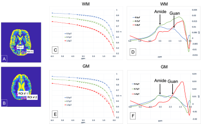 |
101 |
3D Steady-State Golden Angle Radial Sparse Parellel CEST (ssGraspCEST)
MRI of the human brain at 3T.
Rodolphe Leforestier1,
Ding Xia1,
Li Feng1,
and Xiang Xu1
1Biomedical Engineering and Imaging Institute, Icahn School of Medicine at Mount Sinai, New York, NY, United States Keywords: CEST & MT, CEST & MT The purpose of this project was to study the applicability of a fast 3D CEST MRI technique called GraspCEST to extract amdie, guanidinium and rNOE CEST contrasts in human brain at 3T. |
|
3157.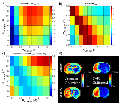 |
102 |
Optimization of Saturation Power for pH-enhanced MRI in Stroke
Rodents
Julius Juhyun Chung1 and
Tao Jin1
1Radiology, University of Pittsburgh, Pittsburgh, PA, United States Keywords: CEST & MT, CEST & MT The sensitivity of CEST to pH makes it a potential modality for the assessment of intracellular pH alterations under pathologic conditions. We previously introduced a method for pH-enhanced MRI using amide and guanidyl protons. In this study, we determine saturation powers for imaging both exchangeable protons to optimize either contrast or contrast-to-variation ratio (CVR) between healthy and infarcted tissue in stroke rats. The contrast across parameters remained steady at ~2-3%, while CVR was dominated by variation across ROIs due to residual non-specific contrast. Optimizing CVR minimizes contrast within the lesion and contralesional tissue while preserving contrast between the two tissues. |
|
3158.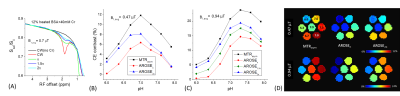 |
103 |
Adjustment of Saturation and Rotation Effects (AROSE) for CEST
Imaging
Tao Jin1 and
Julius Juhyun Chung1
1Radiology, University of Pittsburgh, Pittsburgh, PA, United States Keywords: CEST & MT, CEST & MT Endogenous CEST signal usually has low specificity due to contamination from the magnetization transfer (MT) effect and from other labile protons with close Larmor frequencies. We propose to improve CEST signal specificity with AROSE which measures the difference between CEST signals acquired with similar average saturation power but largely different duty cycles (DC), e.g., a continuous wave or a high DC pulse train versus a low DC one. Simulation and creatine phantom studies showed that AROSE can improve the specificity of slow to intermediate exchanging CEST signals with relatively limited loss of sensitivity. |
|
3159.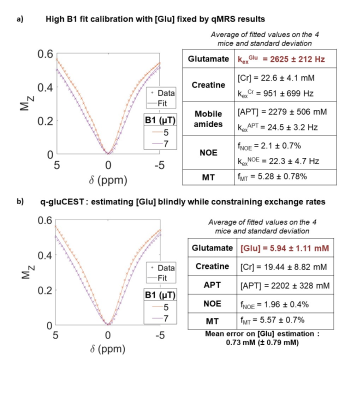 |
104 |
Development of in vivo quantitative gluCEST fitting using
quantitative 1H-MRS as a reference in the mouse brain
Cécile Maguin1,
Eloïse Mougel1,
Julien Valette1,
and Julien Flament1
1Université Paris-Saclay, CEA, CNRS, MIRCen, Laboratoire des Maladies Neurodégénératives, Fontenay-aux-roses, France Keywords: CEST & MT, Modelling, Quantitative imaging In this work, we propose to quantify glutamate with CEST imaging. We use quantitative magnetic resonance spectroscopy as a standard method to measure glutamate concentration in the mouse striatum, and build a CEST model optimized for high saturation power to properly fit glutamate concentration. The CEST estimator developed in this way allowed us to recover glutamate concentration with an average error of 0.7 mM. |
|
3160.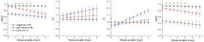 |
105 |
Comparison of pH-weighted and conventional MRI signal in
ischemic stroke
Tao Jin1,
Jicheng Wang2,
T. Kevin Hitchens3,
Dandan Sun4,
Andriy Bandos5,
Joseph Mettenburg1,
Ping Wang1,
and Julius Juhyun Chung1
1Department of Radiology, University of Pittsburgh, Pittsburgh, PA, United States, 2Department of Urology, University of Pittsburgh, Pittsburgh, PA, United States, 3Department of Neurobiology, University of Pittsburgh, Pittsburgh, PA, United States, 4Department of Neurology, University of Pittsburgh, Pittsburgh, PA, United States, 5Department of Biostatistics, University of Pittsburgh, Pittsburgh, PA, United States Keywords: CEST & MT, Stroke, APT, Penumbra The sensitivity of APT MRI in the evaluation of ischemic tissue status was evaluated in stroke rat brains and compared with conventional MRI methods. APT can detect the different levels of tissue acidosis in the ischemic core, penumbra, and oligemia regions. Additionally, APT can also differentiate the severity of tissue acidosis associated with different blood glycemic levels. Our results indicate that APT MRI is a sensitive pH-weighted imaging marker with great potential for the evaluation of the ischemic tissue status and prediction of the stroke outcome. |
|
3161.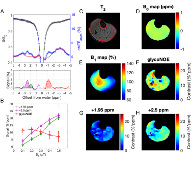 |
106 |
Simultaneous mapping of glycogen and phosphocreatine in human
skeletal muscle by saturation transfer MRI
Chongxue Bie1,2,
Chao Zhou1,
Peter C. M. van Zijl3,4,
Nirbhay N. Yadav3,4,
Yin Wu1,
Hairong Zheng1,
and Yang Zhou1
1Shenzhen Institute of Advanced Technology, Chinese Academy of Sciences, Shenzhen, China, 2Department of Information Science and Technology, Northwest University, Xi’an, China, 3F.M. Kirby Research Center for Functional Brain Imaging, Kennedy Krieger Institute, Baltimore, MD, United States, 4The Russell H. Morgan Department of Radiology, The Johns Hopkins University School of Medicine, Baltimore, MD, United States Keywords: CEST & MT, Metabolism Glycogen and phosphocreatine (PCr) are essential metabolites for maintaining cellular ATP levels. Recent reports show the possibility of PCr mapping in skeletal muscle using CEST MRI but glycogen levels measured using this approach have been inconclusive. Here we report the high-resolution mapping of glycogen and PCr in human skeletal muscle based on relayed nuclear Overhauser effects (glycoNOE) and CEST, respectively. Results at 5T show homogeneous distributions of glycogen and PCr in the calf at levels corresponding to biopsy. Exciting possible applications include localized studies of cellular energetics and noninvasive in situ evaluation of exercise and muscle metabolism in humans. |
|
3162.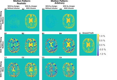 |
107 |
The impact of motion and motion correction in dynamic glucose
enhanced MRI
Patrick M. Lehmann1,
Christos Papageorgakis2,
Stefano Casagranda2,
Anina Seidemo1,
Xiang Xu3,4,
Nirbhay N. Yadav4,5,
Xu Li4,5,
Ronnie Wirestam1,
Patrick Liebig6,
Frederik Testud7,
Pia C. Sundgren8,9,10,
Peter C.M. Van Zijl4,5,
and Linda Knutsson1,4,5
1Medical Radiation Physics, Lund University, Lund, Sweden, 2Department of R&D Advanced Applications, Olea Medical, La Ciotat, France, 3BioMedical Engineering and Imaging Institute, Icahn School of Medicine at Mount Sinai, New York, NY, United States, 4Russell H. Morgan Department of Radiology and Radiological Science, Johns Hopkins University School of Medicine, Baltimore, MD, United States, 5F.M. Kirby Research Center for Functional Brain Imaging, Kennedy Krieger Institute, Baltimore, MD, United States, 6Siemens Healthcare GmbH, Erlangen, Germany, 7Siemens Healthcare AB, Malmö, Sweden, 8Department of Radiology, Lund University, Lund, Sweden, 9Lund University Bioimaging Centre, Lund University, Lund, Sweden, 10Department of Medical Imaging and Physiology, Skane University Hospital, Lund, Sweden Keywords: CEST & MT, Motion Correction Dynamic glucose-enhanced (DGE) MRI is a dynamic chemical exchange saturation transfer (CEST) method that can provide information about D-glucose uptake in tissue. DGE signal changes are small and so-called pseudo-DGE effects can appear as true DGE effects. In this study, we investigated how motion and motion correction influenced the DGE effects using both a realistic (measured) motion pattern and an arbitrary motion pattern. We observed that pseudo-DGE effects are governed by the head motion pattern and originate either from tissue mixing at tissue interfaces or B0-shifts. Although motion correction can reduce these effects, new pseudo-DGE effects can also be introduced. |
|
3163.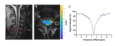 |
108 |
Reproducibility of pH-Weighted Chemical Exchange Saturation
Transfer (CEST) Contrast in the Healthy Cervical Spinal Cord
Alicia Cronin1,2,
Patrick Liebig3,
Sarah Detombe4,
Neil Duggal1,4,
and Robert Bartha1,2
1Medical Biophysics, Western University, London, ON, Canada, 2Centre for Functional and Metabolic Mapping, Robarts Research Institute, London, ON, Canada, 3Siemens Healthcare GmbH, Erlangen, Germany, 4Clinical Neurological Sciences, University Hospital, London Health Sciences Centre, London, ON, Canada Keywords: CEST & MT, Spinal Cord Ischemia and hypoxia can occur in the spinal cord due to several conditions, including compression and injury; however, in-vivo measurements of ischemia have been challenging. Chemical Exchange Saturation Transfer (CEST) can produce pH-weighted contrast, which is an indicator of tissue hypoxia. The purpose of this study was to optimize pH-weighted CEST contrast in the healthy cervical spinal cord using a prototype 3D CEST sequence on a 3T Siemens Prisma Fit MRI and determine the reproducibility of measurements at various levels along the cervical spinal cord. |
|
3164.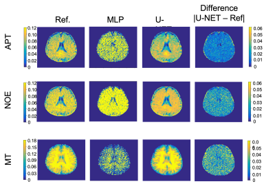 |
109 |
CEST Mapping from Undersampled Z-spectra in the Brain Using Deep
Learning
Karandeep S Cheema1,2,
Pei Han1,
Hsu-Lei Lee1,
Hui Han1,
Yibin Xie1,
Anthony Christodoulou1,2,
and Debiao Li1,2
1Cedars Sinai Medical Center, Los Angeles, CA, United States, 2Bioengineering, University of California, Los Angeles (UCLA), Los Angeles, CA, United States Keywords: CEST & MT, Machine Learning/Artificial Intelligence, CEST, frequency offsets, Fisher Information gain Chemical exchange saturation transfer (CEST) imaging uses radio frequency pulses at different frequency offsets to generate CEST maps. In this work, we used deep learning to calculate CEST maps from steady-state CEST (ss-CEST) images at undersampled frequency offsets, reducing the total scan time by a factor of 3.5. The Z-spectrum was undersampled by selecting the top 15 frequency offsets from Fisher information gain analysis. Fitting results from the proposed method were compared with those from multi-pool fitting with fully sampled Z-spectrum. We showed that it is feasible to reconstruct CEST maps from undersampled, field uncorrected ss-CEST images. |
|
3165.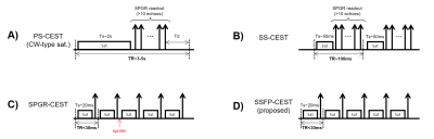 |
110 |
A Steady State Free Precession CEST Sequence with Short
Saturation at 3T Human Scanner
Chuyu Liu1,
Yifan Li1,
Zhongsen Li1,
and Xiaolei Song1
1Center for Biomedical Imaging Research, Department of Biomedical Engineering, Tsinghua University, Beijing, China Keywords: CEST & MT, CEST & MT In this study, we proposed a steady state free precession CEST sequence with short saturation at 3T scanner. Our contributions are: 1) We investigated the SSFP process containing chemical saturation pulses and gave an approximate analytic solution. 2) Simulation and phantom results revealed that the proposed SSFP-CEST sequence had higher SNR and higher saturation efficiency than previous steady-state CEST, while the acquisition time was relatively short. |
|
3166.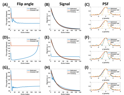 |
111 |
SNR-boosted whole-brain chemical exchange saturation transfer
imaging by optimized variable flip angles
Xingwang Yong1,
Yi-Cheng Hsu2,
Yi Sun2,
and Yi Zhang1
1Key Laboratory for Biomedical Engineering of Ministry of Education, Department of Biomedical Engineering, College of Biomedical Engineering & Instrument Science, Zhejiang University, Hangzhou, Zhejiang, China, 2MR Collaboration, Siemens Healthcare Ltd., Shanghai, China Keywords: CEST & MT, CEST & MT It is well-known that the refocusing flip angles (FAs) of the SPACE sequence can be optimized to generate different image contrasts, such as T1-weighted or T2-weighted. However, the existing SPACE variable FA scheme is unsuitable for CEST imaging whose utmost aim is to increase the SNR during signal readout instead of enforcing contrast. Here, we derived a model to describe the signal-to-noise ratio (SNR) of the SPACE sequence, and maximized SNR by varying refocusing flip angles. Compared to the original constant FA protocol, the optimized variable flip angles yielded both SNR and resolution improvement. |
|
3167.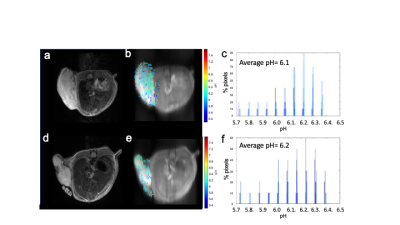 |
112 |
Can pH can be a marker of hypoxia? Role of MRI CEST pH imaging
in differentiating hypoxic from non-hypoxic tumors.
Aruna Singh1,2,
Julia Stabinska1,2,
Balaji Krishnamachary1,
Kuldeep Gupta1,
Farzad Sedaghat3,
Sridhar Nimmagadda1,
Jeff W. M. Bulte1,
Zaver M. Bhujwalla1,
and Michael T. Mcmahon1,2
1Russell H. Morgan Department of Radiology and Radiological Science, Johns Hopkins University School of Medicine, Baltimore, MD, United States, 2F.M. Kirby Research Center for Functional Brain Imaging, Kennedy Krieger Institute, Baltimore, MD, United States, 3James Buchanan Brady Urological Institute and Department of Urology, Johns Hopkins University School of Medicine, Baltimore, MD, United States Keywords: CEST & MT, CEST & MT, CEST MRI, hypoxia, acidosis, pH, Iopamidol, breast cancer, HIF-1 alpha Acidosis and hypoxia play a key role in developing metastasis and chemoresistance in tumors. pH measurement can be used as indicator of hypoxia and can provide insights into the metastatic potential of tumors. This study have explored chemical exchange-dependent saturation transfer (CEST) MRI based pH imaging to differentiate between two types of breast tumor models differing in their Hypoxia Inducible Factor-1α (mediator for hypoxia related adaptations in tumors) expression. Tumor acidification was observed in both with lower average extracellular pH (pHe = 6.1) in tumors with intact HIF-1α expression compared to those with HIF-1α silenced group (pHe = 6.2). |
|
3168.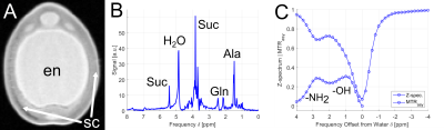 |
113 |
Metabolite detection in plants: A new application field for
CEST?
Simon Mayer1,2,
Fabian Tobias Gutjahr1,
Ljudmilla Borisjuk2,
and Peter Michael Jakob1
1Experimental Physics 5, University of Wuerzburg, Wuerzburg, Germany, 2Leibniz-Institute of Plant Genetics and Crop Plant Research, Gatersleben, Germany Keywords: CEST & MT, Metabolism, Technology transfer Chemical shift imaging (CSI) is rather challenging for the in vivo detection of low concentrated metabolites in plants due to its intrinsic low detection sensitivity and the need for accurate water suppression in the presence of magnetically inhomogeneous plant structures. As an alternative solution, we propose Chemical Exchange Saturation Transfer (CEST) for spectroscopic imaging. Within the work, several plant models were identified where CEST can be used as a versatile alternative for the detection of sugar and amino acids with increased spatial resolution and sensitivity, which demonstrates that CEST has a high potential for plant examinations. |
|
3169.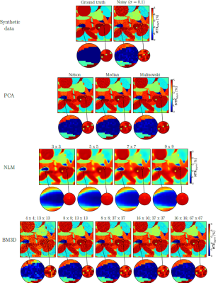 |
114 |
No denoising, no diamonds? - In silico analysis of denoising
methods for CEST MR imaging
Karl Ludger Radke1,
Vibhu Adriaenssens1,
Benedikt Kamp1,
Patrik Jan Gallinnis1,
Eric Bechler1,
Hans-Jörg Wittsack1,
Gerald Antoch1,
and Anja Müller-Lutz1
1University Dusseldorf, Medical Faculty, Department of Diagnostic and Interventional Radiology, Düsseldorf, Germany, Düsseldorf, Germany Keywords: CEST & MT, In Silico CEST imaging requires a high SNR to detect low metabolite concentrations, especially at clinical used magnetic field strength (3 Tesla). In recent years, various approaches have been used to denoise CEST data. Our study investigated the performance of different denoising algorithms using 1000 synthetic CEST MR data. Our results showed that PCA produced the best denoising results for CEST MR imaging in a simulation study. |
|
3170.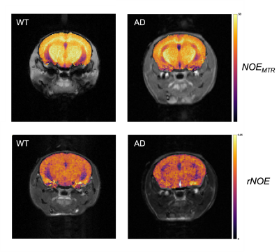 |
115 |
Early-stage mapping of macromolecular tissue content in the
brain of APPNL-F mouse model using nuclear Overhauser enhanced
(NOE) MRI
Anshuman Swain1,
Narayan Datt Soni2,
Neil Wilson2,
Halvor Juul2,
Blake Benyard1,
Dushyant Kumar2,
Ravi Prakash Reddy Nanga2,
Mohammad Haris2,
and Ravinder Reddy2
1Bioengineering, University of Pennsylvania, Philadelphia, PA, United States, 2Radiology, University of Pennsylvania, Philadelphia, PA, United States Keywords: CEST & MT, Alzheimer's Disease This study utilizes nuclear Overhauser effect (NOE) MRI to map early-stage changes in macromolecular brain content of an APPNL-F model of Alzheimer's disease. NOEMTR and rNOE are the quantitative metrics used to assess lipids and proteins in major regions of the mouse brain. Following ROI analysis, there is a statistically significant decrease in NOEMTR contrast between wild-type and AD mice in the hippocampus, with rNOE showing a similar trend with a strong suggestion of statistical significance. Overall, this study shows that NOE MRI can be used to successfully detect changes in the macromolecular content of mouse brain tissue through NOEMTR. |
|
3171.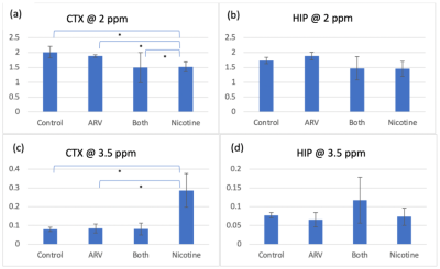 |
116 |
Probing metabolites in antiretroviral and nicotine treated mouse
brains using MRS and CEST-MRI
Gabriel Gauthier1,
Aditya Bade1,
and Yutong Liu1
1University of Nebraska Medical Center, Omaha, NE, United States Keywords: CEST & MT, CEST & MT, MRI, MRS, HIV, Antiretroviral, nicotine, glutamate, myo-inositol, choline To better understand synergistic effects of antiretroviral drugs (ARV) and nicotine on neuroimmune functions, we observed drug-associated metabolites in mice utilizing CEST MRI and MRS. Increased CEST signal was found at 3.5 ppm suggesting increased glutamate in mice treated with nicotine. MRS results showed increased myo-inositol in both ARV and nicotine treated mice. glutamate is an excitatory neurotransmitter and myo-inositol is a biomarker of glial activation. The imaging results suggested elevated neuronal activation and neuroimmune dysfunction. |
|
3172.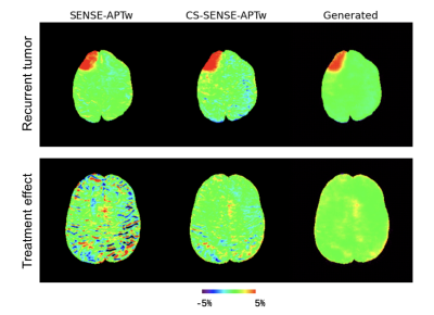 |
117 |
Using a deep residual learning framework to enhance APT-weighted
(APTw) MR images of brain tumors acquired by SENSE with
compressed sensing
Jingpu Wu1,2,
Yiqing Shen1,3,
Pengfei Guo1,3,
Qianqi Huang3,
Babak Moghadas1,
Hye-Young Heo1,
Jinyuan Zhou1,
and Shanshan Jiang1
1Department of Radiology, School of Medicine, Johns Hopkins University, Baltimore, MD, United States, 2Department of Applied Mathematics and Statistics, Whiting School of Engineering, Johns Hopkins University, Baltimore, MD, United States, 3Department of Computer Science, Whiting School of Engineering, Johns Hopkins University, Baltimore, MD, United States Keywords: CEST & MT, CEST & MT Sensitivity encoding (SENSE) is often adopted to accelerate image acquisition for various MRI sequences, including APTw. This is further accelerated with SENSE with compressed sensing (called CS-SENSE), but the image quality degrades to some extent. We collected both SENSE- and CS-SENSE-APTw images and trained a generative model with residual learning to generate SENSE images from CS-SENSE images. The generated results were proved to be highly similar to SENSE-APTw images and less noisy than both SENSE- and CS-SENSE-APTw images. With a larger dataset, we can train more robust models and eventually replace SENSE- with CS-SENSE for a speedup of ~50%. |
|
3173.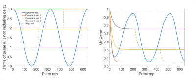 |
118 |
Simulation of a dynamic saturation CEST technique using MR
fingerprinting for quantitative amide proton transfer imaging at
3 T
Jason Ostenson1,2 and
Zhongliang Zu1,2
1Vanderbilt University Institute of Imaging Science, Vanderbilt University Medical Center, Nashville, TN, United States, 2Department of Radiology and Radiological Sciences, Vanderbilt University Medical Center, Nashville, TN, United States Keywords: CEST & MT, MR Fingerprinting Conventionally, quantitative CEST imaging requires steady-state acquisitions so that the obtained signals can be fitted to simple analytical models, which takes a long scan time. Recently, transient-state techniques such as MR fingerprinting were developed to shorten the scan time. This study evaluates the potential of using a new pulse-by-pulse modulation of the saturation power acquisition in conjunction with an MRF reconstruction and fitting approach, termed dynamic saturation CEST, for quantitative APT imaging. Based on signal simulations, sensitivity analysis, and image simulations, we find it is plausible to estimate amide and macromolecular pool concentration, as well as water R1. |
|
3174.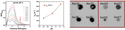 |
119 |
Characterization and Optimization of New CEST Reporter Proteins
with Increased Detection Specificity
David E. Korenchan1,
Adam Fillion2,
Leo L. Cheng3,
Assaf A. Gilad2,
and Christian T. Farrar1
1Martinos Center for Biomedical Imaging, Massachusetts General Hospital, Charlestown, MA, United States, 2Department of Chemical Engineering and Materials Science, Michigan State University, East Lansing, MI, United States, 3Department of Pathology, Massachusetts General Hospital, Charlestown, MA, United States Keywords: CEST & MT, Cell Tracking & Reporter Genes CEST reporter genes could play a critical role in the optimization of novel biological therapeutics. However, existing amide proton based CEST reporter genes have poor specificity as the amide protons of the reporter gene have the same chemical shift as amide protons from endogenous proteins. Here we report on a new class of reporter proteins with CEST contrast at 5-ppm, based on tyrosine hydroxyl ring protons. |
|
3175.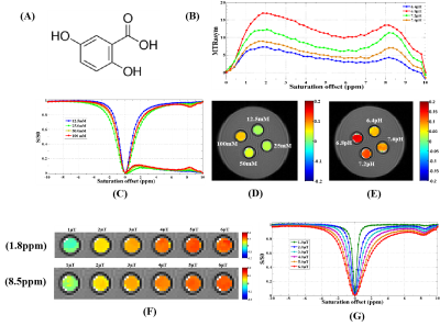 |
120 |
Novel contrast agents for MR based on chemical exchange
saturation transfer and its potential applications
Yanlong Jia1,
Feng Wu1,
Lin Yang2,
Beibei Chen2,
Yazhi Zhong3,
Renhua Wu2,
and Feng Li1
1Department of Radiology, Xiangyang Central Hospital, Affiliated Hospital of Hubei University of Arts and Science, Xiangyang, China, 2Department of Radiology, Second Affiliated Hospital, Shantou University Medical College, Shantou, China, 3Department of Radiology, Huizhou central people's hospital, Huizhou, China Keywords: CEST & MT, Molecular Imaging This study aimed to investigate some small molecular compounds with an appropriate chemical exchange site that can be employed as negative contrast agents for magnetic resonance imaging. 17 kinds of small molecular substances were selected and the imaging were performed on 7.0T MR system. Except for biotin, a series of small molecular substances have the characteristics of CEST. Salicylic acid (9.3ppm), 5-aminosalicylic acid (8.0ppm), and olsalazinesodium (9.8ppm) have large frequency offset, with optimal pH value 6.4~6.8, which are ideal candidates for CEST contrast agents. Due to the differences of molecular structure, the CEST characteristics were different. |
|
 Back to Meeting Home
 Back to the Program-at-a-Glance
The International Society for Magnetic Resonance in Medicine is accredited by the Accreditation Council for Continuing Medical Education to provide continuing medical education for physicians.
 Back to Meeting Home
 Back to the Program-at-a-Glance
 View Presentation Video