Digital Poster
Multicontrast MRI: Applications & Phantoms
ISMRM & ISMRT Annual Meeting & Exhibition • 03-08 June 2023 • Toronto, ON, Canada

Digital Poster
Multicontrast MRI: Applications & Phantoms
| Computer # | |||
|---|---|---|---|
4889.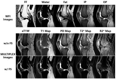 |
121 |
MULTIPLEX with water-fat imaging (WFI) capacity for body imaging
Yongquan Ye1,
Zhongqi Zhang1,
and Jian Xu1
1United Imaging, Houston, TX, United States Keywords: Multi-Contrast, Body A multi-parametric method with WFI imaging capacity, i.e. MULTIPLEX-WFI, was developed and demonstrated. Routine two-point Dixon WFI images are additionally achieved, and a fat-suppression mask is extracted for use on the MULTIPLEX images to improve the overall image quality for body imaging. The cost of adding the WFI capacity to the MULTIPLEX method was negligible. The MULTIPLEX-WFI method was tested on phantom and in vivo knee and pelvis scans, showing much improved imaging quality for body imaging. |
|
4890.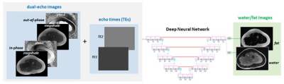 |
122 |
Deep Learning-based Flexible Echo Time Dual-Echo Water-Fat
Separation
Yan Wu1,
Zhitao Li1,
Marcus Alley1,
Zhifei Wen2,
Zheng Zhong1,
Fan Zhang3,
John Pauly1,
and Shreyas Vasanawala1
1Radiology, Stanford University, Stanford, CA, United States, 2Hoag Hospital, Newport Beach, CA, United States, 3Radiology, Stanford Children's Hospital, Stanford, CA, United States Keywords: Fat, Fat, deep learning, dual-echo water-fat separation, flexible echo time We designed a deep learning-based dual-echo water-fat separation method with capability to support flexible echo times. A densely connected hierarchical network was employed, where input included dual-echo images and echo times, and ground truth images were produced using the projected power method. The model was trained and tested using 78 contrast enhanced image sets acquired with optimal echo times, and further validated on 15 non-contrast enhanced image sets obtained with different imaging parameter values. The proposed water-fat separation method has demonstrated high accuracy when dual-echo images were acquired with optimal or non-optimal echo times. |
|
4891.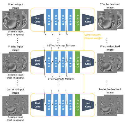 |
123 |
HiFNet: Hierarchical feature sharing network for multi-echo GRE
denoising
Juhyung Park1,
Chungseok Oh1,
Sooyeon Ji1,
Jonghyo Youn1,
and Jongho Lee1
1Seoul National University, Seoul, Korea, Republic of Keywords: Susceptibility, Susceptibility A new denoising network, HiFNet, for complex valued multi-echo GRE image denoising, is proposed. This network shared network features hierarchically from the first echo image to the last echo image, utilizing the redundancy in multi-echo GRE images along the echo dimension. When tested with synthetic noise denoising and real-world denoising experiments, the proposed network shows better performance than a network with no featuring or feature sharing in reverse order, demonstrating the effectiveness of the proposed feature sharing. |
|
4892.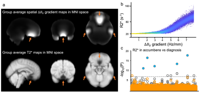 |
124 |
Correction of macroscopic field variation confounding R2* maps
in UK Biobank: simulations and preliminary results in phantom
Chaoyue Wang1,2,
Aaron T. Hess1,
William T. Clarke1,
Stuart Clare1,
Diego Hernando3,
Scott B. Reeder3,
Karla L. Miller1,
and Benjamin C. Tendler1
1Wellcome Centre for Integrative Neuroimaging, FMRIB, Nuffield Department of Clinical Neurosciences, University Of Oxford, Oxford, United Kingdom, 2SJTU-Ruijin-UIH Institute for Medical Imaging Technology, Shanghai, China, 3Departments of Radiology, University of Wisconsin, Madison, WI, United States Keywords: Electromagnetic Tissue Properties, Electromagnetic Tissue Properties Our results suggest that R2* maps in UK Biobank are confounded by the macroscopic field variation which leads to spurious associations driven by field variations due to the air/tissue interfaces. This effect can be modeled as a signal modulation which can be removed through a voxel-wise correction. |
|
4893.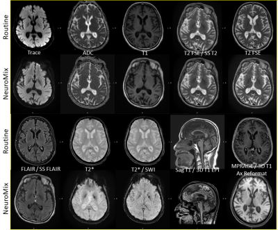 |
125 |
Comparing Diagnostic Performance and Quality of NeuroMix (NM) to
Routine Brain MRI Sequences
Eugene Milshteyn1,
Harry Griffin2,
Yi Shuen Chang2,
Tim Sprenger3,4,
Stefan Skare4,5,
Christopher J. Maclellan2,
and Salil Soman2
1GE Healthcare, Boston, MA, United States, 2Department of Radiology, Beth Israel Deaconess Medical Center, Harvard Medical School, Boston, MA, United States, 3GE Healthcare, Stockholm, Sweden, 4Karolinska Institutet, Stockholm, Sweden, 5Karolinska University Hospital, Stockholm, Sweden Keywords: Multi-Contrast, Brain The focus of this study is to assess the diagnostic performance of NeuroMix, a single push-button, novel, fast multi-contrast MRI sequence, to that of routine brain scans that typically consist of multiple sequences. NeuroMix has the ability to provide several different contrasts in about 3.5 minutes with only one prescription and prescan. While the sequence has initially shown to be fast and motion-robust, more evaluation is needed across the various contrasts compared to gold standard, optimized sequences routinely used in the clinic. Here we present initial qualitative and quantitative comparisons between NeuroMix and routine scans across a variety of patients. |
|
4894.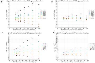 |
126 |
Characterizing the Temperature Dependence of T2 in Phantoms
using an Ethylene Glycol Reference
Adrienne G. Siu1,
Boyan Ivanov1,
Alex K. Smith1,
Matthew D. Robson1,
and Roberto Salvati1
1Perspectum, Oxford, United Kingdom Keywords: Relaxometry, Phantoms, Temperature Phantom vials of varying T2 were calibrated as a function of temperature, where the temperature was measured via multi-echo gradient echo imaging of ethylene glycol in a phantom vial. The temperature calibrations were used to adjust T2 to a reference temperature, achieving T2 deviations to within [-3.2, 2.0] ms [-3.1%, 2.9%] on Siemens 3T (16.7ºC – 25.1ºC) and [-3.5, 1.4] ms [-3.4%, 0.8%] on GE 1.5T (16.5ºC – 23.7ºC). This demonstrates that corrections for T2 due to temperature can be performed while measuring temperature from gradient-echo imaging of ethylene glycol. |
|
4895.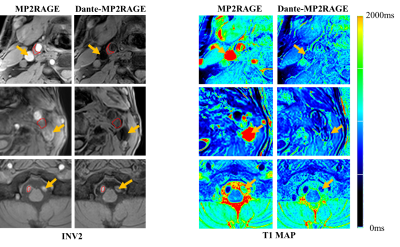 |
127 |
Quantitative MR T1 Value of Arterial Plaque using 3D Black-blood
MP2RAGE:A Feasibility Study
Qizeng Ruan1,
Zehe He1,
Zeping Liu2,
Yuhui Nie2,
Liping Liao1,
Qingchun Li1,
Mingxia Tan1,
Lanbin Huang1,
Guoxi Xie2,
YuanLi Wang1,
and Minglu Zhou1
1The First People’s Hospital of Qinzhou, Qinzhou, China, 2Guangzhou Medical University, Guangzhou, China Keywords: Relaxometry, Cardiovascular, T1 mapping Carotid plaque is an important cause of stroke. The T1 value of the plaque could provide potential information for the diagnosis. In this work, we compared the performance of T1 mapping of BB-MP2RAGE and conventional MP2RAGE in carotid plaque. Experiments demonstrated BB-MP2RAGE could achieve more accurate measurement of T1 value for diagnosis of carotid plaque. |
|
4896.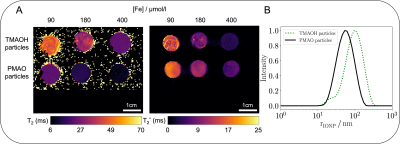 |
128 |
Monte Carlo simulations of transverse relaxation for assessing
physico-chemical properties of novel superparamagnetic iron
oxide particles
Lauritz Klünder1,
Bastian Maus1,
Maria Belen Rivas Aiello2,
Thomas Kirse3,4,
Cristian Strassert3,4,
and Cornelius Faber1
1Clinic of Radiology, University of Münster, Münster, Germany, 2Instituto de Investigaciones Fisicoquímicas Teóricas y Aplicadas, Universidad Nacional de La Plata - CONICET, La Plata, Argentina, 3Institut für Anorganische und Analytische Chemie, University of Münster, Münster, Germany, 4CeNTech, CiMIC, SoN, University of Münster, Münster, Germany Keywords: Relaxometry, Contrast Agent, Simulations Physico-chemical properties of superparamagnetic iron oxide nanoparticles (IONP) strongly affect their impact on transverse relaxation and their efficiency as MR contrast agents. Monte Carlo simulations were developed to model relaxation in silico for two novel IONPs with distinct physico-chemical properties. The IONPs comprised an identical magnetite core but differed in preparation strategy, defining their coating and size. Simulations were compared to in vitro relaxometry measurements and reproduced experimentally measured relaxation times. Using lognormally distributed particle radii in the simulations was required to obtain correct R2* values. Together, simulations and measurements unveiled additional particle aggregation in the MR samples. |
|
4897. |
129 |
In Vivo Single Voxel Relaxation-Diffusion Correlation Spectra
Revealed the Microstructure of Human Brain
Lixian Wang1,2,
Tomohisa Okada3,
Takashi Hanakawa1,3,
Baogui Zhang2,4,
Huilou Liang2,
Jing An5,
Rong Xue2,4,6,
and Fangrong Zong2,7
1Department of Integrated Neuroanatomy and Neuroimaging, Kyoto University, Kyoto, Japan, 2Institute of Biophysics, Chinese Academy of Sciences, Beijing, China, 3Human Brain Research Center, Kyoto University, Kyoto, Japan, 4University of Chinese Academy of Sciences, Beijing, China, 5Siemens Shenzhen Magnetic Resonance Ltd, Shenzhen, China, 6Beijing Institute for Brain Disorders, Beijing, China, 7School of Artificial Intelligence, Beijing University of Posts and Telecommunications, Beijing, China Keywords: Multi-Contrast, Diffusion/other diffusion imaging techniques Multidimensional MRI resolves the correlations between physical parameters and relaxation properties and has become an increasingly important protocol in biomedical engineering. It has sub-voxel resolution but most research still uses multi-voxel ROIs. In this study, we obtained the single voxel T1-D Relaxation-Diffusion correlation spectra from in vivo human brain tissue boundaries. The significant variance in these spectra might help to identify tissue microstructure at the sub-voxel resolution. Our research disclosed the feasibility of applying multidimensional MRI techniques to detect and segment the human brain. |
|
4898.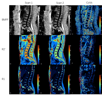 |
130 |
Repeatability of R1, R2* and fat fraction in Human Lumbar
Vertebrae using a simultaneous multi-relaxation-time Imaging
(TXI) method
Jie Yang1,
Hao Feng1,
Meining Chen2,
Xu Yan2,
Jianqi Li3,
Yinqiao Yi3,
Haodong Zhong3,
and Jianquan Zhong1
1Department of Radiology, Zigong First People's Hospital, Zigong, China, 2MR Scientific Marketing, Siemens Healthineers, Shanghai, China, 3Shanghai Key Laboratory of Magnetic Resonance, East China Normal University, Shanghai, China Keywords: Multi-Contrast, Multi-Contrast The quantitative MRI mapping techniques showed great potential in evaluating intervertebral disc degeneration (IVD) and diagnosing lower back pain. The repeatability of the quantitative methods is very important for clinical evaluation. A simultaneous multi-relaxation-time mapping (TXI) method was evaluated here, which can quantify the bone marrow fat fraction (BMFF), R2* and R1 value by a single scan and fast speed. The quantitative parameters of TXI showed high repeatability with high consistency and low CoVs, and could be applied in future clinical applications of IVD degeneration disease. |
|
4899.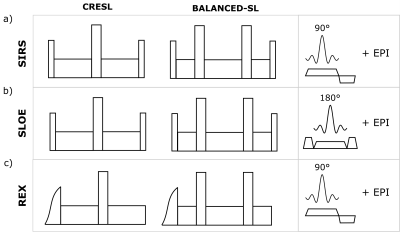 |
131 |
Analysis of spin-lock techniques for the non-invasive detection
of neuronal currents with magnetic resonance
Milena Capiglioni1,
Federico Turco1,
Claus Kiefer1,
and Roland Wiest1
1Support Center for Advanced Neuroimaging (SCAN), University of Bern, Bern, Switzerland Keywords: Electromagnetic Tissue Properties, Simulations, Spin-lock, Neuronal Current Imaging Different SL techniques have been proposed to generate images based with a contrast based on neuronal activity. We analyzed the contrast of different SL techniques for the detection of oscillatory magnetic fields and their dependence with the duration of the SL and the initial phase of the target field. |
|
4900. |
132 |
Optimisation of T1 measurement sensitivity for Oxygen-Enhanced
MRI assessment of hypoxia in patients with head and neck cancers
Maira Tariq1,2,
Alison Macdonald2,
Mathew R Orton1,2,
Michael J Dubec3,
David J Collins1,2,
Jessica M Winfield1,2,
and James P B O’Connor1,3,4
1Division of Radiotherapy and Imaging, The Institute of Cancer Research, London, United Kingdom, 2MRI Unit, The Royal Marsden NHS Foundation Trust, London, United Kingdom, 3Division of Cancer Sciences, University of Manchester, Manchester, United Kingdom, 4Radiology, The Christie NHS Foundation Trust, Manchester, United Kingdom Keywords: Relaxometry, Quantitative Imaging, Oxygen-Enhanced MRI Hypoxia is an important prognostic factor for head and neck cancers (HNC). Oxygen-Enhanced MRI (OE-MRI) can map hypoxia, by quantifying change in longitudinal relaxation time, T1, but the technique suffers from low signal-to-noise ratio (SNR), which reduces the sensitivity of hypoxia detection. We optimised standard T1 mapping methods on a 1.5T scanner, to select an accurate, precise, and high SNR sequence, derived in a phantom and in healthy volunteers. 3D Variable Flip Angle spoiled gradient-echo acquisition with view-sharing, flip angles 2o and 8-10o and with B1 correction applied provided suitable protocol for clinical application. |
|
4901.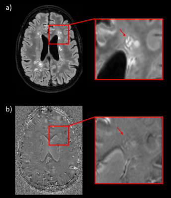 |
133 |
FLAIR3Phase: a new synthetic MRI contrast for paramagnetic rim
lesions detection in multiple sclerosis
Colin Vanden Bulcke1,2,
Nicolas Delinte2,
and Benoît Macq2
1Institute of Neurosciences, Université Catholique de Louvain, Brussels, Belgium, 2Institute for Information and Communication Technologies, Electronics and Applied Mathematics, Université Catholique de Louvain, Louvain-la-Neuve, Belgium Keywords: Multi-Contrast, Multiple Sclerosis, Paramagnetic Rim Lesions Chronic active multiple sclerosis (MS) lesions are visible on susceptibility-based MRI as paramagnetic rim lesions (PRL); these lesions are characterized by severe tissue damage and strongly correlate with disease severity. PRL assessment is usually performed manually on unwrapped-filtered phase images. However, PRL assessment is associated with high intra/inter-rater variability due to the poor visibility of PRL and to the lack of standardized imaging protocols/guidelines for their detection. Here, we propose a new synthetic contrast called FLAIR3Phase to augment PRL assessment workflow and to reduce rater variability. |
|
4902.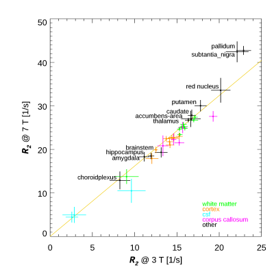 |
134 |
Brain R2 dependence on field strength
Yicun Wang1,
Peter van Gelderen1,
Jacco A de Zwart1,
and Jeff H Duyn1
1AMRI, LFMI, NINDS, NIH, Bethesda, MD, United States Keywords: Relaxometry, Brain Gradient Echo Sampling of Spin Echo signal was used to obtain R2 maps in nine adults at 3 T and 7 T. These were analyzed using anatomically defined ROIs, across most of the brain. Comparison between the two field strengths shows in most brain regions the R2 increases by about a factor of 1.62, with the notable exception of regions high in iron. Linear fits of R2 versus estimated iron concentration yielded 50.6 and 132 1/s/(mg/g) for 3 T and 7 T respectively, in line with prior reports at lower fields for the field dependence of iron related relaxation. |
|
4903.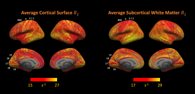 |
135 |
Whole Brain R2 Maps at 7T Highlight Iron-Rich Cortical Gray and
Sub-cortical White Matter
Yicun Wang1,
Peter van Gelderen1,
Jacco de Zwart1,
and Jeff H. Duyn1
1AMRI, LFMI, NINDS, NIH, Bethesda, MD, United States Keywords: Relaxometry, Relaxometry, T2 mapping; Iron Gradient Echo Sampling of Spin Echo signal was used to obtain whole-brain R2 maps from 8 young and 1 senior healthy volunteers at 7T. Cortical surface reconstruction showed increased R2 in areas of high iron content as known from histology. Furthermore, high R2 in sensory-motor and visual cortices was accompanied by low R2 in underlying white matter. Highest R2 in subcortical white matter was found in frontal and temporal lobes. The low sensitivity of R2 to fiber orientation and venous vasculature may render it more suitable than R2* and $$$\chi$$$ for iron quantification, especially near the brain surface. |
|
4904.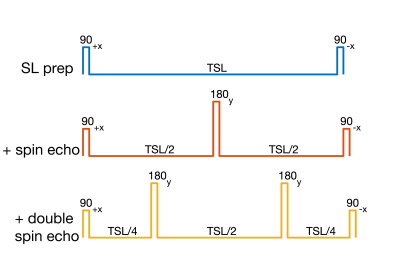 |
136 |
B1 and B0 insensitive R1σ preparation pulses can reduce
sensitivity to R1σ dispersion
Kevin D Harkins1,2,3,
Jason Ostenson1,2,
Fatemeh Adelnia1,2,
Feng Wang1,2,
Zhongliang Zu1,2,
and John C Gore1,2,3
1Radiology and Radiological Sciences, Vanderbilt University Medical Center, Nashville, TN, United States, 2Institute of Imaging Science, Vanderbilt University Medical Center, Nashville, TN, United States, 3Biomedical Engineering, Vanderbilt University, Nashville, TN, United States Keywords: Relaxometry, Simulations R1σ dispersion imaging can be used to uncover specific relaxation mechanisms in soft tissues, but is sensitive to B0 and B1 inhomogeneity. Several varieties of R1σ preparation pulses have been developed to reduce sensitivity to B0 and B1, typically by introducing a spin echo or double spin echo as part of the preparation pulse. This simulation study shows that such pulses can actually reduce the sensitivity to R1σ dispersion. |
|
4905.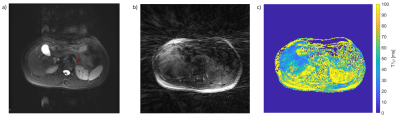 |
137 |
Free-breathing T1ρ Quantification in the Pancreas
Hannah J. S. Ehler1,
Sharon Clarke2,3,
Geoff Williams4,5,
and James Rioux1,2,3
1Department of Physics & Atmpspheric Science, Dalhousie University, Halifax, NS, Canada, 2Department of Diagnostic Radiology, Dalhousie University, Halifax, NS, Canada, 3BIOTIC, Nova Scotia Health, Halifax, NS, Canada, 4Department of Digestive Care & Endoscopy, Dalhousie University, Halifax, NS, Canada, 5Nova Scotia Health, Halifax, NS, Canada Keywords: Relaxometry, Pancreas, T1rho T1ρ quantification in the pancreas is largely unexplored due to the difficulty of compensating for breathing motion. In this study we demonstrate a free-breathing T1ρ technique for use in the abdomen which is able to quantify T1ρ in the pancreas without respiratory gating, through the use of golden-angle radial sampling. The technique was validated in phantoms and tested in three healthy volunteers. T1ρ values in the liver and spleen agreed with recent literature, while differences in T1ρ values in the pancreas may be related to the use of free breathing instead of respiratory gating. |
|
4906.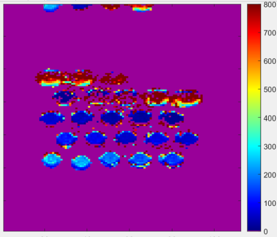 |
138 |
Sampling Time Considerations for T1 map acquisition on the 64mT
Hyperfine Swoop system: A Phantom Evaluation Study
Vivian S. Nguyen1,2,
Sudarshan Ragunathan3,
Timothy J. Carroll4,
Adil Javed5,
Marcella K. Vaicik1,
John G. Georgiadis1,
and Keigo Kawaji1,2
1Biomedical Engineering, Illinois Institute of Technology, Chicago, IL, United States, 2Medicine - Cardiology, University of Chicago Medical Center, Chicago, IL, United States, 3Hyperfine Inc., Guilford, CT, United States, 4Radiology, University of Chicago Medical Center, Chicago, IL, United States, 5Neurology, University of Chicago Medical Center, Chicago, IL, United States Keywords: Relaxometry, Low-Field MRI T1 maps provide quantitative relaxometric measures of tissue responses that may carry correlative indices of specific disease indications. On the recently commercialized 64mT system (Hyperfine Inc.), little has been reported regarding both clinical T1 relaxometric normal values in presence of Gadolinium-based contrast and relevant acquisition schemes with Gd contrast enhancement. In this study, we examine an array of Gadolinium-doped vials that represent 10-5000 fold dilution in the vasculature using an Inversion-Recovery FSE acquisition to gain key insights on: a) anticipated normal values in presence of Gd, b) conventional fitting algorithm performance, and c) potential inferences on inversion timing selection. |
|
 Back to Meeting Home
 Back to the Program-at-a-Glance
The International Society for Magnetic Resonance in Medicine is accredited by the Accreditation Council for Continuing Medical Education to provide continuing medical education for physicians.
 Back to Meeting Home
 Back to the Program-at-a-Glance
 View Presentation Video