Digital Poster
Spectroscopy: Deuterium
ISMRM & ISMRT Annual Meeting & Exhibition • 03-08 June 2023 • Toronto, ON, Canada

| Computer # | |||
|---|---|---|---|
3855.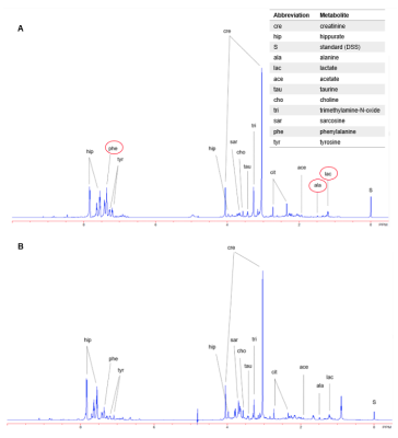 |
101 |
A Magnetic Resonance Spectroscopy Method in Characterization of
Urine Metabolomics for Prostate Cancer
Leo L Cheng1,
Anna-Laura M Hasubek1,
and Adam S Feldman2
1Massachusetts General Hospital, Harvard Medical School, Boston, MA, United States, 2Massachusetts General Hospital, Boston, MA, United States Keywords: Spectroscopy, Prostate, Metabolomics Currently, Prostate Cancer (PC) diagnosis is achieved either through invasive measures, like biopsies, or imaging techniques, such as sonography, CT, and MRI. The biomarker PSA in blood is used for screening, however its use is considered controversial. It doesn’t show to reduce PC all-cause mortality and PSA screening comes with a high number of false-positive results and associated risks. Here, using HRMAS MRS, we studied human urine samples obtained from prostate cancer patients and healthy controls to reveal potential PC-associated metabolomic changes, which may assist with early and non-invasive PC diagnosis and screening. |
|
3856.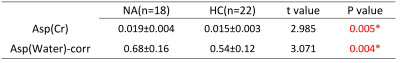 |
102 |
Changes in Aspartate Metabolism in the medial-prefrontal cortex
of Nicotine Addicts Based on J-edited Magnetic resonance
spectroscopy
Miaomiao Yu1,
Liangjie Lin2,
Ke Xu1,
Man Xu1,
Jianxin Ren1,
Xiaoyu Niu1,
Xinyu Gao1,
Mengzhe Zhang1,
Zhengui Yang1,
Jinghan Dang1,
Qiuying Tao1,
Shaoqiang Han1,
Weijian Wang1,
Jingliang Cheng1,
and yong zhang1
1Magnetic Resonance Imaging, The First Affiliated Hospital of Zhengzhou University, Zhengzhou, China, 2Clinical and Technical Support, Philips Healthcare, Beijing, China Keywords: fMRI, Metabolism, Hydrogen proton magnetic resonance spectroscopy (1H MRS) Cigarette smoking harms nearly every organ of the body, causes many diseases, and reduces the health of smokers in general. Our study aims to explore the changes of aspartate (Asp) levels in the medial prefrontal cortex of patients with nicotine addiction using the J-edited 1H MRS technique. Results showed that the Asp level in medial prefrontal of nicotine addicts is relatively increased, suggesting that the metabolism of aspartate may play a key role in nicotine dependence. |
|
3857.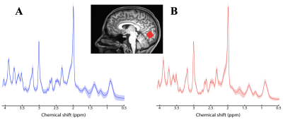 |
103 |
Neurochemical profile of major depressive disorder in
adolescents: 7 T MRS study
Guglielmo Genovese1,
Paul E. Croarkin2,
Can Ozger2,
and Małgorzata Marjańska1
1Center for Magnetic Resonance Research, University of Minnesota, Minneapolis, MN, United States, 2Department of Psychiatry and Psychology, Mayo Clinic, Rochester, MN, United States Keywords: Spectroscopy, Psychiatric Disorders, Ultra-High Field 7 T, Adolescents, Neuro, Major Depressive Disorder Major depressive disorder in adolescents (MDD) is a substantial public health problem. Existing treatments are often ineffective and do not target relevant neurobiological markers. It has been hypothesized that neurobiological mechanism underlying depression involves a dysfunction of the excitatory/inhibitory neurotransmitters. Here, the neurochemical profile from the occipital lobe of twenty adolescents with MDD and twenty-four age-matched healthy volunteers (HV) were acquired at 7 T. Larger SDs were observed for some metabolites in MDD versus HV and suggest heterogeneity of depression severity in the MDD cohort. Positive correlation in MDD between aspartate and GABA could be indicative of a neurotransmitter imbalance. |
|
3858. |
104 |
Efficient Myocardial Spectroscopy combining Metabolite-Cycling
with Respiratory Bellows at 3T
Fabian Bschorr1,
Tobias Speidel1,
Niklaus Zoelch2,3,
Andreas Hock4,
Alireza Abaei5,
and Volker Rasche1
1Ulm University Hospital, Ulm, Germany, 2Zurich University Hospital of Psychiatry, University of Zurich, Zurich, Switzerland, 3Institute of Forensic Medicine, University of Zurich, Zurich, Switzerland, 4Zurich University of Applied Sciences, Wädenswil, Switzerland, 5Core Facility Small Animal Imaging (CF-SANI), University of Ulm, Ulm, Germany Keywords: Spectroscopy, Cardiovascular Despite its diagnostic values1-4, the application of cardiac spectroscopy (CMRS) in clinical routine is generally limited by technical challenges and long scan times. A protocol for CMRS is proposed using metabolite-cycling, local, ECG-triggered shimming, ECG-triggering and a respiratory bellows. The protocol is evaluated in a volunteer cohort and provides myocardial triglyceride content (MTGC) quantification results that correlate well intrasession- (r=0.98, p<0.001) and intersession-wise (r=0.95, p<0.001). Thus, a good repeatibility and technical stability for the quantification of MTGC could be achieved with an average scan time of 440s at 3T. |
|
3859.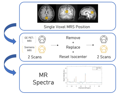 |
105 |
Comparison of Magnetic Resonance Spectroscopy Data Acquired with
Hybrid PET-MRI and Standalone MRI Scanners
Aditya Bhattacharya1,
Benjamin B. Risk2,
and Candace C. Fleischer3,4
1Neuroscience, Georgia Institute of Technology, Atlanta, GA, United States, 2Biostatistics and Bioinformatics, Emory University, Atlanta, GA, United States, 3Radiology and Imaging Sciences, Emory University School of Medicine, Atlanta, GA, United States, 4Biomedical Engineering, Georgia Institute of Technology and Emory University, Atlanta, GA, United States Keywords: Spectroscopy, PET/MR Magnetic resonance spectroscopy (MRS) is a powerful tool for quantifying metabolite concentrations, but spectral quality has not been thoroughly explored in hybrid MRI scanners. Using repeated measurements in a brain phantom and healthy volunteer, we determined the reproducibility and accuracy of MRS data on two systems, a hybrid GE PET-MRI (3.0 T) and Siemens 3.0 T MRI. Spectra acquired using the PET-MRI scanner produced metabolite concentrations with higher accuracy and lower variance across repeated measurements compared to the standalone MRI, suggesting spectral quality is not hindered by the hybrid system. |
|
3860.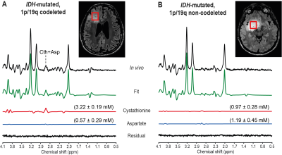 |
106 |
Neurochemical differences between 1p/19q codeleted and
non-codeleted gliomas assessed by in vivo magnetic resonance
spectroscopy
Francesca Branzoli1,
Roberto Liserre2,
Dinesh Deelchand3,
Pietro Luigi Poliani4,
Franck Bielle5,
Lucia Nichelli6,
Marc Sanson1,7,
Stéphane Lehéricy1,6,8,
and Małgorzata Marjańska3
1Sorbonne University, UMR S1127, INSERM U1127, CNRS UMR 7225, Paris Brain Institute - ICM, Paris, France, 2Department of Radiology, Neuroradiology Unit, ASST Spedali Civili University Hospital, Brescia, Italy, 3Center for Magnetic Resonance Research, Department of Radiology, University of Minnesota, Minneapolis, MN, United States, 4Pathology Unit, Department of Molecular and Translational Medicine, University of Brescia, Brescia, Italy, 5Laboratory R Escourolle, University Hospital La Pitié Salpêtrière, Paris, France, 6Department of neuroradiology, University Hospital La Pitié Salpêtrière, Paris, France, 7Department of neurology 2, University Hospital La Pitié Salpêtrière, Paris, France, 8Center for Neuroimaging Research (CENIR), ICM, Paris, France Keywords: Spectroscopy, Cancer Mutations in the genes encoding for isocitrate dehydrogenase (IDH), and 1p/19q codeletion are genetic alterations that can be often found in low-grade gliomas and are associated with better prognosis and response to treatment. Nevertheless, the biological effects of the 1p/19q codeletion are still not well known.
In this study, we compared the neurochemical profile of IDH-mutated,
1p/19q codeleted gliomas and their non-codeleted
counterparts using in
vivo magnetic resonance spectroscopy. We showed that
the only metabolite that differed significantly between the
two groups was cystathionine, pointing to this metabolite
as the most useful biomarker for the identification of
1p/19q codeleted gliomas.
|
|
3861.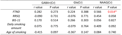 |
107 |
Correlation of NAAG level in the bilateral medial prefrontal
cortex with nicotine dependence score in adult cigarette smokers
Liangjie Lin1,
Miaomiao Yu2,
Ke Xu2,
Man Xu2,
Jianxin Ren2,
Xiaoyu Niu2,
Xinyu Gao2,
Mengzhe Zhang2,
Zhengui Yang2,
Jinghan Dang2,
Qiuying Tao2,
Shaoqiang Han2,
Weijian Wang2,
Jingliang Cheng2,
and yong zhang2
1Clinical and Technical Support, Philips Healthcare, Beijing, China, 2Magnetic Resonance Imaging, The First Affiliated Hospital of Zhengzhou University, Zhengzhou, China Keywords: Spectroscopy, Metabolism, Hydrogen proton magnetic resonance spectroscopy (1H MRS) Smoking can cause cancer, heart disease, stroke, lung diseases, diabetes, and chronic obstructive pulmonary disease including emphysema and chronic bronchitis. Our study aims to explore the correlation between neurotransmitter levels in the medial prefrontal cortex of patients with nicotine addiction (NA) using the J-edited 1H MRS technique. Results showed that the N-acetylaspartylglutamate (NAAG) level in medial prefrontal of nicotine addicts was significantly correlated with the score of Fagerstrom Test for Nicotine Dependence, suggesting that the metabolism of NAAG may play a key role in nicotine dependence patients. |
|
3862.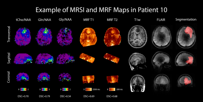 |
108 |
Comparing 7 T FID-CRT-MRSI with 3 T MRF in High-Grade Gliomas
Philipp Lazen1,
Sukrit Sharma2,
Cornelius Cadrien1,
Wolfgang Marik3,
Thomas Roetzer-Pejrimovsky4,
Eva Niess2,
Lukas Hingerl2,
Stephan Gruber2,
Bernhard Strasser2,
Barbara Kiesel1,
Adelheid Wöhrer4,
Matthias Preusser5,
Julia Furtner3,6,
Wolfgang Bogner2,7,
Georg Widhalm1,
Siegfried Trattnig2,7,8,
Karl Rössler1,7,
and Gilbert Hangel1,2,7
1Department of Neurosurgery, Medical University of Vienna, Vienna, Austria, 2High-field MR Center, Department of Biomedical Imaging and Image-guided Therapy, Medical University of Vienna, Vienna, Austria, 3Division of Neuroradiology and Musculoskeletal Radiology, Department of Biomedical Imaging and Image-guided Therapy, Medical University of Vienna, Vienna, Austria, 4Division of Neuropathology and Neurochemistry, Department of Neurology, Medical University of Vienna, Vienna, Austria, 5Division of Oncology, Department of Internal Medicine I, Medical University of Vienna, Vienna, Austria, 6Medical Image Analysis and AI, Danube Private University, Krems, Austria, 7Christian Doppler Laboratory for MR Imaging Biomarkers, Vienna, Austria, 8Institute for Clinical Molecular MRI, Karl Landsteiner Society, St. Pölten, Austria Keywords: Spectroscopy, Brain We compared 7T MRSI-derived metabolic ratios with T1 and T2 maps from MR fingerprinting in a cohort of glioma patients by defining hotspots and calculating Dice similarity coefficients (DSCs) between them and a segmentation. Notable high DSCs were 0.799 for tCho/NAA vs T2 and 0.753 for Gln/NAA vs T1. We also investigated values for T1, T2, and metabolite ratios in tumors, hotspots, and a control region, with the median relaxation times being T1=1610 ms and T2=79 ms in the tumor, and T1=964 ms and T2=46 ms in NAWM. |
|
3863.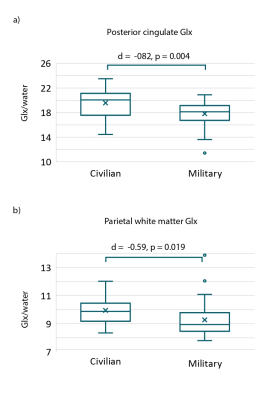 |
109 |
Altered cortical metabolism contributes to cognitive deficits in
a healthy military cohort
Julie M Joyce1,2,
Kristin Heaton3,
Huijun J Liao2,
and Alexander P Lin2
1cBRAIN, Department of Child and Adolescent Psychiatry, Psychosomatics, and Psychotherapy, Ludwig-Maximilians-Universität, Munich, Germany, 2Center for Clinical Spectroscopy, Department of Radiology, Brigham and Women's Hospital, Harvard Medical School, Boston, MA, United States, 3Military Performance Division, United States Army Research Institute of Environmental Medicine, Natick, MA, United States Keywords: Spectroscopy, Neuro, Military The selection of control groups is of increasing interest in military research. Here, we used single voxel magnetic resonance spectroscopy (MRS) to probe cortical metabolism in the anterior and posterior cingulate gyri, temporal lobe and parietal white matter in healthy military service members and civilian controls. Computerized neuropsychological tests were administered to evaluate neurocognitive abilities. We identified higher incidence of cognitive deficits in military service members relative to civilian controls as well as associations between neurometabolite levels and poor cognitive performance. |
|
3864.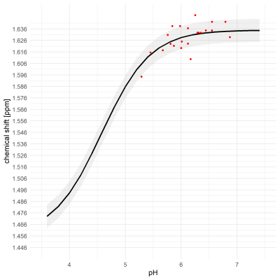 |
110 |
In situ determination of pH in brain tissue using postmortem
1H-MRS
Sabina Frese1,
Dominic Gascho2,
Michael Thali2,
Sebastian Kozerke1,
and Niklaus Zölch2,3
1Institute for Biomedical Engineering, ETH Zürich, Zürich, Switzerland, 2Institute for Forensic Medicine, University of Zurich, Zürich, Switzerland, 3University Hospital for Psychiatry, University of Zurich, Zürich, Switzerland Keywords: Spectroscopy, Spectroscopy Postmortem, relatively low pH values have been described and reported to be dependent on the cause of death and the duration of the agonal phase. Using 1H-MRS on decedents and subsequent control pH measurements in collected samples, we investigated to what extent the pH dependence of peak positions of acetate and lactate are suitable to detect such low pH values non-invasively. We found that the measured chemical shift of acetate and corresponding pH value follow a simulated titration curve. Hence, acetate is a promising candidate to detect low postmortem brain pH for forensic investigations. |
|
3865.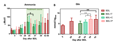 |
111 |
Novel urease inhibitor as potential treatment of hepatic
encephalopathy led to brain glutamine decrease
Dunja Simicic1,2,
Diana Evstafeva3,
Filip Ilievski3,
Yinyin Bao3,
Sunghyun Kang3,
Dario Sessa4,
Stefanita-Octavian Mitrea1,2,
Katarzyna Pierzchala1,2,5,
Jean-Christophe Leroux3,
and Cristina Cudalbu1,2
1CIBM Center for Biomedical Imaging, Lausanne, Switzerland, 2Animal Imaging and Technology, EPFL, Lausanne, Switzerland, 3Institute of Pharmaceutical Sciences, ETH Zurich, Zurich, Switzerland, 4Swiss Center for Liver Disease in Children, University Hospitals Geneva, Geneva, Switzerland, 5Laboratory of Functional and Metabolic Imaging, EPFL, Lausanne, Switzerland Keywords: Spectroscopy, Brain, Metabolism, Hepatic Encephalopathy, proton Type C hepatic encephalopathy (HE) is a severe neuropsychiatric disorder associated with chronic liver disease. Ammonia (partially produced in the gut by bacterial urease activity) has been pinpointed to explain the observed neurological alterations in HE and connected with the increase in brain Gln measured using 1H-MRS. As such current treatment strategies focus on either reducing ammonia production and absorption or on promoting its elimination. We showed that targeting urease activity using the urease inhibitor 2-octynoHA has a beneficial effect in reducing blood ammonia levels and reducing brain Gln measured using 1H-MRS at 9.4T in the cerebellum. |
|
3866.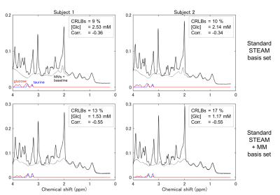 |
112 |
Cerebral glucose quantification using 1H short-TE STEAM
spectroscopy at 7T: Influence of macromolecule signals
Hideto Kuribayashi1,
Yuta Urushibata1,
Thuy Ha Duy Dinh2,
Hirohiko Imai3,
Sinyeob Ahn4,
Ravi Teja Seethamraju5,
Tadashi Isa2,
and Tomohisa Okada2
1Siemens Healthcare K.K., Tokyo, Japan, 2Human Brain Research Center, Kyoto University Graduate School of Medicine, Kyoto, Japan, 3Kyoto U Graduate School of Informatics, Kyoto, Japan, 4Siemens Medical Solutions, Berkeley, CA, United States, 5Siemens Medical Solutions, Boston, MA, United States Keywords: Spectroscopy, Neuro, glucose The ability of conventional 1H short-TE STEAM spectroscopy at 7T was investigated to quantitate cerebral glucose. Spectra were analyzed using LCModel with the standard 7T STEAM basis set and with that plus macromolecule basis set. With the evaluation of Cramér-Rao lower bounds, the precision of glucose quantification reduced with the macromolecule basis set. Moreover, estimated glucose and taurine concentrations were shown to correlate for both analytical conditions. Thus, the quantification is not improved using the macromolecule basis set and may be more precise via detecting H1-α-glucose, which is free from spectral overlap. |
|
3867.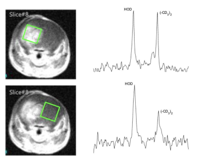 |
113 |
2H MR measurement of amino acid uptake in glioblastoma
multiforme
Samantha McClendon1,
Kyu-Ho Song2,
Xia Ge2,
Joseph J.H. Ackerman2,3,4,5,
Joel R. Garbow2,5,
and Scott Beeman1
1School of Biological and Health Systems Engineering, Arizona State University, Tempe, AZ, United States, 2Department of Radiology, Washington University in St.Louis, St.Louis, MO, United States, 3Department of Chemistry, Washington University in St.Louis, St.Louis, MO, United States, 4Department of Internal Medicine, Washington University in St.Louis, St.Louis, MO, United States, 5Alvin J. Siteman Cancer Center, Washington University School of Medicine in St.Louis, St.Louis, MO, United States Keywords: Deuterium, Tumor, Amino Acid Glioblastoma (GBM) brain tumors are among the most lethal of all human cancers, with a median survival of ~15 months. Standard-of-care radiologic methods fail to detect infiltrating GBM, which leads to undertreatment. We report a 2H MR method to better resolve/characterize GBM based on its enhanced uptake of branched-chain amino acids (e.g., leucine). We measure relaxation time constants for deuterated leucine in agarose phantoms and show the ability to detect d10-leucine at ~200 mM in a 4x4x4 mm3 voxel in phantoms. Finally, we demonstrated 2H MR measurement of enhanced deuterated leucine uptake in a rodent model of GBM. |
|
3868.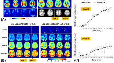 |
114 |
High spatiotemporal resolution whole-brain 2H MRS imaging
(DMRSI) to differentiate grey and white matter metabolic
dynamics in human brain at 7T
Xin Li1,
Xiao-Hong Zhu1,
Yudu Li2,3,
Hannes M. Wiesner1,
Zhi-Pei Liang2,4,
and Wei Chen1
1Department of Radiology, University of Minnesota, Minneapolis, MN, United States, 2Beckman Institute for Advanced Science and Technology, University of Illinois at Urbana-Champaign, Urbana, IL, United States, 3National Center for Supercomputing Applications, University of Illinois at Urbana-Champaign, Urbana, IL, United States, 4Department of Electrical and Computer Engineering, University of Illinois at Urbana-Champaign, Urbana, IL, United States Keywords: Deuterium, Brain Deuterium MRS imaging (DMRSI) is a promising tool to quantitatively study brain glucose metabolism, but it is challenging to simultaneously achieve high spatial and temporal resolution to capture metabolite dynamics in the human brain. In this study, we applied advanced RF head coil and post-processing techniques to perform high spatiotemporal-resolution (0.7cc nominal voxel and 2.5 min) DMRSI covering entire human brain at 7T with oral administration of deuterated glucose. The results show superior DMRSI sensitivity for mapping and differentiating the TCA cycle activity in grey and white matters. This capability is critical for disease applications including brain tumor. |
|
3869.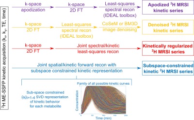 |
115 |
Improving SNR in deuterium metabolic imaging of cancer: spatial
denoising, joint spectral/metabolic-kinetics processing, and
beyond
Elton Montrazi1,
Qingjia Bao2,
Ricardo Martinho3,
Dana Peters4,
Talia Harris1,
Keren Sasson1,
Lilach Agemy1,
Avigdor Scherz1,
and Lucio Frydman1
1Weizmann, Rehovot, Israel, 2Chinese Academy of Sciences, Wuhan, China, 3University of Twente, Enschede, Netherlands, 4Yale School of Medicine, New Haven, CT, United States Keywords: Deuterium, Cancer, MRSI As DMI is a promising cancer screening approach but is challenged by low sensitivity, this study assesses multiple approaches to increase its SNR. Some of these –apodization, Compressed Sensing Multiplicative (CoSeM), Block-matching/3D filtering (BM3D)– involve image denoising. Others take into account the metabolic kinetics, and include it as dimension to be denoised. This can be achieved by smoothing the kinetic data axis via regularization, or by using subspace-constrained representations to concurrently solve for the 4D spatial/spectral/kinetics set. These methods can be further denoised –e.g., by CoSeM– leading to much clearer observations of lactate generated by in vivo tumor models. |
|
3870.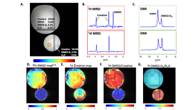 |
116 |
Parallel Proton and Deuterium Metabolic Imaging
Yanning Liu1,
Chathura Kumaragamage1,
Terry W. Nixon1,
Scott McIntyre1,
Henk De Feyter1,
and Robin A. de Graaf1
1Yale University, New Haven, CT, United States Keywords: Deuterium, Deuterium, DMI 1H MRSI and DMI can provide crucial metabolic information to facilitate evaluations of neurological diseases. In this work, we applied interleaved 1H/2H methodology to achieve time-efficient, parallel 1H MRSI and DMI acquisition. We first describe the necessary sequence adjustments for both nuclei, including the re-optimization of the VAPOR water suppression scheme and the incorporation of a 2H equilibrium pulse. Phantoms were then used to demonstrate that high quality 1H MRSI and DMI data can be acquired in parallel. The interleaved 1H MRSI-DMI sequence is compatible with other interleaved MRI-DMI sequences and can be implemented for a complete MRI-DMI protocol. |
|
3871.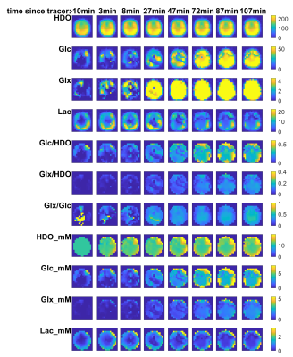 |
117 |
Time-resolved deuterium metabolic imaging of the human brain at
7T
Minghao Zhang*1,
Jabrane Karkouri*1,
Daniel Atkinson1,
and Christopher T. Rodgers1
1Wolfson Brain Imaging Centre, University of Cambridge, Cambridge, United Kingdom Keywords: Deuterium, Deuterium Deuterium metabolic imaging (DMI) is a new way to track brain metabolism. We present initial results from 3 volunteers scanned with a new 18-element 2H + 2-element 1H array coil on a Siemens 7T Terra scanner. We utilised our array’s high SNR to track metabolism by whole-brain 3D DMI after drinking 6,6’-[2H2]-glucose (Glc). Signals from HDO, Glc, Glx and Lac were visible over 2hrs at 5min temporal and 6.9mL spatial resolution. We also ran 2.9mL 3D DMI. We achieved HDO SNR of 43 in 13min, which compares favourably published SNR=13 in 10min at 9.4T1 and ~50 in 28min at 7T.2 |
|
3872.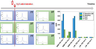 |
118 |
Direct comparison of deuterium 2H MRSI at 3T vs 7T
Jabrane Karkouri *1,
Mary McLean *2,
Minghao Zhang *1,
Joshua D Kaggie2,
Ashley Grimmer2,
Alixander Khan2,
Tomasz Matys2,
Daniel Atkinson1,
Ferdia Gallagher *2,
and Christopher Rodgers *1
1University of Cambridge, Cambridge, United Kingdom, 2Radiology, University of Cambridge, Cambridge, United Kingdom Keywords: Deuterium, Deuterium Deuterium metabolic imaging (DMI) is a new method to probe brain metabolism. Theory predicts higher SNR at higher field strength, but clinical translation is easier at lower field strength. We undertook 2H MRSI in 3 healthy volunteers at 3T and 7T sequentially. Acquisition was undertaken at baseline and after 11.1g oral D2O consumption. Data quality was acceptable in all scans. SNR was 3x higher at 7T, in agreement with theory. D2O cerebral uptake rate was similar between field strengths (0.13 min-1 at 3T vs 0.07 min-1 at 7T). |
|
3873.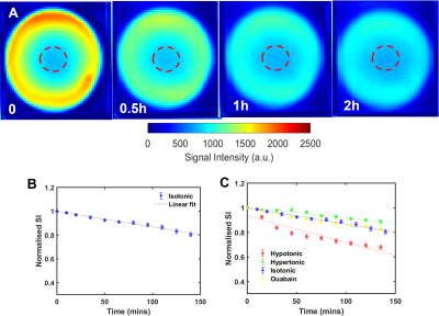 |
119 |
Deuterium oxide MRI to image the water inflow in the bovine lens
of the eye
Xingzheng Pan1,
Emily MacFarlane1,
and Paul Donaldson1
1Physiology, University of Auckland, Auckland, New Zealand Keywords: Deuterium, Aging, Human eye In the absence of a blood supply the lens operates an internal microcirculation to deliver nutrients, remove metabolic wastes, and controll the lens volume, which together to maintain the optical properties of the lens. While this microcirculation is generated by circulating fluxes of ions and water, visualising water flow throughout the whole lens and especially into the central of lens in real time has proven challenging. To address this, we developed and optimised new deuterium oxide (D2O) MRI protocols to image water flow within multiple organ-cultured bovine lenses that can be routinely performed using 3T clinical MRI. |
|
3874.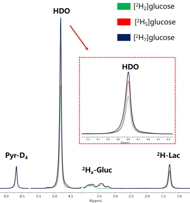 |
120 |
Comparison the metabolism of [2H2]glucose,[2H7]glucose and
[2H5]glucose in rat C6 glioma cells
Jiawen Yuan1,2,
Yi Fang1,
Qian Wan1,
Chao Zou1,3,
Xiaoliang Zhang4,
Xin Liu1,3,
Hairong Zheng1,3,
and Ye Li1,3
1Lauterbur Imaging Research Center, Shenzhen Institute of Advanced Technology, Chinese Acamedy of Sciences, Shenzhen, China, 2Southern University of Science and Technology, Shenzhen, China, 3Key Laboratory for Magnetic Resonance and Multimodality Imaging of Guangdong Province, Shenzhen, China, 4Department of Biomedical Engineering, State University of New York at Buffalo, Buffalo, NY, United States Keywords: Deuterium, Deuterium Deuterium MRS(I) has emerged as a novel metabolic imaging method, which can effectively reflect the increased aerobic glycolysis of tumors. Recently, [2,3,4,6,6-2H5] glucose has been shown to be a cost-effective deuterium-labeled substrate for studying glycolysis in tumors. However, the effects of different deuterium-labeled substrates on metabolism require further comparison. In this study, we compared the differences between [6,6-2H2] glucose, [1,2,3,4,5,6,6’-2H7] glucose, and [2,3,4,6,6-2H5] glucose in rat C6 glioma cells. |
|
 Back to Meeting Home
 Back to the Program-at-a-Glance
The International Society for Magnetic Resonance in Medicine is accredited by the Accreditation Council for Continuing Medical Education to provide continuing medical education for physicians.
 Back to Meeting Home
 Back to the Program-at-a-Glance
 View Presentation Video