Digital Poster
ASL: Applications
ISMRM & ISMRT Annual Meeting & Exhibition • 03-08 June 2023 • Toronto, ON, Canada

| Computer # | |||
|---|---|---|---|
2568.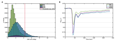 |
41 |
7T spin-echo dynamic susceptibility contrast MRI to assess
cerebral microvascular perfusion
Elles P. Elschot1,2,
Marieke van den Kerkhof1,2,
Robert J. van Oostenbrugge2,3,4,
Abraham A. Kroon4,5,
Walter H. Backes1,2,4,
and Jacobus F. A. Jansen1,2,6
1Radiology & Nuclear Medicine, Maastricht University Medical Center +, Maastricht, Netherlands, 2School for Mental Health and Neuroscience, Maastricht University, Maastricht, Netherlands, 3Neurology, Maastricht University Medical Center +, Maastricht, Netherlands, 4School for Cardiovascular Diseases, Maastricht University, Maastricht, Netherlands, 5Internal Medicine, Maastricht University Medical Center +, Maastricht, Netherlands, 6Electrical Engineering, Eindhoven University of Technology, Eindhoven, Netherlands Keywords: Contrast Mechanisms, Perfusion Ultra-high-field exploits the possibilities of MRI techniques, which are less sensitive at lower field strength, such as spin-echo (SE) perfusion MRI. We assessed the feasibility of SE dynamic susceptibility contrast MRI at 7T and investigated the ability to measure variations in perfusion in an elderly, partially hypertensive, population (n=36). Microvascular cerebral blood flow (CBF) was higher in grey matter compared to white matter, corresponding with biological expectations, serving as a first validation step. Furthermore, CBF significantly decreased with age. |
|
2569.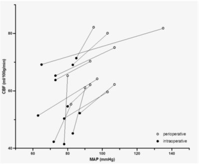 |
42 |
Effect of general anesthesia on cerebral blood flow measured by
Arterial Spin Labeling: A retrospective study
Thomas Lindner1,
Monika Huhndorf2,
Christine Eimer3,
Tobias Becher3,
Hajrullah Ahmeti4,
Olav Jansen2,
Michael Synowtiz4,
Michael Helle5,
and Stephan Ulmer2,6
1UKE Hamburg, Hamburg, Germany, 2Department of Radiology and Neuroradiology, University Hospital Schleswig-Holstein, Kiel, Germany, 3Department of Anesthesia, University Hospital Schleswig-Holstein, Kiel, Germany, 4Department of Neurosurgery, University Hospital Schleswig-Holstein, Kiel, Germany, 5Philips Gmbh Innovative Technologies, Hamburg, Germany, 6neurorad.ch, Zurich, Switzerland Keywords: Arterial spin labelling, Brain, Anesthesia In this study, we non-invasively evaluated the change in cerebral blood flow (CBF) and correlation with mean arterial pressure (MAP) before and during anesthesia using Arterial Spin Labeling. It could be shown that both CBF and MAP are reduced comparing the awake and anesthesia-induced state in patients. |
|
2570.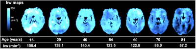 |
43 |
Age dependent changes of water exchange rate across the
blood-brain barrier (BBB) in 115 subjects with age ranging from
8 to 87 years
Xingfeng Shao1,
Qinyang Shou1,
Kimberly Felix2,
Brandon Ojogho1,
Xuejuan Jiang2,3,
Brian T. Gold4,
Megan M. Herting2,
and Danny JJ Wang1
1Laboratory of FMRI Technology (LOFT), Mark & Mary Stevens Neuroimaging and Informatics Institute, Keck School of Medicine, University of Southern California, Los Angeles, CA, United States, 2Department of Preventive Medicine, Keck School of Medicine, University of Southern California, Los Angeles, CA, United States, 3Department of Ophthalmology, Keck School of Medicine, University of Southern California, Los Angeles, CA, United States, 4Department of Neuroscience, College of Medicine, University of Kentucky, Lexington, KY, United States Keywords: Arterial spin labelling, Permeability, BBB water exchange, permeability, ageing We studied age-related BBB water exchange rate (kw) changes in a wide range of age groups with 115 subjects using diffusion prepared pseudo-continuous arterial spin labeling (DP-pCASL). We observed a significant trend of decreasing kw with age (slow when ≤50 years old and fast when >50 years old) and large spread of kw values in subjects >50 years, which may indicate compromised BBB function in some ageing populations. The inverted U-shaped correlations between kw versus age suggest a more complex age dependent changes in hippocampus and PHG regions. |
|
2571.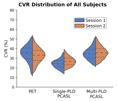 |
44 |
Comparing the Repeatability of Cerebrovascular Reserve Measured
by PET and ASL using Simultaneous PET/MRI
Moss Y Zhao1,
Audrey P Fan2,
Mohammad Mehdi Khalighi1,
Dawn Holley1,
Kim Halbert1,
Maria Jovin1,
Bin Shen1,
Guido A Davidzon1,
Elizabeth Tong1,
Michael Moseley1,
and Greg Zaharchuk1
1Department of Radiology, Stanford University, Stanford, CA, United States, 2Department of Biomedical Engineering, University of California Davis, Davis, CA, United States Keywords: Arterial spin labelling, Perfusion Cerebrovascular reserve (CVR) is an important biomarker to assess vascular hemodynamics and link to the risk for acute strokes. Whilst several imaging modalities have been applied to measure CVR, their reproducibility remains to be elucidated. Here we compare the reproducibility of CVR measurements in 22 normal subjects between single and multi-delay ASL using 15O-water PET as the reference. Results implied that multi-delay ASL achieved a higher reproducibility and should be the preferred non-invasive modality for clinical applications. |
|
2572.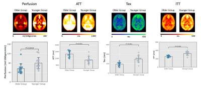 |
45 |
Change in Blood-brain Barrier Permeability with Age – Comparing
a Physiologically Informed Biophysical Model with a
Triexponential Decay Model
Amnah Mahroo1 and
Matthias Günther1,2,3
1Imaging Physics, Fraunhofer Institute for Digital Medicine MEVIS, Bremen, Germany, 2MR-Imaging and Spectroscopy, University of Bremen, Bremen, Germany, 3mediri GmbH, Heidelberg, Germany Keywords: Arterial spin labelling, Brain, Blood-brain barrier Permeability, Aging, Multi-TE ASL With age the integrity of blood-brain barrier (BBB) slowly deteriorates. Multi-TE ASL, being a water-based MRI method, offers detection of subtle changes in the BBB by probing T2 relaxation. In this study, we evaluated the sensitivity of multi-TE ASL method in detecting changes in the BBB occurring due to age using two models; a physiologically informed biophysical (PIB) model by estimating exchange time and a simpler tri-exponential decay (TD) model by determining the rate of change of tissue fraction. We found that both models were able to detect age-based changes in BBB. Moreover, PIB model showed higher robustness. |
|
2573.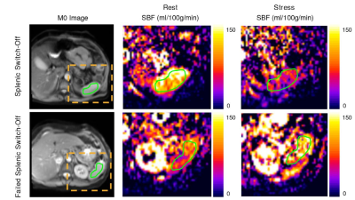 |
46 |
Assessment of Splenic Switch-Off With Arterial Spin Labeling in
Adenosine Perfusion Cardiac MRI
Verónica Aramendia-Vidaurreta1,2,
Sergio M. Solís-Barquero1,2,
Ana Ezponda1,2,
Marta Vidorreta3,
Rebeca Echeverria-Chasco1,2,
Marina Pascual4,
Gorka Bastarrika1,2,
and María A. Fernández-Seara1,2
1Radiology, Clínica Universidad de Navarra, Pamplona, Spain, 2IdiSNA, Instituto de Investigación Sanitaria de Navarra, Pamplona, Spain, 3Siemens Healthineers, Madrid, Spain, 4Cardiology, Clínica Universidad de Navarra, Pamplona, Spain Keywords: Arterial spin labelling, Perfusion Splenic switch-off, defined as the stress to rest splenic blood flow (SBF) attenuation in response to adenosine, is an indicator of stress adequacy. This study aims to explore the ability of pseudo-continuous arterial spin labeling (PCASL) to identify splenic switch-off in patients with suspected CAD. In healthy subjects, multi-delay PCASL data were acquired to quantify SBF and determine the adequate postlabeling delay. In patients, single-delay PCASL and first-pass perfusion images were acquired under rest and adenosine conditions. This study could demonstrate the feasibility of PCASL to identify splenic switch-off during adenosine perfusion MRI. |
|
2574.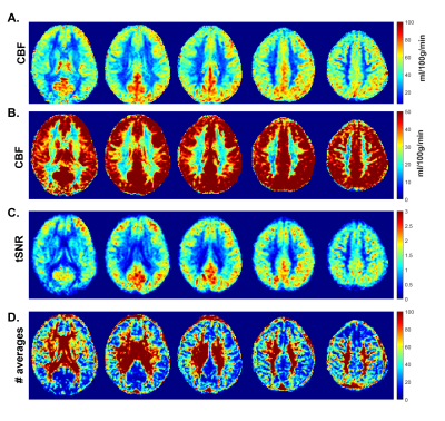 |
47 |
Optimal PLD selection for White Matter perfusion measurements at
7T.
Emiel C.A. Roefs1,
Lydiane Hirschler1,
Natalia Petridou2,
and Matthias J.P. van Osch1
1Radiology, C.J. Gorter MRI Center, Leiden Univeristy Medical Center, Leiden, Netherlands, 2Radiology, Center for Image Sciences, Univeristy Medical Center Utrecht, Utrecht, Netherlands Keywords: Arterial spin labelling, High-Field MRI White matter (WM) perfusion measurements are challenging because of the generally lower perfusion in combination with a longer bolus arrival time (BAT). We investigated the use of long-PLD perfusion measurements at high-field (7T) to compensate for longer BAT in WM. We showed that 3D background suppressed FAIR at 7T with a PLD of 2300ms provides robust perfusion measurement in almost 80% of deep WM within 4min of scan time. |
|
2575.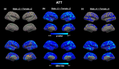 |
48 |
APOE genotype-dependent sex differences in cortical hemodynamics
measured with arterial spin labeling MRI
Nikou Louise Damestani1,2,
John Jacoby1,
Barnaly Rashid1,3,
Allison E Lovely1,
Shrikanth M Yadav1,
Aurea Michael1,
Marziye Eshghi4,
Melissa Terpstra5,
Carlos Cruchaga6,7,8,
David H Salat1,2,9,
and Meher R Juttukonda1,2
1Athinoula A. Martinos Center for Biomedical Imaging, Massachusetts General Hospital, Boston, MA, United States, 2Department of Radiology, Harvard Medical School, Boston, MA, United States, 3Department of Neurology, Harvard Medical School, Boston, MA, United States, 4MGH Institute of Health Professions, Boston, MA, United States, 5MU School of Medicine, University of Missouri, Columbia, MO, United States, 6Department of Psychiatry, Washington University School of Medicine, St. Louis, MO, United States, 7NeuroGenomics and Informatics Center, Washington University School of Medicine, St. Louis, MO, United States, 8Hope Center for Neurologic Diseases, Washington University, St. Louis, MO, United States, 9Neuroimaging Research for Veterans Center, VA Boston Healthcare System, Boston, MA, United States Keywords: Arterial spin labelling, Aging Identifying biomarkers that could characterize typical from atypical aging is crucial for understanding the aging process. One of the most significant genetic risk factors for Alzheimer’s Disease, an age-related neurodegenerative disorder, is the presence of an apolipoprotein-E (APOE) ε4 allele. Here, we investigate the impact of APOE genotype on cortical hemodynamics in a large cohort of participants across the lifespan, accounting for interacting effects of age, sex, and cardiovascular risk. We found unique spatial patterns of cerebral blood flow and arterial transit time between males and females within distinct APOE genotypes, potentially indicating the presence of a complex interaction. |
|
2576.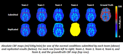 |
49 |
The ASL Challenge Reproduction Study: Replicating Findings from
The OSIPI-ISMRM ASL MRI Challenge
Channelle Tham1,
Ahmed Abdalle2,
Joseph G Woods3,
Maria-Eleni Dounavi4,
Andre Pascoal5,
and Udunna Anazodo6
1Department of Cognitive Neuroscience, Radboud University, Nijmegen, Netherlands, 2Schulich School of Medicine and Dentistry, Western University, London, ON, Canada, 3Wellcome Centre for Integrated Neuroimaging, FMRIB, Nuffield Department of Clinical Neuroscience, University of Oxford, Oxford, United Kingdom, 4Department of Psychiatry, University of Cambridge, Cambridge, United Kingdom, 5Instituto de Radiologia e Departamento de Radiologia e Oncologia, University of Sao Paulo, Sao Paulo, Brazil, 6Montreal Neurological Institute, McGill University, Montreal, QC, Canada Keywords: Arterial spin labelling, Challenges, Reproducibility While the recommendation for ASL acquisition is widely adopted to standardize quantification of cerebral blood flow (CBF), ASL analysis still produces wide variability in CBF estimates. The recent OSIPI-ISMRM ASL MRI Challenge highlighted how differences in image processing contribute to CBF variability. Here, we explored the replicability of ASL image processing approaches when used as documented to quantify CBF. The results of 5 of the 8 ASL Challenge teams were replicated and compared to their submitted results. The overall errors in global mean CBF were 0.03-5 times larger in the replicated results, demonstrating the need for ASL analysis standards. |
|
2577.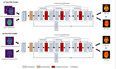 |
50 |
Extracting CBF and ATT from One or Two Post-labeling
Delays-based Arterial Spin Labeled Perfusion MRI via Deep
Learning
Yiran Li1,
Xiufeng Li2,
John A Detre3,
and Ze Wang1
1University of Maryland School of Medicine, Baltimore, MD, United States, 2University of Minnesota, Minneapolis, MN, United States, 3University of Pennsylvania Perelman School of Medicine, Philadelphia, PA, United States Keywords: Arterial spin labelling, Arterial spin labelling Cerebral blood flow (CBF) and arterial transit time (ATT) can be quantified through fitting the arterial spin labeling (ASL) perfusion MRI signal acquired at different post-labeling delays (PLDs) into a kinetic model. Acquiring multiple-PLD ASL MRI needs exponentially prolonged total scan time compared to the single-PLD acquisition, making it highly sensitive to motions and impractical for clinical use. We proposed a deep neural network that can reliably estimate ATT and CBF maps from significantly fewer PLD ASL MRI acquisitions without image quality loss. |
|
2578.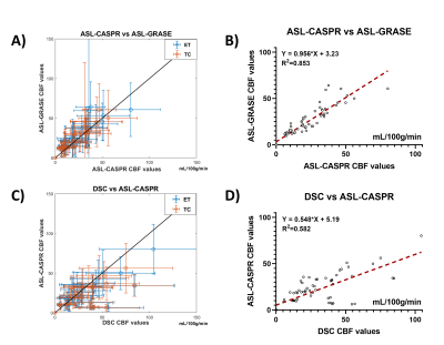 |
51 |
DSC-MRI and ASL-MRI Measured Perfusion in Glioblastoma: Are they
Competitive or Complementary?
Limin Zhou1,
Yiming Wang2,
Marco C. Pinho1,3,
Durga Udayakumar1,3,
Michael Youssef4,5,
Joseph A. Maldjian1,3,
and Ananth J. Madhuranthakam1,3
1Department of Radiology, University of Texas Southwestern Medical Center, DALLAS, TX, United States, 2Philips Healthcare, Shanghai, China, 3Advanced Imaging Research Center, University of Texas Southwestern Medical Center, DALLAS, TX, United States, 4Department of Neurology, University of Texas Southwestern Medical Center, DALLAS, TX, United States, 5Department of Hematology and Oncology, University of Texas Southwestern Medical Center, DALLAS, TX, United States Keywords: Arterial spin labelling, Perfusion, Dynamic susceptibility contrast (DSC), Brain, Cancer, Glioblastoma (GBM) |
|
2579.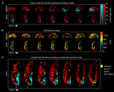 |
52 |
Investigating border zone hemodynamics with time-encoded
super-selective pCASL in internal carotid and basilar artery
perfusion territories
Lena Vaclavu1 and
Matthias JP van Osch1
1C.J. Gorter MRI Center, Department of Radiology, Leiden University Medical Center, Leiden, Netherlands Keywords: Arterial spin labelling, Perfusion, super-selective, time-encoded, borderzones The borders between cerebral perfusion territories are regions of the brain that are susceptible to infarction. In this study, time-encoded super-selective pCASL was implemented to study the hemodynamic properties of the border zones between the left internal carotid artery and basilar artery perfusion territories. Perfusion territories were well-defined with little-to-no detectable overlap. The longest arrival times were found at the border zones, compared to 'inland' of the perfusion region. Combining time-encoded with super-selective pCASL enables the simultaneous investigation of border zones and their arrival times. |
|
2580.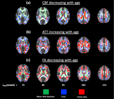 |
53 |
Impact of age and sex on regional white matter hemodynamics
across the adult lifespan
Nikou Louise Damestani1,2,
John Jacoby1,
Qiyuan Tian1,2,
Allison E Lovely1,
Shrikanth M Yadav1,
David H Salat1,2,3,
and Meher R Juttukonda1,2
1Athinoula A. Martinos Center for Biomedical Imaging, Massachusetts General Hospital, Boston, MA, United States, 2Department of Radiology, Harvard Medical School, Boston, MA, United States, 3Neuroimaging Research for Veterans Center, VA Boston Healthcare System, Boston, MA, United States Keywords: Arterial spin labelling, Aging White matter perfusion has been difficult to measure with arterial spin labeling (ASL) MRI due to the elongated arterial transit times of white matter. Here, we identified changes in white matter cortical hemodynamics associated with age and between sex for the first time using multi-delay pseudo-continuous ASL with tract-based analyses in a large typically aging cohort. We found reductions in white matter CBF and increases in white matter ATT with advancing age, as well as widespread sex differences across tracts. This work serves as the first step towards understanding the relationship between white matter physiology and age-related structural decline. |
|
2581.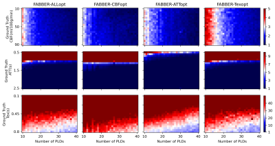 |
54 |
Detectability of changes in the blood-brain barrier permeability
to water using optimised multi-TE pseudo-continuous arterial
spin labelling
Logan X. Zhang1,2 and
Michael Chappell1,3
1Sir Peter Mansfield Imaging Centre and Mental Health & Clinical Neurosciences, School of Medicine, University of Nottingham, Nottingham, United Kingdom, 2Institute of Biomedical Engineering, Department of Engineering Science, University of Oxford, Oxford, United Kingdom, 3Wellcome Centre for Integrative Neuroimaging, FMRIB, Nuffield Department of Clinical Neurosciences, University of Oxford, Oxford, United Kingdom Keywords: Arterial spin labelling, Arterial spin labelling The blood-brain barrier integrity can be reflected by the pre-exchange lifetime of water molecules, Tex, which can be measured using multi-TE multi-delay ASL. Nevertheless, the low SNR of ASL limits the detectability of subtle Tex changes within a feasible scan time for clinical applications. In this study, optimised protocols were evaluated for the theoretical detectability of 10% changes in haemodynamic parameters. Simulation shows that while changes in CBF and arterial transit time (ATT) could be detected on individual level in a five-minute scan, the same detectability of Tex would require a minimum of 6 subjects within ten minutes of scanning. |
|
2582.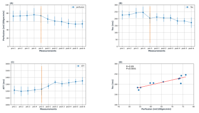 |
55 |
Increased Blood-brain Barrier Permeability in Response to
Caffeine Challenge
Amnah Mahroo1,
Simon Konstandin1,2,
Daniel Christopher Hoinkiss1,
Jochen Hirsch1,
and Matthias Günther1,2,3
1Imaging Physics, Fraunhofer Institute for Digital Medicine MEVIS, Bremen, Germany, 2mediri GmbH, Heidelberg, Germany, 3MR-Imaging and Spectroscopy, University of Bremen, Bremen, Germany Keywords: Arterial spin labelling, Brain, Blood-brain Barrier (BBB) Permeability, caffeine, multi-TE ASL Caffeine is a commonly used stimulant drug and is known to change brain physiology by constricting vessels leading to decreased perfusion. We tested the impact of caffeine ingestion on blood-brain barrier (BBB) permeability by measuring exchange time using multi-TE ASL technique. Six healthy volunteers were examined for five pre-caffeine sets and six post-caffeine sets. The multi-TE two-compartment model was used to estimate exchange time (Tex) along with perfusion and ATT. We found that perfusion and Tex decreased over time while ATT increased. Decreased Tex indicates that the labelled water transferred at a faster rate reflecting increased BBB permeability. |
|
2583.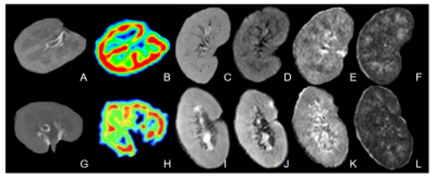 |
56 |
The value of ASL combined with DTI for detection of impaired
renal allograft function
Bin Jiang1,
Yangyang Tian2,
Ximing Wang1,
Peng Wu3,
Jiayi Wan1,
Mengjiao Yin1,
Ning Wang1,
Rui Xu1,
Linkun Hu2,
and Mo Zhu1
1Department of Radiology, The First Affiliated Hospital of Soochow University, Suzhou, China, 2Department of Urology, The First Affiliated Hospital of Soochow University, Suzhou, China, 3Philips Healthcare, Shanghai, China Keywords: Arterial spin labelling, Kidney Allograft injury continues to be a major problem in renal allograft recipients, which leads to eventual graft loss. Non-invasive and reliable detection of impaired renal allograft function is crucial to preventing irreversible nephron loss and graft failure. This study aims to investigate the value of ASL combined with DTI for the detection of impaired renal allograft function. Results showed that a higher diagnostic efficacy could be achieved through the combined use of ASL and DTI. |
|
2584.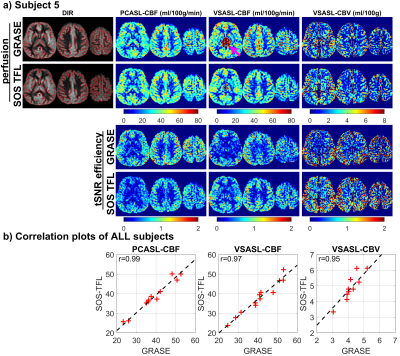 |
57 |
Evaluation of 3D Stack-of-Spiral Turbo FLASH Acquisitions for
PCASL- and VSASL-Derived Brain Perfusion Mapping
Dan Zhu1,2,
Feng Xu1,2,
Dapeng Liu1,2,
Doris Lin2,
Peter van Zijl1,2,
and Qin Qin1,2
1F.M. Kirby Research Center for Functional Brain Imaging, Kennedy Krieger Institute, Baltimore, MD, United States, 2The Russell H. Morgan Department of Radiology and Radiological Science, Johns Hopkins School of Medicine, Baltimore, MD, United States Keywords: Arterial spin labelling, Arterial spin labelling The most-used 3D acquisition for ASL at 3T is GRASE or stack-of-spiral (SOS) based FSE, which requires multiple shots to cover the full k-space. Alternatively, turbo FLASH (TFL) acquisition allows longer echo trains with slower T1 (than T2) relaxation, and 3D SOS-TFL has the potential to reduce the number of shots to even single-shot, thus improving the temporal resolution for ASL. Here we demonstrated comparable performance of 3D SOS-TFL with 3D GRASE on PCASL- and VSASL-derived CBF mapping and VSASL-derived CBV mapping at 3T on 12 healthy subjects, and the utility of 3D SOS-TFL on a stroke patient. |
|
2585.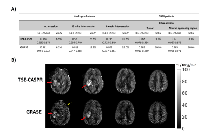 |
58 |
Reproducibility of Pseudocontinuous Arterial Spin Labeling
Measured Perfusion in Healthy Volunteers and Glioblastoma
Patients
Limin Zhou1,
Yiming Wang2,
Marco C. Pinho1,3,
Durga Udayakumar1,3,
Michael Youssef4,5,
Joseph A. Maldjian1,3,
and Ananth J. Madhuranthakam1,3
1Department of Radiology, University of Texas Southwestern Medical Center, DALLAS, TX, United States, 2Philips Healthcare, Shanghai, China, 3Advanced Imaging Research Center, University of Texas Southwestern Medical Center, DALLAS, TX, United States, 4Department of Neurology, University of Texas Southwestern Medical Center, DALLAS, TX, United States, 5Department of Hematology and Oncology, University of Texas Southwestern Medical Center, DALLAS, TX, United States Keywords: Arterial spin labelling, Precision & Accuracy, Reproducibility, Precision Accuracy, Perfusion, Cancer, Glioblastoma (GBM), Translational studies We evaluated the intra-session repeatability and inter-session reproducibility of brain and tumor perfusion measured using 3D pseudo-continuous Arterial spin labeled (PCASL) MRI with TSE based Cartesian acquisition with spiral profile reordering (CASPR) in comparison to 3D pCASL with GRASE in healthy volunteers and glioblastoma (GBM) patients at 3T. 3D pCASL with TSE-CASPR or GRASE provided high intra-session repeatability and 3 weeks inter-session reproducibility in both volunteers and GBM patients. While both readouts generated robust images, TSE-CASPR provided images with reduced distortion particularly in GBM patients and could be a better readout for pCASL measured perfusion in GBM patients. |
|
2586.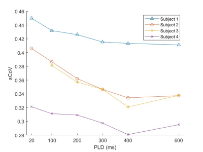 |
59 |
Microvascular specificity in velocity-selective arterial spin
labeling: Effects of the post-labeling delay
Ryan A. Barnes1,2,
Conan Chen1,2,3,
Eric C. Wong1,3,4,
Thomas T. Liu1,3,4,5,
and Divya S. Bolar1,3
1Center for fMRI, University of California San Diego, La Jolla, CA, United States, 2Department of Electrical and Computer Engineering, UC San Diego, La Jolla, CA, United States, 3Department of Radiology, UC San Diego, La Jolla, CA, United States, 4Department of Psychiatry, UC San Diego, La Jolla, CA, United States, 5Department of Bioengineering, UC San Diego, La Jolla, CA, United States Keywords: Arterial spin labelling, Arterial spin labelling, Velocity-selective ASL, CBF Velocity-selective ASL labels arterial blood spins close to the microvasculature if a sufficiently low velocity cutoff (Vcut) is used. Lowering Vcut, however, introduces diffusion and eddy current effects that confound the CBF measurement. A Vcut of 2 cm/s is typical to minimize these effects, but labels blood slightly upstream from the microvasculature; in this scenario, a non-zero post-labeling delay (PLD) allows time for additional microvascular delivery. We evaluate microvascular specificity (using the spatial coefficient of variation of CBF) as a function of PLD. We find that macrovascular specificity increases with PLD up to around 500ms for a typical ASL resolution. |
|
2587.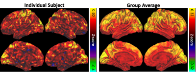 |
60 |
Resting state neurovascular coupling patterns estimated using
advanced multiband multi-echo BOLD/ASL imaging
Alexander D. Cohen1 and
Yang Wang1
1Radiology, Medical College of Wisconsin, Milwaukee, WI, United States Keywords: Arterial spin labelling, Data Analysis, Coupling, Simultaneous BOLD/ASL Impaired neurovascular coupling (NVC) plays a critical role in many neurovascular pathological processes. Several resting state fMRI methods have been used to estimate NVC including BOLD/CBF coupling and, to a lesser extent, ALFF and fALFF. In this study, resting state BOLD/CBF coupling was evaluated using advanced multiband multi-echo BOLD/ASL sequence and correlated with ALFF and fALFF in healthy volunteers. Significant correlation between BOLD/CBF coupling and ALFF and fALFF was seen in major brain network hubs. These results indicate MBME BOLD/ASL may provide similar, but complimentary measures of NVC compared to traditional RS metrics. |
|
 Back to Meeting Home
 Back to the Program-at-a-Glance
The International Society for Magnetic Resonance in Medicine is accredited by the Accreditation Council for Continuing Medical Education to provide continuing medical education for physicians.
 Back to Meeting Home
 Back to the Program-at-a-Glance
 View Presentation Video