Digital Poster
Perfusion: Technical Developments
ISMRM & ISMRT Annual Meeting & Exhibition • 03-08 June 2023 • Toronto, ON, Canada

Digital Poster
Perfusion: Technical Developments
| Computer # | |||
|---|---|---|---|
2744.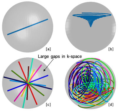 |
41 |
Efficient 3D cone trajectory design for improved combined
angiographic, structural and perfusion imaging using arterial
spin labelling
Qijia Shen1,
Mark Chiew1,2,3,
Wenchuan Wu1,
and Thomas Okell1
1Wellcome Centre for Integrative Neuroimaging, FMRIB, Nuffield Department of Clinical Neurosciences, University of Oxford, Oxford, United Kingdom, 2Physical Sciences, Sunnybrook Research Institute, Toronto, ON, Canada, 3Department of Medical Biophysics, University of Toronto, Toronto, ON, Canada Keywords: Arterial spin labelling, Arterial spin labelling 4D combined angiographic, structural and perfusion radial imaging using arterial spin labelling (CASPRIA) provides a tool to simultaneously acquire information about brain structure and blood flow. However, the radial trajectory used limits the resolution and SNR, especially when the data is highly undersampled. In this work, an optimised 3D cone trajectory is presented with 3D golden means rotation to improve sampling at the k-space periphery whilst maintaining flexibility in spatiotemporal resolution. A locally low rank reconstruction was used to leverage spatiotemporal correlations and improve image quality. In vivo results showed considerable improvements in resolution and SNR over the radial approach. |
|
2745.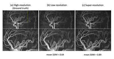 |
42 |
Multi-2D ASL-MRA and super-resolution convolutional neural
network for improved intracranial peripheral arteries
visualization
Yuriko Suzuki1,
Ioannis Koktzoglou2,3,
Peter Jezzard1,
and Thomas Okell1
1Wellcome Centre for Integrative Neuroimaging, FMRIB, Nuffield Department of Clinical Neurosciences, University of Oxford, Oxford, United Kingdom, 2Department of Radiology, NorthShore University HealthSystem, Evanston, IL, United States, 3Pritzker School of Medicine, University of Chicago, Chicago, IL, United States Keywords: Arterial spin labelling, Blood vessels, MRA The non-invasive nature of Arterial Spin Labeling (ASL) technique makes ASL-based intracranial dynamic MR angiography (MRA) a potential alternative to diagnostic X-ray digital subtraction angiography. In elderly and diseased patients with slower blood flow, however, the vessel visualization of distal peripheral arteries tends to be poor, as the repeatedly applied RF excitation pulses decrease ASL blood signal rapidly. In this study, we address such a limitation by using multiple 2D slice acquisition to reduce the saturation of arterial blood signal. Additionally, to avoid losing vessel conspicuity and sharpness with 2D slice acquisition, we apply a super-resolution convolutional neural network method. |
|
2746.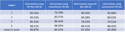 |
43 |
Weakly supervised learning improves vascular territorial mapping
of random vessel-encoded ASL
Yining He1 and
Lirong Yan1,2
1Radiology, Northwestern University, Chicago, IL, United States, 2Neurology, University of Southern California, Los Angeles, CA, United States Keywords: Arterial spin labelling, Perfusion This study proposed a weakly supervised learning algorithm for vascular territorial mapping with rVE-ASL. The territory maps generated by the proposed deep learning (DL) method was compared with the territories from the conventional rVE-ASL method by visual inspection and F1 score. Our initial results showed that the DL method outperformed the conventional rVE-ASL method in the vascular territory mapping with improved detection of VA territory. The DL method also provided reliable vascular territorial maps with reduced numbers of encodings, significantly reducing rVE-ASL scan time. These findings suggest that DL could be an effective approach for vascular territorial mapping of rVE-ASL. |
|
2747.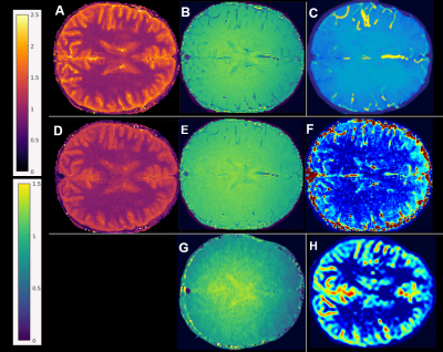 |
44 |
Towards Look-Locker encoding as an arterial spin labeling
mechanism: T1/B1+/perfusion mapping with MR Multitasking
Fardad M Serry1,
Hsu-Lei Lee1,
Shihan Qiu1,2,
Yibin Xie1,
Fei Han1,3,
Hui Han1,
and Anthony G Christodoulou1,2
1Biomedical Imaging Research Institute, Cedars-Sinai Medical Center, Los Angeles, CA, United States, 2Bioengineering, University of California, Los Angeles, Los Angeles, CA, United States, 3Siemens Medical Solutions, Los Angeles, CA, United States Keywords: Arterial spin labelling, Arterial spin labelling, multitasking, Look-Locker, B1+, spin history, T1, blood, brain, IR-FLASH, inversion efficiency, white matter, gray matter We hypothesized that the Look-Locker (LL) effect can label arterial spins without the need for subtraction between different inversion schemes. We developed a multiple-flip-angle, non-selective-inversion MR multitasking brain scan to produce co-registered T1, B1+, and perfusion-weighted maps. Flow-sensitive LL fitting homogenized B1+ maps and produced flow maps combining features of time-of-flight angiography images and PCASL perfusion-weighted images. GM/WM flow ratios were similar to PCASL and the literature range. |
|
2748.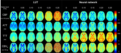 |
45 |
Multiparameter estimation from DANTE-prepared multi-delay ASL
using artificial neural network
Shota Ishida1,
Yasuhiro Fujiwara2,
Naoyuki Takei3,
Yuki Matta4,
Masayuki Kanamoto4,
Hirohiko Kimura5,6,
and Tetsuya Tsujikawa7
1Department of Radiological Technology, Faculty of medical sciences, Kyoto College of Medical Science, Nantan, Japan, 2Department of Medical Image Sciences, Faculty of Life Sciences, Kumamoto University, Kumamoto, Japan, 3GE Healthcare, Hino, Japan, 4Radiological center, University of Fukui Hospital, Eiheiji, Japan, 5Faculty of Medical Sciences, University of Fukui, Eiheiji, Japan, 6Radiology section, National Health Insurance Echizen-cho Ota Hospital, Unyu, Japan, 7Department of Radiology, Faculty of Medical Sciences, University of Fukui, Eiheiji, Japan Keywords: Arterial spin labelling, Arterial spin labelling A simulation-based supervised neural network was developed for simple and robust parameter estimation from multi-delay DANTE-prepared arterial spin labeling (ASL). The network was trained using 15 million simulation data points. Accuracy and precision were compared between the proposed and conventional methods. The neural-network-based estimation presented higher accuracy and precision than the conventional method that used table lookup. A higher noise immunity was also observed with the proposed method. A simulation-based supervised neural network simplifies the estimation process of multiparametric ASL. The estimation performance of cerebral blood flow and arterial cerebral blood volume was particularly improved by the proposed method. |
|
2749.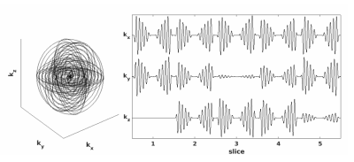 |
46 |
Fast, isotropic, whole head perfusion imaging using a Spin Echo
Rotating In-Out Spiral with velocity selective arterial spin
labeling
David Joseph Frey1,
Jeffrey A. Fessler2,
Douglas C. Noll1,
and Luis Hernandez-Garcia1
1Radiology, University of Michigan, Ann Arbor, MI, United States, 2Electrical Engineering and Computer Science, University of Michigan, Ann Arbor, MI, United States Keywords: Arterial spin labelling, New Trajectories & Spatial Encoding Methods We investigate the performance of a new and efficient k-space sampling scheme that presents several advantages for velocity selective ASL imaging by rotating 2D spirals about the kx and ky axis. This paper compares the new readout to a traditional Stack-of-spirals readout by evaluating SNR for perfusion weighted images and the accuracy of z-scores for ASL FMRI experiments. |
|
2750.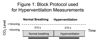 |
47 |
Investigating systematic error in pseudocontinuous Arterial Spin
Labelling estimates of cerebrovascular reactivity
Colette Clare Milbourn1 and
Nicholas Paul Blockley1
1School of Life Sciences, University of Nottingham, Nottingham, United Kingdom Keywords: Arterial spin labelling, Arterial spin labelling New clinical tools are needed for the diagnosis and prognosis of cerebrovascular diseases. Cerebrovascular Reactivity (CVR) is a potential marker for brain health and can be induced using stressors to the brain like a hypercapnia challenge and quantitatively mapped using pseudocontinuous Arterial Spin Labelling (pCASL). However, measurements of CVR using pCASL have been shown to vary depending on the parameters of the unbalanced pCASL preparation scheme used. In this study the effect of a diminished B1 at the labelling plane and changes in B0 during hyperventilation were investigated as the origin of this discrepancy. |
|
2751.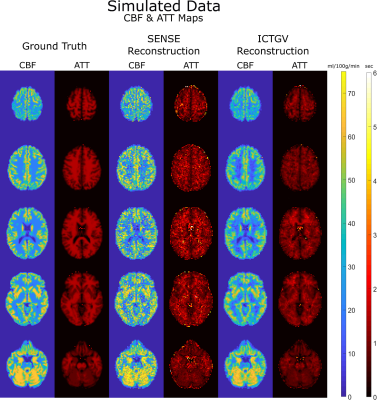 |
48 |
Dedicated ICTGV-regularized Reconstruction for time-encoded ASL
Ingmar S. Sorgenfrei1,
Koen Baas2,
Stefan Spann1,
Martin Uecker1,
and Rudolf Stollberger1
1Institute of Biomedical Imaging, TU Graz, Graz, Austria, 2Department of Radiology and Nuclear Medicine, Amsterdam UMC, Amsterdam, Netherlands Keywords: Arterial spin labelling, Brain Time-encoded Arterial Spin Labeling (teASL) enables time-efficient acquisition of multiple averages of multiple post-labeling-delay (PLD) perfusion-weighted images (PWIs). We developed a dedicated teASL reconstruction that employs Infimal Convolution of Total Generalized Variation (ICTGV) regularization directly on the PWIs. Using simulated and healthy volunteer data, the ICTGV reconstruction produced inherently denoised images and improved cerebral blood flow and arterial transit time maps compared to a SENSE reconstruction. Also, for an 8-fold accelerated in-vivo dataset, the ICTGV reconstruction still produced reasonable PWIs, which the SENSE reconstruction could not. Therefore, ICTGV reconstruction is promising for teASL and allows high acceleration with inherent denoising. |
|
2752.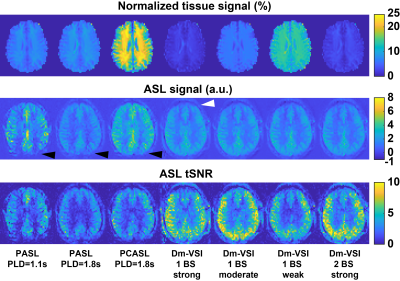 |
49 |
Optimizing background suppression for dual-module
velocity-selective ASL and characterizing the temporal noise
Jia Guo1
1Bioengineering, University of California Riverside, Riverside, CA, United States Keywords: Arterial spin labelling, Brain Background suppression (BS) was optimized for dual-module velocity-selective arterials spin labeling (dm-VSASL) using VS inversion (dm-VSI). Compared with the Siemens product pulsed ASL (PASL) and pseudo-continuous ASL (PCASL) labeling, dm-VSI with optimized BS produced significantly higher temporal SNR; and a better suppression of the temporal noise from background tissues was observed with dm-VSI labeling by analyzing the temporal noise level with respect to the BS level. Further investigation is needed to verify and understand these findings to take full advantage of the improved SNR performance of dm-VSASL. |
|
2753.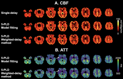 |
50 |
Theoretical and experimental evaluation of the test and retest
reliability of single- and multi-delay 3D pCASL
Xingfeng Shao1,
Zixuan Liu1,
Qinyang Shou1,
Kay Jann1,
and Danny JJ Wang1
1Laboratory of FMRI Technology (LOFT), Mark & Mary Stevens Neuroimaging and Informatics Institute, Keck School of Medicine, University of Southern California, Los Angeles, CA, United States Keywords: Arterial spin labelling, Perfusion, test and retest reliability, multi-delay ASL The goal of this study is to evaluate the quantification accuracy and measurement reliability of single- and multi-PLD pCASL via both simulation and in-vivo test and retest data. CBF calculated by single-delay ASL achieved highest test and retest reliability as compared to 5-PLD ASL. However, CBF values may be under-estimated due to prolonged ATT. 5-PLD ASL can be used to quantify ATT and CBF simultaneously with slightly reduced test and retest reliability. Model-fitting approach is more preferrable than the weighted-delay approach in terms of both quantification error and reliability. |
|
2754.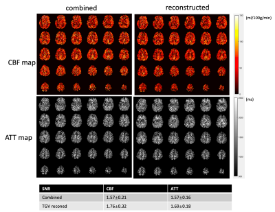 |
51 |
Total Generalized Variation (TGV) Constrained Reconstruction
Improves Test-retest Reliability of High Resolution 3D pCASL in
Children
Qinyang Shou1,
Xingfeng Shao1,
Kimberly Felix2,
Rudolf Stollberger3,
Megan M Herting2,
and Danny JJ Wang1
1Laboratory of Functional MRI Technology (LOFT), Stevens Neuroimaging and Informatics Institute, University of Southern California, Los Angeles, CA, United States, 2Department of Population and Public Health Sciences, Keck School of Medicine, University of Southern California, Los Angeles, CA, United States, 3Institute of Biomedical Imaging, Graz University of Technology, Graz, Austria Keywords: Arterial spin labelling, Brain We developed an accelerated multi-delay 3D pCASL scheme with high-resolution (iso-2mm), CAIPI acceleration and TGV reconstruction to achieve a ~9-minute protocol with 5 delays with 8 averages per delay. We tested the feasibility of this protocol on 19 pediatric subjects and found the proposed imaging protocol with TGV constrained reconstruction can improve the test-retest reliability of high-resolution 3D pCASL perfusion imaging in typically developing children compared to a standard segmented acquisition. |
|
2755.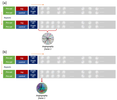 |
52 |
Subspace reconstruction of high temporal resolution arterial
spin labeling angiography using a kinetic model
Qijia Shen1,
Mark Chiew2,3,4,
Wenchuan Wu4,
and Thomas Okell4
1University of Oxford, Oxford, United Kingdom, 2Physical Sciences, Sunnybrook Research Institute, Toronto, ON, Canada, 3Department of Medical Biophysics, University of Toronto, Toronto, ON, Canada, 4Wellcome Centre for Integrative Neuroimaging, FMRIB, Nuffield Department of Clinical Neurosciences, University of Oxford, Oxford, United Kingdom Keywords: Arterial spin labelling, Arterial spin labelling 4D combined angiography and perfusion using radial imaging and arterial spin labelling (CAPRIA) allows dynamic angiograms to be reconstructed. However, the current approach uses a relatively long temporal window to ensure sufficient k-space coverage within each frame, resulting in abrupt changes between adjacent frames, and additional artefacts. In this work, we use a kinetic model of the angiographic signal to constrain the reconstruction of highly undersampled data. A subspace reconstruction was developed for compressed representation of signal during reconstruction for computational efficiency. In-vivo results showed smoother variation of the angiographic signal in the temporal dimension compared to the original approach. |
|
2756.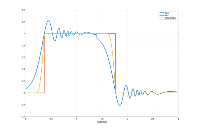 |
53 |
A Mathematical Model for Velocity-Selective Arterial Spin
Labeling
Thomas Liu1,2,3,4,
Eric Wong1,2,3,
Divya Bolar1,2,
Conan Chen1,2,5,
and Ryan Barnes1,5
1Center for fMRI, University of California San Diego, La Jolla, CA, United States, 2Department of Radiology, UCSD, La Jolla, CA, United States, 3Department of Psychiatry, UCSD, La Jolla, CA, United States, 4Department of Bioengineering, UCSD, La Jolla, CA, United States, 5Department of Electrical and Computer Engineering, UCSD, La Jolla, CA, United States Keywords: Arterial spin labelling, Arterial spin labelling A mathematical model is presented that describes the arterial delivery function and cerebral blood volume components in velocity-selective arterial spin labeling. The model incorporates physiologically valid approximations of changes in acceleration and velocity along the vascular system. Applications of the model are shown using a set of example labeling and saturation profiles. |
|
2757.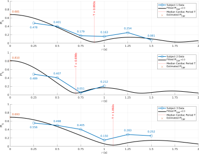 |
54 |
Measuring Microvascular Pulsatility with Short Bolus Duration
(τ) VSASL
Conan Chen1,2,
Ryan A. Barnes1,2,
Eric C. Wong1,3,
Thomas T. Liu1,3,4,
and Divya S. Bolar1
1Center for fMRI, Department of Radiology, University of California San Diego, La Jolla, CA, United States, 2Department of Electrical and Computer Engineering, University of California San Diego, La Jolla, CA, United States, 3Department of Psychiatry, University of California San Diego, La Jolla, CA, United States, 4Department of Bioengineering, University of California San Diego, La Jolla, CA, United States Keywords: Arterial spin labelling, Arterial spin labelling Pulsatile blood flow has been linked to structural damage to the cerebral microvasculature. Previous methods have successfully measured pulsatility in the arteries, but few methods exist to target the microvasculature where the damage is occurring. In this current work, we present a simple theoretical model for CBF pulsatility, apply the model to experimental data acquired in human subjects, and report in vivo microvascular pulsatility using VSASL. |
|
2758.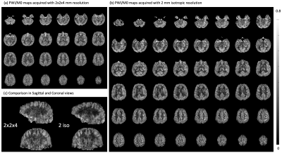 |
55 |
Whole Brain Distortion-free 3D pseudo-Continuous Arterial Spin
Labeling at 7T with Turbo FLASH, Optimized Labeling and
Background Suppression
Chenyang Zhao1,
Xingfeng Shao1,
Qinyang Shou1,
Samantha Ma2,
Sayim Gokyar1,
Christina Graf3,
Rudolf Stollberger3,
and Danny JJ Wang1,4
1Mark & Mary Stevens Neuroimaging and Informatics Institute, Keck School of Medicine, University of Southern California, Los Angeles, CA, United States, 2Siemens Medical Solutions USA, Los Angeles, CA, United States, 3Institute of Biomedical Imaging, Graz University of Technology, Graz, Austria, 4Department of Neurology, Keck School of Medicine, University of Southern California, Los Angeles, CA, United States Keywords: Arterial spin labelling, Arterial spin labelling, Perfusion The potential of 7T has not been fulfilled for pseudo-Continuous Arterial Spin Labeling (pCASL) because of challenges in labeling, background suppression (BS), and readouts. This study proposed a new labeling parameter, a superior BS pulse, and an accelerated and segmented 3D Turbo-FLASH (TFL) readouts. Despite the field inhomogeneity at 7T, the new labeling parameter achieves high labeling efficiency without signal interference, and OPTIM as BS pulse robustly achieves high inversion efficiency. 3D TFL pCASL provides whole brain coverage, abundant anatomical information without distortion, high resolution, and sufficient SNR in 11 mins. |
|
2759.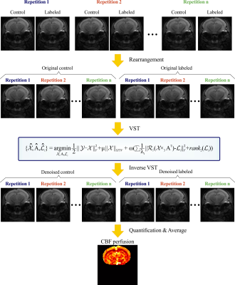 |
56 |
Spatiotemporal redundancy based denoising method in arteprial
spin labeling MRI: A ticket to free sensitivity improvement
Xinran Chen1,
Liangjie Lin2,
Zhiliang Wei3,
Lin Chen1,
and Zhong Chen1
1Department of electronic science, Xiamen University, Xiamen, China, 2Clinical & Technical Support, Philips Healthcare, Beijing, China, 3Russell H. Morgan Department of Radiology and Radiological Science, Johns Hopkins University School of Medicine, Baltimore, MD, United States Keywords: Arterial spin labelling, Data Processing, Denoising Arterial-spin-labeling (ASL) MRI has been widely used in neurological studies for investigating brain activity via focusing on regional perfusion differences. However, sensitivity is a general issue in ASL MRI to prevent the high-resolution comparisons of perfusion heterogeneity, which are associated with pathological process, or lead to prolonged scan time, which inhibits the temporal resolution in monitoring acute-stage perfusion changes (e.g., perfusion recovery after spontaneous resuscitation in cardiac arrest). Therefore, in this study, a post-processing method exploring spatiotemporal redundant information of ASL data is proposed to enhance the sensitivity without extending scan durations. |
|
2760. |
57 |
Feasibility Study to Spatially Map the Filtration Function of
the Kidneys with Multi-TE Arterial Spin Labeling
Lena Sommer1,
Daniel Christopher Hoinkiss1,
Jörn Huber1,
Simon Konstandin1,2,
and Matthias Günther1,2,3
1Fraunhofer Institute for Digital Medicine MEVIS, Bremen, Germany, 2mediri GmbH, Heidelberg, Germany, 3University of Bremen, Bremen, Germany Keywords: Arterial spin labelling, Kidney A spatial mapping of the renal filtration function of the blood might be helpful in identifying parts of the kidney that only work with reduced function and are therefore impaired the most. This work shows an approach to a non-invasive method to map the filtration rate of the kidney. |
|
2761.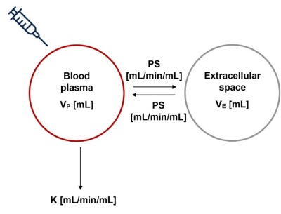 |
58 |
A novel approach to derive robust arterial input functions for
DCE-MRI in small animals
Ebony Rosalind Gunwhy1,
Sirisha Tadimalla2,
John C. Waterton3,4,
Paul D. Hockings5,6,
Gunnar Schütz7,
Claudia Green7,
J. Gerry C. Kenna4,
and Steven Sourbron1
1Department of Infection, Immunity and Cardiovascular Disease, The University of Sheffield, Sheffield, United Kingdom, 2Institute of Medical Physics, The University of Sydney, Sydney, Australia, 3Centre for Imaging Sciences, Division of Informatics Imaging & Data Sciences, School of Health Sciences, Faculty of Biology Medicine & Health, University of Manchester, Manchester, United Kingdom, 4Bioxydyn Ltd, Manchester Science Park, Manchester, United Kingdom, 5BioVentureHub, Antaros Medical, Mölndal, Sweden, 6MedTech West, Chalmers University of Technology, Gothenburg, Sweden, 7MR & CT Contrast Media Research, Bayer AG, Berlin, Germany Keywords: Validation, Animals, Arterial input functions, small animals, spleen, liver Accurate biomarker quantification is hindered in small animal MRI due to difficulties in reliably deriving arterial input functions (AIFs). This study provides a robust alternative to commonly used approaches by deriving AIFs from a simple, whole-body circulation model. This method is compared with individual and population spleen-derived AIFs by evaluating performance in gadoxetate DCE-MRI of the rat liver. Results demonstrated that the whole-body circulation model-derived AIF yields greater repeatability, reproducibility, and goodness-of-fit to observed data, indicating that it provides more accurate biomarker quantification than the individual or population spleen-derived AIFs. |
|
2762. |
59 |
The choroid plexus as a source for selecting an arterial input
function for resting cerebral perfusion measures
Olivia Sobczyk1,
Ece Su Sayin2,
Julien Poublanc1,
Harrison Toby Levine2,
James Duffin2,
Joseph Arnold Fisher2,
and David John Mikulis1
1University Health Network, Toronto, ON, Canada, 2Physiology, University of Toronto, Toronto, ON, Canada Keywords: Susceptibility, Perfusion The choroid plexus is composed of arteries and stroma with negligible metabolic activity allowing its vessels to remain arterialized throughout its structure. We assessed the choroid plexus as a suitable alternate arterial input function rather than the middle cerebral artery in DSC imaging in individuals with both normal vasculature and pathology affecting the middle cerebral artery. |
|
2763.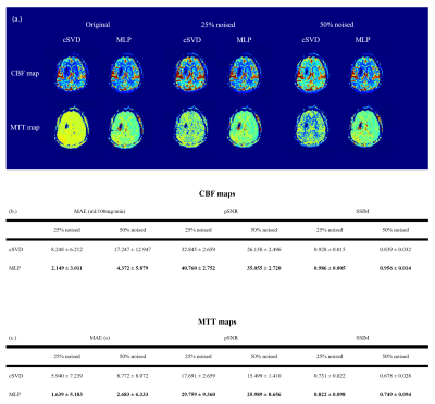 |
60 |
A deep learning approach for robust and accurate deconvolution
of DSC MRI perfusion calculation
Muhammad Asaduddin1,
Eung Yeop Kim2,
and Sung-Hong Park1
1Bio and Brain Engineering, Korea Advanced Institute of Science and Technology, Daejeon, Korea, Republic of, 2Department of Radiology, Samsung Medical Center, Sungkyunkwan University College of Medicine, Seoul, Korea, Republic of Keywords: Contrast Agent, DSC & DCE Perfusion The conventional deconvolution method in DSC perfusion MRI suffers from sensitivity to noise and threshold level. Regularization methods to mitigate the noise issue also suffers from other issues. In this study, we present a deep learning approach to perform deconvolution more robustly and accurately. Our result showed multi layers perceptron (MLP) performed deconvolution more accurately in synthetic data compared to the traditional regularization method. We also showed that MLP performed more robustly in patient data with varying levels of noise. This study provides a strong argument for using MLP as a stable and accurate deconvolution method for DSC perfusion calculation. |
|
 Back to Meeting Home
 Back to the Program-at-a-Glance
The International Society for Magnetic Resonance in Medicine is accredited by the Accreditation Council for Continuing Medical Education to provide continuing medical education for physicians.
 Back to Meeting Home
 Back to the Program-at-a-Glance
 View Presentation Video