Digital Poster
Diffusion: Applications
ISMRM & ISMRT Annual Meeting & Exhibition • 03-08 June 2023 • Toronto, ON, Canada

| Computer # | |||
|---|---|---|---|
4149.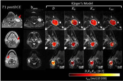 |
61 |
Measuring cellular-interstitial water exchange time in patients
with head and neck cancer using time-dependent diffusion
experiments
Eddy Solomon1,
Gregory Lemberskiy2,
Steven Baete2,
Kenneth Hu3,
Dariya Malyarenko4,
Scott Swanson4,
Amita Shukla-Dave5,
Stephen E Russek6,
Elcin Zan2,
and Sungheon Gene Kim1
1Radiology, Weill Cornell Medicine, New York, NY, United States, 2Radiology, New York University, New York, NY, United States, 3Radiation Oncology, New York University, New York, NY, United States, 4Radiology, University of Michigan, Ann Arbor, MI, United States, 5Medical Physics and Radiology, Memorial Sloan Kettering Cancer Center, New York, NY, United States, 6National Institute of Standards and Technology, Boulder, CO, United States Keywords: Diffusion/other diffusion imaging techniques, Cancer Cellular-interstitial water exchange time has been suggested to be associated with a number of important cellular properties such as membrane permeability, tumor aggressiveness and treatment response. In this study we investigated the reliability of measuring water exchange times based on diffusivity and diffusional kurtosis at long diffusion times. We used two well-established diffusion phantoms and found that diffusion and kurtosis show stable values over a wide range of diffusion times. In head and neck cancer patients, we found that the Kärger model is a valid model for measuring water exchange time in metastatic lymph node voxels. |
|
4150.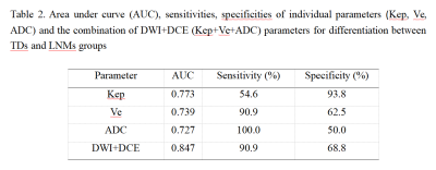 |
62 |
Preliminary study of DCE-MRI combined with DWI in discriminating
tumor deposits from lymph node metastasis in rectal cancer
WEN JUN HU1,
Anliang Chen1,
and AIlian Liu1
1The First Affiliated Hospital of Dalian Medical University, Dalian, China Keywords: Diffusion/other diffusion imaging techniques, Cancer Tumor deposits (TDs) in rectal cancer have been shown to be an important marker of poor prognosis. Although very similar to lymph node metastasis (LNMs), TDs have unique features in terms of biology and outcome, suggesting that distinguishing between these two entities may be of great importance. Results of this study indicate DWI and DCE-MRI can effectively differentiate TDs and LNMs in rectal cancer, Combination of DWI and DCE-MRI imaging may serve as an effective noninvasive method for differentiation of TDs from LNMs in rectal cancer. |
|
4151.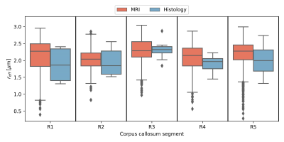 |
63 |
Effective axon radii across the human corpus callosum for a
group of subjects: comparability between MRI and histology
Laurin Mordhorst1,
Mohammad Ashtarayeh1,
Maria Morozova2,3,
Luke J. J Edwards2,
Carsten Jäger2,
Henriette Rusch3,
Tobias Streubel1,
Nikolaus Weiskopf2,4,
Markus Morawski2,3,
and Siawoosh Mohammadi1,2
1Institute of Systems Neuroscience, University Medical Center Hamburg-Eppendorf, Hamburg, Germany, 2Department of Neurophysics, Max Planck Institute for Human Cognitive and Brain Sciences, Leipzig, Germany, 3Paul Flechsig Institute - Center of Neuropathology and Brain Research, Medical Faculty University of Leipzig, Leipzig, Germany, 4Felix Bloch Institute for Solid State Physics, Faculty of Physics and Earth Sciences, Leipzig University, Leipzig, Germany, Leipzig, Germany Keywords: Diffusion/other diffusion imaging techniques, Validation, Histology Connectome Axon radius Deep learning Robust MRI-based axon radius estimation is sensitive to a tail-weighted estimate of the ensemble-average axon radius, i.e., the effective axon radius ($$$r_{eff}$$$). Existing validation studies of $$$r_{eff}$$$ in the human brain are confounded because the histological gold standard cannot representatively sample the tail of the axon radii distribution due to limited sample size. We compare in vivo, MRI-based $$$r_{eff}$$$ of five healthy adults against a representative histological gold standard of three donors in the human corpus callosum and demonstrate that spatial patterns of the $$$r_{eff}$$$ along the anterior-posterior axis agree between in vivo MRI and ex vivo histology. |
|
4152.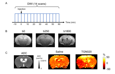 |
64 |
Aquaporin4 inhibitor alters the apparent diffusion coefficients
in mouse brain
Tomokazu Tsurugizawa1,2,
Cuong Pham3,
Anna Deàs Just3,
Benjamin Le Gac3,
Dongdong Li3,
Bruno Cauli3,
and Yuji Komaki4
1National Institute of Advanced Industrial Science and Technology (AIST), Tsukuba, Japan, 2Faculty of Engineering, University of Tsukuba, Tsukuba, Japan, 3Sorbonne University, Paris, France, 4Central Institute for Experimental Animals, Kawasaki, Japan Keywords: Diffusion/other diffusion imaging techniques, Diffusion/other diffusion imaging techniques The glymphatic system seems to be involved to protect the brain pathologies like Alzheimer’s disease, by the clearance of brain waste1. A previous study showed the apparent diffusion coefficients (ADC) in several brain regions were increased by aquaporin 4 (AQP4) inhibitor2. However, the time course of ADC change has not been investigated. In this study, we showed the time course of ADC change in multiple mouse brain regions following injection of the TGN020 and the effect of TGN020 on volume-dependent intrinsic optical signals in brain slices. |
|
4153.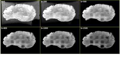 |
65 |
Evaluating histotripsy treatment dosage in the brain using MRI
Dinank Gupta1,
Tarana P Kaovasia1,
Dave Choi1,
Steven P Allen2,
Timothy L Hall1,
Zhen Xu1,
and Douglas C Noll1
1Biomedical Engineering, University of Michigan, Ann Arbor, MI, United States, 2Brigham Young University, Provo, UT, United States Keywords: Diffusion/other diffusion imaging techniques, Focused Ultrasound, Histotripsy Histotripsy is a non-invasive, non-thermal focused ultrasound ablation method that mechanically breaks down the target tissue into acellular debris and has shown promising results for brain surgery. Quantifying histotripsy treatment dosage is required to avoid over/under-treatment of the target region. In this work, we assess whether T1, T2 and ADC maps can be utilized to quantify the ablation effect generated by varying histotripsy dosage post-treatment using an ex-vivo bovine brain. We show that only the ADC map is able to visualize the histotripsy ablation. Increasing histotripsy dosage results in increasing ADC. |
|
4154.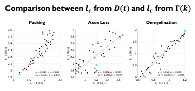 |
66 |
Revealing signatures of demyelination and axonal loss in white
matter extra-axonal space using time dependent diffusion
Ricardo Coronado-Leija1,
Hong-Hsi Lee2,
Els Fieremans1,
and Dmitry S Novikov1
1Radiology, NYU Grossman School of Medicine, New York, NY, United States, 2Radiology, Massachusetts General Hospital, Harvard Medical School, Boston, MA, United States Keywords: Microstructure, Simulations In this work, we relate a specific feature of brain microstructure, the axon packing correlation length $$$l_c$$$, with parameters of time-dependent diffusion $$$D(t)$$$ obtained from Monte Carlo simulations. We observe that diffusion correlation length estimated from $$$D(t)$$$ shows good correspondence with $$$l_c$$$ estimated from geometries of packed disks with several densities, simple models for axon loss and demyelination, as well as for realistic substrates obtained from segmented electron microscopy. |
|
4155. |
67 |
Probing gray matter microstructure with diffusion MRI in
preclinical Alzheimer’s disease: A comparison of methods
Teresa Scheidt1,
Markus Nilsson1,
Danielle van Westen1,2,
Erik Stomrud3,4,
Oskar Hansson3,4,
and Nicola Spotorno3
1Diagnostic Radiology, Department of Clinical Sciences, Lund University, Lund, Sweden, 2Image and Function, Skåne University Hospital, Lund, Sweden, 3Clinical Memory Research Unit, Department of Clinical Sciences, Lund University, Lund, Sweden, 4Memory Clinic, Skåne University Hospital, Malmö, Sweden Keywords: Diffusion/other diffusion imaging techniques, Alzheimer's Disease Gray matter microstructural changes appear prior to macrostructural changes in Alzheimer’s disease, these changes can be probed by diffusion MRI. However, different methods can be used to extract metrics in the thin cortical ribbon. The influence of the choice of method on the results has not been investigated yet. In this work, two different methods of extracting diffusion metrics in the cortex are compared, a surface-based approach and gray matter based spatial statistics (GBSS). We improve upon GBSS and show it now yields comparable results to a surface-based method. |
|
4156. |
68 |
Time-dependent diffusion MRI study of chemoradiation treatment
response in patients with HPV positive oropharyngeal carcinoma
Eddy Solomon1,
Steven H. Baete2,
Joseph K. Kim3,
Moses Tam3,
Zujun Li4,
Kenneth Hu3,
Elcin Zan2,
and Sungheon Gene Kim1
1Radiology, Weill Cornell Medical College, New York, NY, United States, 2Radiology, New York University Grossman School of Medicine, New York, NY, United States, 3Radiation Oncology, New York University Grossman School of Medicine, New York, NY, United States, 4Medical Oncology, New York University Grossman School of Medicine, New York, NY, United States Keywords: Diffusion/other diffusion imaging techniques, Treatment In this study, we evaluated the time-dependence of diffusivity and kurtosis in HPV-positive oropharyngeal squamous cell carcinoma patients before and during chemo-radiation treatment, as part of a study for adaptive de-escalation of the therapy. The non-deescalated patients with less than 40% nodal shrinkage had significantly higher diffusivity and lower kurtosis at pre-treatment than the de-escalated patients with more than 40% nodal volume shrinkage. The water exchange times were longer in the de-escalated patients than in the non-deescalated patients, although not significant. The prognostic accuracy of the pre-treatment imaging parameters was between 0.7 and 0.85. |
|
4157.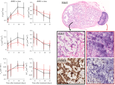 |
69 |
Multidimensional diffusion MRI for monitoring radiotherapy
response in human prostate cancer xenografts in mice: A
longitudinal pilot study
Filip Szczepankiewicz1,
Marcella Safi2,
Crister Ceberg1,
Michael Gottschalk3,
Evangelia Sereti4,
Anders Bjartell4,
Oskar Vilhelmsson Timmermand5,
Linda Knutsson1,6,
Sven-Erik Strand1,7,
and Joanna Strand2,7
1Medical Radiation Physics, Lund University, Lund, Sweden, 2Hematology, Oncology, Radiation Physics, Lund University, Lund, Sweden, 3Lund University Bioimaging Centre, Lund University, Lund, Sweden, 4Translational Medicine, Lund University, Lund, Sweden, 5King's College London, London, United Kingdom, 6F.M. Kirby Research Center for Functional Brain Imaging, Kennedy Krieger Institute, Baltimore, MD, United States, 7Dept of Oncology, Lund University, Lund, Sweden Keywords: Diffusion/other diffusion imaging techniques, Microstructure, Prostate Cancer, Multidimensional MRI, Radiotherapy We use multidimensional diffusion MRI to monitor longitudinal effect of external radiotherapy in human prostate cancers in mice. The measured dimensions are “diffusion time” and “shape of the b-tensor,” enabling a probe of tissue heterogeneity, microscopic anisotropy and restriction sizes. We show that the diffusivity is highly time dependent in all tumors, and that it is significantly altered by radiotherapy, already within 1-15 days of treatment. The largest effect is seen for the diffusivity and its time dependence, but the isotropic diffusional variance is also impacted. Throughout, histology is used qualitatively to provide plausible interpretations to the observations. |
|
4158.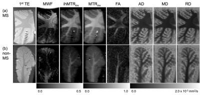 |
70 |
Detection of multiple sclerosis by extraction of quantitative
parameters of brain tissue microstructure in ex-vivo cerebellum
samples
Gopal Varma1,
Aaron K Grant1,
Olivier M Girard2,
Guillaume Duhamel2,
and David C Alsop1
1Radiology, Division of MR Research, Beth Israel Deaconess Medical Center, Harvard Medical School, Boston, MA, United States, 2CNRS, CRMBM, Aix-Marseille Univ, Marseille, France Keywords: Microstructure, Quantitative Imaging Extraction of quantitative parameters of brain tissue microstructure is possible with DTI, inhomogeneous MT (ihMT), and MWF MRI. We examine the ability of the parameters from these techniques to distinguish ex-vivo specimens of the cerebellum from donors with MS relative to non-MS tissue. Significant differences were found for DTI and MT/ihMT metrics in WM, but only for MT/ihMT in GM. Differences between quantitative MT parameters from fits to ihMT data were not significant. |
|
4159.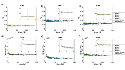 |
71 |
Longitudinal assessment of MD and FA in four whole human brains
using the DTI model
Nina Lüthi1,
Francisco J. Fritz1,
Björn Fricke1,
Tobias Streubel1,
Herbert Mushumba2,
Klaus Püschel2,
and Siawoosh Mohammadi1,3
1Department of Systems Neurosciences, University Medical Center Hamburg-Eppendorf, Hamburg, Germany, 2Institute of Legal Medicine, University Medical Centre Hamburg-Eppendorf, Hamburg, Germany, 3Department of Neurophysics, Max Planck Institute for Human Cognitive and Brain Sciences, Leipzig, Germany Keywords: Diffusion/other diffusion imaging techniques, Ex-Vivo Applications, fixation, PFA, in-situ MRI, whole human brain We investigated the longitudinal fixation effect of 4% paraformaldehyde and phosphate-buffered saline on diffusion imaging-derived mean diffusivity (MD) and fractional anisotropy (FA) across deep gray, cortical gray and white matter in human brains using suboptimal and optimal diffusion protocols with varying maximal b-values.We had two main findings: (1) a suboptimal diffusion protocol led to increased bias in the FA and MD up to 36.26% and 38.11%, the same trend was also observed for the across-brain variation; (2) MD changed by a maximum of 26.42% within the first 14 days in fixative and saturated afterward, whereas FA remained almost constant. |
|
4160.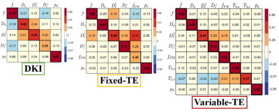 |
72 |
White matter microstructure mapping using diffusion-only or
diffusion-relaxation: protocol design and comparison of accuracy
and precision
Ying Liao1,
Santiago Coelho1,
Filip Szczepankiewicz2,
Dmitry S. Novikov1,
and Els Fieremans1
1Radiology, NYU School of Medicine, NEW YORK, NY, United States, 2Diagnostic Radiology, Clinical Sciences, Lund University, Lund, Sweden Keywords: Diffusion/other diffusion imaging techniques, Diffusion/other diffusion imaging techniques Combining diffusion and relaxation is promising in probing the tissue microstructure in brain white matter. We designed an optimal diffusion-relaxation protocol with varying b-values, b-tensor shapes and echo times by minimizing the error of parameter estimation. We compared the Standard Model parameters estimated by diffusion-relaxation and diffusion-only data using sensitivity-specificity matrices and test-retest data on volunteers and found excellent agreement. With a comprehensive acquisition protocol, the Standard Model can accurately capture the signal content in white matter with diffusion MRI. |
|
4161.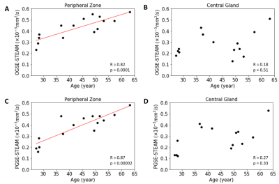 |
73 |
Age-related time-dependent diffusivity changes within healthy
human prostate
Xiao Ma1,
Peter Seres1,
Adam Kinnaird2,
Christopher Fung3,
Thorsten Feiweier4,
and Christian Beaulieu1
1Department of Biomedical Engineering, University of Alberta, Edmonton, AB, Canada, 2Department of Urology, University of Alberta, Edmonton, AB, Canada, 3Department of Radiology, University of Alberta, Edmonton, AB, Canada, 4Siemens healthineers, Erlangen, Germany Keywords: Diffusion/other diffusion imaging techniques, Prostate Diffusion MRI is used daily in prostate cancer diagnosis. Modification of the diffusion time can probe alterations in prostate microstructure dimensions that may occur with aging or cancer. The mean diffusivity (MD) difference between ‘long’ diffusion time stimulated echo acquisition mode (STEAM) and ‘short’ diffusion time oscillating gradient spin echo (OGSE), and between STEAM and ‘medium’ diffusion time pulsed gradient spin echo (PGSE) showed significant positive linear correlation versus age in peripheral zone of healthy adult prostate (n=15); however, this correlation was not identified in the central gland. Diffusion time sensitivity could reveal age-related prostate tissue microstructural remodeling. |
|
4162.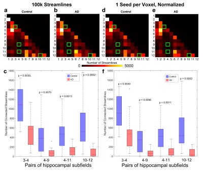 |
74 |
Comparison of Intra-Hippocampal Fiber Tracks and Connectomes
between Alzheimer’s Patients and Age-Matched Healthy Controls
Devon Karl Overson1,2,
Yixin Ma3,
Trong-Kha Truong1,2,4,
David J. Madden1,5,
Jeffrey R. Petrella1,2,4,
and Allen Song1,2,4
1Duke-UNC Brain Imaging & Analysis Center, Duke University, Durham, NC, United States, 2Duke Medical Physics Graduate Program, Duke University, Durham, NC, United States, 3Athinoula A. Martinos Center for Biomedical Imaging, Massachusetts General Hospital, Harvard Medical School, Boston, MA, United States, 4Department of Radiology, Duke University, Durham, NC, United States, 5Department of Psychiatry and Behavioral Sciences, Duke University, Durham, NC, United States Keywords: Tractography & Fibre Modelling, Alzheimer's Disease, DTI, Hippocampus, Hippocampal Subfield, Connectome Changes in intra-hippocampal connectivity caused by early neurodegeneration in specific hippocampal subfields could serve as a biomarker for the early diagnosis of Alzheimer’s disease (AD), years before the onset of symptoms, when the neurodegeneration may still be reversed by treatments. Here, we use high-resolution DTI to compare intra-hippocampal fiber tracks and connectomes across different hippocampal subfields in AD patients and age-matched healthy controls. A significant (p < 0.01) decrease in number of streamlines was found in the AD group compared to the control group for four pairs of subfields, independently of a reduced hippocampal volume affecting nearly all subfields. |
|
4163.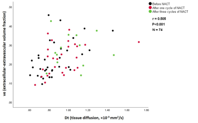 |
75 |
Does intravoxel incoherent motion MRI measure tumor perfusion? A
comparison with DCE-MRI in patients with breast cancer.
Zyad M. Almutlaq1,2,
Sarah E. Bacon3,
Daniel J. Wilson3,
Nisha Sharma4,
and David L. Buckley1
1Biomedical Imaging, University of Leeds, Leeds, United Kingdom, 2Radiological Sciences Department, College of Applied Medical Sciences, King Saud bin Abdulaziz University for Health Sciences, Riyadh, Saudi Arabia, 3Department of Medical Physics & Engineering, Leeds Teaching Hospitals NHS Trust, Leeds, United Kingdom, 4Department of Radiology, Leeds Teaching Hospitals NHS Trust, Leeds, United Kingdom Keywords: Diffusion/other diffusion imaging techniques, DSC & DCE Perfusion, IVIM DCE-MRI can provide quantitative estimates of perfusion-related parameters in tumors, such as blood flow and blood volume fraction. It has also been proposed that IVIM MRI can be used to characterize perfusion in addition to microstructure. This proposal remains controversial and requires further investigation. In this study, we investigate the relationship between perfusion-related parameters measured by IVIM and DCE-MRI in a cohort of patients with breast cancer imaged before and after one and two cycles of neoadjuvant chemotherapy. |
|
4164.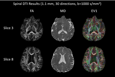 |
76 |
Feasibility of spiral diffusion imaging on a clinical 3T MR
system
Lars Kasper1,
Zhe Wu1,
Alexander Jaffray1,2,
Sriranga Kashyap1,
and Kamil Uludag1,3,4
1BRAIN-TO Lab, Techna Institute, University Health Network, Toronto, ON, Canada, 2UBC MRI Research Centre, Vancouver, BC, Canada, 3Department of Medical Biophysics, University of Toronto, Toronto, ON, Canada, 4Center for Neuroscience Imaging Research, Institute for Basic Science & Department of Biomedical Engineering, Sungkyunkwan University, Suwon, Korea, Republic of Keywords: Diffusion/other diffusion imaging techniques, Diffusion Tensor Imaging, Spiral DWI Spiral diffusion imaging has been shown to provide substantial SNR advantages (>50%) over state-of-the-art echo-planar imaging, due to the attainable shorter echo times. We investigate the feasibility of this approach on a standard 3T MR system without additional instrumentation. All tools for successful implementation are freely made available, i.e., the characterization measurement for the gradient system using a standard phantom, and an open-source, fast image reconstruction suite in Julia correcting for gradient imperfections, eddy currents, and static B0 off-resonance. We show that this low-cost, accessible solution provides high-resolution diffusion images (1.1mm) in-vivo with reduced geometric distortion and improved quantitative maps. |
|
4165.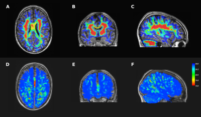 |
77 |
Tractography of the subcortical U-fibers using a
position-dependent maximum angle
Nicolas Delinte1,2,
Quentin Dessain1,
Manon Dausort1,
Colin Vanden Bulcke1,2,
and Benoit Macq1
1ICTEAM, UCLouvain, Louvain-la-Neuve, Belgium, 2IoNS, UCLouvain, Louvain-la-Neuve, Belgium Keywords: Brain Connectivity, Tractography & Fibre Modelling Short association fibers in the subcortical white matter, also known as U-fibers, represent the connections between neighboring gyri. Due to the geometry of the cortical folds and the sharp turns along the cortical surface, tractography of U-fibers remains a challenge since increasing the maximum angle between tractography steps also increases the occurence of false positive streamlines. We propose to replace the fixed maximum angle value in tractography algorithms by an angular map, allowing higher angles at the interface between grey and white matter. This enables a more accurate tracking of U-fibers, while keeping a low number of false positive streamlines. |
|
4166.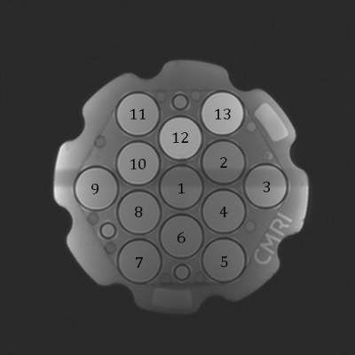 |
78 |
Validation of Intravoxel Incoherent Motion (IVIM) Methods in the
Liver using the NIST Diffusion Phantom
Marissa Brown1,
Juan Vasquez2,
Geoffrey Clarke1,
and John Blangero3
1Radiological Sciences, University of Texas Health Science Center San Antonio, San Antonio, TX, United States, 2School of Medicine, Indiana University, Indianapolis, IN, United States, 3South Texas Diabetes and Obesity Institute, School of Medicine, University of Texas Rio Grande Valley, Brownsville, TX, United States Keywords: Phantoms, Diffusion/other diffusion imaging techniques, intravoxel incoherent motion, liver, fibrosis Intravoxel incoherent motion (IVIM) MRI is used to measure quantitative biomarkers of liver fibrosis, but accuracy and reproducibility of various methods requires validation. Six IVIM methods with different acquisition parameters (motion control techniques, b-values, single shot EPI v. SMS) were assessed using the NIST diffusion phantom. The phantom measurements met conformance standards for percent apparent diffusion coefficient (ADC) bias and ADC linearity using a protocol modified for liver MRI. Given that no standard IVIM liver phantom has been developed, the NIST diffusion phantom is shown to work well for validation of liver IVIM acquisitions. |
|
 Back to Meeting Home
 Back to the Program-at-a-Glance
The International Society for Magnetic Resonance in Medicine is accredited by the Accreditation Council for Continuing Medical Education to provide continuing medical education for physicians.
 Back to Meeting Home
 Back to the Program-at-a-Glance
 View Presentation Video