Digital Poster
Magnetic Susceptibility: Methods & Applications
ISMRM & ISMRT Annual Meeting & Exhibition • 03-08 June 2023 • Toronto, ON, Canada

Digital Poster
Magnetic Susceptibility: Methods & Applications
| Computer # | |||
|---|---|---|---|
4167.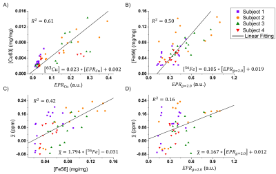 |
81 |
Study of QSM’s contrast sources in the brain using Electron
Paramagnetic Resonance
Fábio Seiji Otsuka1,
Maria Concepción García Otaduy2,
and Carlos Ernesto Garrido Salmon1
1InbrainLab, University of São Paulo, Ribeirão Preto, Brazil, 2LIM44, University of São Paulo, São Paulo, Brazil Keywords: Electromagnetic Tissue Properties, Quantitative Susceptibility mapping QSM is a promising MRI techniques that enables the assessment of magnetic properties of tissue. However, its underlying biophyisical source of contrast is still unknown. It has been shown that iron is well correlated in the basal ganglia, however this hasn't been investigates for other regions. Furthermore, iron can be presented in different forms in the brain, each one having different functions and properties. This work aims to assess the composition of brain structures regarding its paramagnetic ions' and total metal content, comparing their concentrations with magnetic susceptibility assessed by QSM |
|
4168.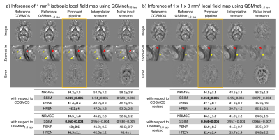 |
82 |
Successful generalization for data with higher or lower
resolution than training data resolution in deep learning
powered QSM reconstruction
Sooyeon Ji1,
Juhyung Park1,
Hyeong-Geol Shin2,3,
Joonhyeok Yoon1,
Minjun Kim1,
and Jongho Lee1
1Department of Electrical Computer Engineering, Seoul National University, Seoul, Korea, Republic of, 2Department of Radiology, Johns Hopkins University School of Medicine, Baltimore, MD, United States, 3F.M. Kirby Research Center for Functional Brain Imaging, Kennedy Krieger Institute, Baltimore, MD, United States Keywords: Susceptibility, Data Processing A pipeline to reconstruct multiple resolution QSM data using a QSM network trained at a single resolution is proposed. The local field map is re-sampled multiple times in different spatial locations, and the re-sampled local field maps are used to reconstruct QSM maps at training data resolution. The reconstructed maps are then combined, and corrected for using a procedure named “dipole compensation”. When compared to two scenarios to reconstruct different resolution data using network trained at a single resolution, the proposed pipeline demonstrated the best performance both qualitatively and quantitatively. |
|
4169.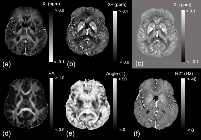 |
83 |
Relationship Between Fractional Anisotropy and Negative
Susceptibility from Single-orientation Magnetic Susceptibility
Separation
Hirohito Kan1,
Yuto Uchida2,
Yoshino Ueki3,
and Harumasa Kasai4
1Department of Integrated Health Scieneces, Nagoya University Graduate School of Medicine, Nagoya, Japan, 2Russell H. Morgan Department of Radiology and Radiological Science, Johns Hopkins University School of Medicine, Baltimore, MD, United States, 3Department of Rehabilitation Medicine, Nagoya City University Graduate School of Medical Sciences, Nagoya, Japan, 4Department of Radiology, Nagoya City University Hospital, Nagoya, Japan Keywords: Electromagnetic Tissue Properties, White Matter The magnetic source separation method can estimate contributions of diamagnetic myelin and paramagnetic iron in the white matter using solely gradient-echo data in white matter. This study determined the relationship between negative susceptibility (χ-) and fractional anisotropy as a myelin-sensitive biomarker and compared with the conventional susceptibility (χ) in young healthy volunteers. There was a significant negative correlation between the χ- and FA. In contrast, the conventional χ has a weaker correlation to the FA than the χ- result. However, the χ- in white matter represented the non-monotonic fiber orientation dependence on the B0 field. |
|
4170.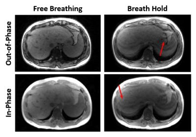 |
84 |
Feasibility and Preliminary Evaluation of Breath Hold and Free
Breathing Quantitative Susceptibility Mapping of the Liver
Julia V Velikina1,
Collin J Buelo1,2,
Yan Wu3,
Marcus T Alley3,
Moniba Nazeef4,
Michael Jeng3,
Alexey A Samsonov1,
Scott B Reeder1,2,4,5,6,
Shreyas S Vasanawala3,
and Diego Hernando1,2
1Radiology, University of Wisconsin - Madison, Madison, WI, United States, 2Medical Physics, University of Wisconsin - Madison, Madison, WI, United States, 3Stanford University, Stanford, CA, United States, 4Medicine, University of Wisconsin - Madison, Madison, WI, United States, 5Biomedical Engineering, University of Wisconsin - Madison, Madison, WI, United States, 6Emergency Medicine, University of Wisconsin - Madison, Madison, WI, United States Keywords: Susceptibility, Liver Quantitative susceptibility mapping (QSM) is a promising non-invasive technique for quantification of liver iron concentration. Abdominal QSM typically requires a breath-hold acquisition since respiration induces liver motion, which leads to blurring artifacts. However, some patients have trouble even with a short breath-hold, which necessitates development of free-breathing approaches. In this work, we report initial results on the feasibility of using the modified “butterfly” navigator approach in multi-echo imaging in conjunction with compressed sensing reconstruction to enable free-breathing liver QSM. |
|
4171.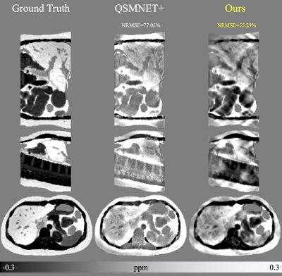 |
85 |
Deep Fourier-Space Inversion
Mathias Lambert1,2,3,
Javier Silva1,2,3,
Carlos Milovic4,
and Cristian Tejos1,2,3
1Department of Electrical Engineering, Pontificia Universidad Católica de Chile, Santiago, Chile, 2Biomedical Imaging Center, Pontificia Universidad Católica de Chile, Santiago, Chile, 3Millennium Institute for Intelligent Healthcare Engineering (iHEALTH), Santiago, Chile, 4School of Electrical Engineering, Pontificia Universidad Católica de Valpaiso, Valparaiso, Chile Keywords: Susceptibility, Quantitative Susceptibility mapping Guiding the network architecture to learn to apply the kernel inversely in Fourier space allows training to be less prone to overfitting. Using simulated images from a single brain image, it is possible to satisfactorily reconstruct a susceptibility map of the abdomen. By having as input the Fourier space of the local field and the kernel of the dipole, the network learned to reduce the noise, to divide the data of the local field by the kernel where possible and to recover the data in the magic cone. |
|
4172.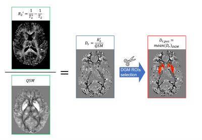 |
86 |
Subject-specific χ-separation method: the effect of introducing
a personalised relaxometric constant Dr
Elena Grosso1,
Antonio Ricciardi2,
Egidio D'Angelo1,3,
Fulvia Palesi1,
and Claudia AM Gandini Wheeler-Kingshott1,2,3
1Department of Brain and Behavioral Sciences, University of Pavia, Pavia, Italy, 2NMR Research Unit, Department of Neuroinflammation, Queen Square Multiple Sclerosis Centre, UCL Queen Square Institute of Neurology, University College London (UCL), London, United Kingdom, 3Brain Connectivity Centre Research Unit, IRCCS Mondino Foundation, Pavia, Italy Keywords: Susceptibility, Modelling, Brain χ-separation is a recently proposed biophysical model to separate the χ-positive and χ-negative contribution of magnetic susceptibility sources in the brain. Such a model relies on assuming a certain relaxometric constant (Dr) calculated as the mean of a group of healthy subjects. Here we demonstrate that Dr is subject-specific and if used in place of the average value, it affects the χ-positive and χ-negative maps in a structured way, while their sum remains similar. |
|
4173.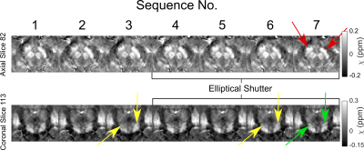 |
87 |
Optimising an Acquisition Protocol and Pipeline for Robust
Clinical Quantitative Susceptibility Mapping to Investigate
Movement Disorders
Oliver C. Kiersnowski1,
David L. Thomas2,
Adam K. Yamamoto2,3,
Mohammed Elgwely2,3,
Anastasia Papadaki2,3,
Tarek Yousry2,3,
John S. Thornton2,3,
and Karin Shmueli1
1Department of Medical Physics and Biomedical Engineering, University College London, London, United Kingdom, 2Neuroradiological Academic Unit, UCL Queen Square Institute of Neurology, University College London, London, United Kingdom, 3Lysholm Department of Neuroradiology, National Hospital for Neurology and Neurosurgery, London, United Kingdom Keywords: Susceptibility, Quantitative Susceptibility mapping Quantitative susceptibility mapping (QSM) has been used to investigate movement disorders but has not been integrated into routine clinical practice. We developed an acquisition protocol and a robust QSM pipeline for neuroradiological investigation of movement disorders. We show that high quality QSMs can be acquired using a multi-echo 3D gradient-echo sequence with partial k-space filling in under 6 minutes with only one of eleven patient QSMs corrupted by motion artifacts. We show that Laplacian phase unwrapping and projection onto dipole fields (PDF) background field removal are robust to artifacts across patients with strong susceptibility sources associated with various pathologies. |
|
4174.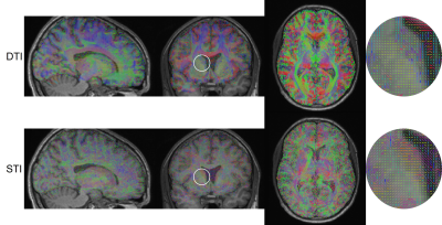 |
88 |
Quantitative comparison of DTI and STI principal eigenvectors
Nestor Munoz1,2,3,
Carlos Milovic3,4,
Christian Langkammer5,
Sergio Uribe2,3,6,
and Cristian Tejos1,2,3
1Electrical Engineering, Pontificia Universidad Catolica de Chile, Santiago, Chile, 2Biomedical Imaging Center, Pontificia Universidad Catolica de Chile, Santiago, Chile, 3Millennium Institute for Intelligent Healthcare Engineering (iHEALTH), Santiago, Chile, 4School of Electrical Engineering, Pontificia Universidad Católica de Valparaiso, Valparaiso, Chile, 5Department of Neurology, Medical University of Graz, Graz, Austria, 6Department of Radiology, Pontificia Universidad Catolica de Chile, Santiago, Chile Keywords: Susceptibility, Susceptibility Diffusion Tensor Imaging (DTI) and Susceptibility Tensor Imaging (STI) are two MRI techniques that produces anisotropy information and fiber direction of biological tissue. For the brain, it has been suggested that both principal eigenvectors (PEV) point towards the same direction (i.e., direction of myelinated axons). However, different resolution of both techniques might produce differences in the PEVs. We proposed a quantitative method to compare DTI and STI PEVs based on the cosine of their angular difference. As expected, we found that PEVs from STI and DTI show similar directions in white matter and that consistency is lost for gray matter and CSF. |
|
4175.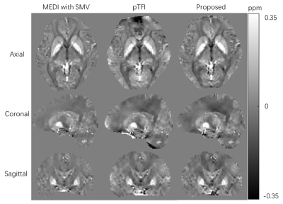 |
89 |
Total Field Inversion for Quantitative Susceptibility Mapping
Based on Boundary Element Modeling
Haodong Zhong1,
Yi Wang2,
and Jianqi Li1
1Shanghai key lab of magnetic resonance, East China Normal University, Shanghai, China, 2Department of Radiology, Weill Medical College of Cornell University, New York, NY, United States Keywords: Susceptibility, Quantitative Susceptibility mapping, Total Field Inversion This study proposes a novel total field inversion method in which the background field is modeled by discrete boundary elements. In this method, the boundary value of background field and local tissue susceptibility are simultaneously estimated in one step. We validated our method with orthogonality numerical simulation and in vivo data. |
|
4176.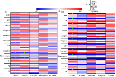 |
90 |
Brain susceptibility and oxygen extraction fraction relate to
cognition altered by white matter hyperintensity in cognitively
normal elderly
Lin Chen1,2,
Anja Soldan3,
Zixuan Lin1,
Kumiko Oishi4,
Kenichi Oishi1,
Andreia Faria1,
Marilyn Albert3,
Peter van Zijl1,2,
and Xu Li1,2
1Department of Radiology and Radiological Sciences, Johns Hopkins University, Baltimore, MD, United States, 2F.M. Kirby Research Center for Functional Brain Imaging, Kennedy Krieger Institute, Baltimore, MD, United States, 3Department of Neurology, Johns Hopkins University, Baltimore, MD, United States, 4Center for Imaging Science, Department of Biomedical Engineering, Johns Hopkins University, Baltimore, MD, United States Keywords: Susceptibility, Aging We investigated associations of brain iron as measured by QSM, global oxygen extraction fraction (OEF) as measured by TRUST and white matter hyperintensity (WMH) as measure by FLAIR, and their possible interactive effects on both global composite and domain-specific cognitive functions in cognitively normal participants. Significant associations were observed between global WMH burden and tissue susceptibility suggesting contributions from small vessel diseases to tissue iron deposition. Negative associations between tissue susceptibility and cognition as well as positive associations between OEF and cognitive performance were observed within participants with low WMH burden, but not as significant in high WMH group. |
|
4177.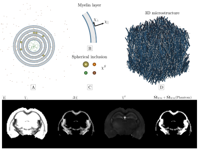 |
91 |
Including mesoscopic frequency shifts from fibrous structure of
white matter improves accuracy of QSM
Anders Dyhr Sandgaard1,
Valerij G. Kiselev2,
Noam Shemesh3,
and Sune Nørhøj Jespersen1,4
1Center for functionally integrative neuroscience, department of clinical medicine, Aarhus University, Aarhus, Denmark, 2Division of Medical Physics, Department of Radiology, University Medical Center Freiburg, Freiburg, Germany, 3Champalimaud Research, Champalimaud Centre for the Unknown, Lisbon, Portugal, 4Department of Phsysics and Astronomy, Aarhus University, Aarhus, Denmark Keywords: Microstructure, Quantitative Susceptibility mapping Quantitative Susceptibility Mapping (QSM) is a highly utilized MRI modality for mapping tissue susceptibility. However, a limitation of QSM is disregarding mesoscopic field effects associated with WM microstructure and anisotropic susceptibility. Here we present a minimal extension of QSM by including frequency shifts due to the fibrous WM microstructure, while still neglecting susceptibility anisotropy and WM spherical inclusions modelling iron complexes. We find that this step already improves the accuracy of QSM as it is shown by comparison with conventional QSM using a digital phantom that includes microstructural frequency shifts from multiple sources. |
|
4178.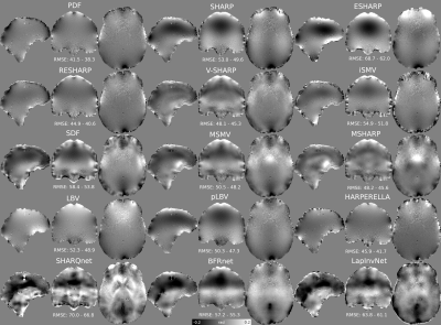 |
92 |
Exhaustive Comparison of QSM Background Field Removal and
Masking using a Realistic Numerical Head Phantom
Carlos Milovic1,2,
Patrick Fuchs3,
Oriana Arsenov3,
Oliver C Kiersnowski3,
Russell Murdoch3,
Laxmi Muralidharan3,
Jannette Nassar3,
and Karin Shmueli3
1School of Electrical Engineering, Pontificia Universidad Catolica de Valparaiso, Valparaiso, Chile, 2iHEALTH, Millennium Institute for Intelligent Healthcare Engineering, Santiago, Chile, 3University College London, London, United Kingdom Keywords: Susceptibility, Susceptibility, QSM Removing background fields is an important preprocessing step in QSM, enabling the reconstruction of fine tissue susceptibility variations in the region of interest (ROI) without being corrupted by susceptibility sources outside of this region. This requires a binary mask of the ROI and most background field removal methods are sensitive to the choice of mask. Here we compared 15 background field removal methods across 4 different masks. We found projection onto dipole fields (PDF) to perform best overall, although it is sensitive to the mask. V-SHARP and RESHARP were more robust to masking and showed good performance. |
|
4179.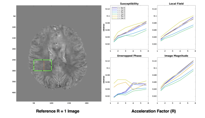 |
93 |
Depiction of Small Vessels in QSM is Influenced by
Regularization Parameter in TGV-Regularized Parallel Image
Reconstruction
Alexander Jaffray1,
Lara Bartels1,
Christian Kames1,
and Alexander Rauscher1,2
1Physics, UBC MRI Research Centre, Vancouver, BC, Canada, 2Department of Pediatrics, UBC, Vancouver, BC, Canada Keywords: Susceptibility, Brain, small vessel, central vein, phase, regularization, vein, qsm, quantitative, mapping Quantitative susceptibility mapping is a possible modality with which to measure and identify diseases involving small vasculature in the brain. However, multi-echo gradient echo images that are a requirement for high quality quantitative susceptibility map reconstruction require long scan durations, limiting clinical utility. This duration can be reduced with the use of parallel imaging strategies, and non-linear reconstruction with total generalized variation regularization is an increasingly popular strategy for undersampled reconstruction. We demonstrate that the depiction of small vessels and surrounding structures in QSM is significantly sensitive to the choice of regularization parameter used in the reconstruction. |
|
4180.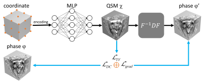 |
94 |
INR-QSM: unsupervised quantitative susceptibility mapping using
implicit neural representation
Ming Zhang1,
Yuyao Zhang2,
and Hongjiang Wei1
1School of Biomedical Engineering, Shanghai Jiao Tong University, Shanghai, China, 2School of Information and Science and Technology, ShanghaiTech University, Shanghai, China Keywords: Susceptibility, Quantitative Susceptibility mapping, implicit neural representation This study introduced an unsupervised deep learning-based method for QSM reconstruction using implicit neural representation (INR-QSM), a training databases-free method for high-quality QSM reconstruction. In INR-QSM, the susceptibility map was represented as a continuous function of the spatial coordinates. A coordinate-based multilayer perceptron (MLP) parameterized this function, took the coordinate as input and predicted the susceptibility value at the corresponding spatial location. The parameters of MLP were updated by minimizing a custom cost function. Preliminary results on two different datasets demonstrated the potential of INR for unsupervised QSM reconstruction. |
|
4181.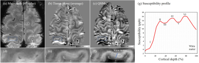 |
95 |
Imaging the cerebral cortex and substantia nigra in vivo using
high-resolution quantitative susceptibility mapping at 5.0-T
Ming Zhang1,
Lanlan Gao2,
Shuheng Zhang2,
and Hongjiang Wei1
1School of Biomedical Engineering, Shanghai Jiao Tong University, Shanghai, China, 2United Imaging Healthcare, Shanghai, China Keywords: Susceptibility, Quantitative Susceptibility mapping, cerebral cortex It is challenging to resolve the human brain microstructure using MRI due to limited SNR and poor image contrast. High-resolution quantitative susceptibility mapping (QSM) at an ultra-high magnetic field could be a useful tool for revealing the subtle structure of the brain due to intrinsic susceptibility differences between tissues. In this study, the six-layer laminae of the cerebral cortex and swallow-tail sign of SN were successfully resolved using high-resolution QSM at 5.0-T. The preliminary result could indicate the potential of high-resolution QSM for investigating the subtle structures of the human brain and related brain disorders. |
|
4182.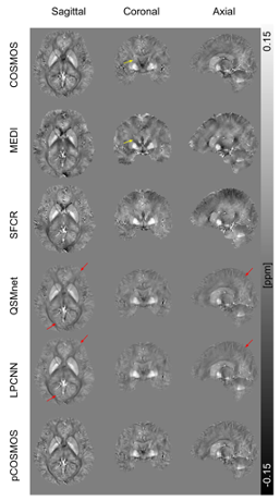 |
96 |
Pseudo COSMOS: Efficient deep learning quantitative
susceptibility mapping based on local field transformation
De-Rong Huang1,
Jhih-Shan Cheng1,
Hsiao-Wen Chung2,
and Ming-Long Wu1,3
1Department of Computer Science and Information Engineering, National Cheng Kung University, Tainan, Taiwan, 2Graduate Institute of Biomedical Electronics and Bioinformatics, National Taiwan University, Taipei, Taiwan, 3Institute of Medical Informatics, National Cheng Kung University, Tainan, Taiwan Keywords: Susceptibility, Machine Learning/Artificial Intelligence A deep learning framework termed pCOSMOS for quantitative susceptibility mapping (QSM) was proposed, which employed a local-field-to-local-field transformation to generate multiple orientation local field maps from single orientation data, followed by COSMOS reconstruction using physical model. 3T multi-orientation data from 10 healthy subjects (7 for training, 3 for testing) were used to investigate performance. Quantitative results compared with MEDI, SFCR, QSMnet, and LPCNN demonstrated superior performance of pCOSMOS comparable to the amongst best algorithms using single orientation data, while showing drastically reduced training time from tens of hours to 3.2 hours. |
|
4183.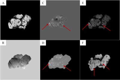 |
97 |
Design of Quadrature RF Coil for Calcium Susceptibility Imaging
in Small Tissue Samples
Jacob Degitz1,
Edith Valle2,
Steven M Wright2,
and Mary P McDougall1
1Biomedical Engineering, Texas A&M University, College Station, TX, United States, 2Electrical Engineering, Texas A&M University, College Station, TX, United States Keywords: Susceptibility, Non-Array RF Coils, Antennas & Waveguides One of the earliest significant events in Duchenne muscular dystrophy (DMD) pathology is an elevated influx of calcium ions into afflicted cells. To study this event and its correlation with disease progression, a custom 22mm diameter quadrature volume coil was designed and used to obtain 1H images at 4.7T, the processing protocol was established, and comparisons were made to μCT images. |
|
4184.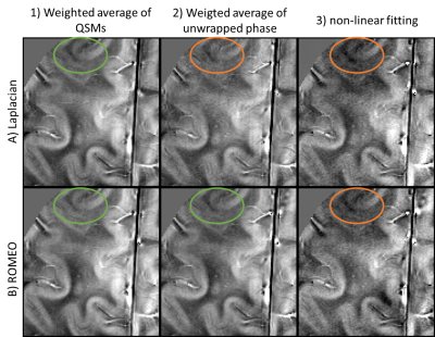 |
98 |
Computationally efficient multi-echo QSM
Korbinian Eckstein1,
Thanh Thuy Dao2,
Ashley Stewart2,3,
Simon Daniel Robinson4,5,6,7,
Markus Barth2,3,4,
and Steffen Bollmann2,3,4
1School of Information Technology and Electrical Engineering, The University of Queensland, Dutton Park, Australia, 2School of Information Technology and Electrical Engineering, The University of Queensland, Brisbane, Australia, 3ARC Training Centre for Innovation in Biomedical Imaging Technology, The University of Queensland, Brisbane, Australia, 4Centre for Advanced Imaging, The University of Queensland, Brisbane, Australia, 5Department of Neurology, Medical University of Graz, Graz, Austria, 6Karl Landsteiner Institute for Clinical Molecular MR in Musculoskeletal Imaging, Vienna, Austria, 7High Field MR Center, Medical University of Vienna, Vienna, Austria Keywords: Electromagnetic Tissue Properties, Quantitative Susceptibility mapping, Multi-echo, ROMEO, Phase With increasingly popular multi-echo QSM, the combination of echoes becomes important in terms of accuracy, SNR and computation time. We compared 6 different pipelines with quantitative and Laplacian unwrapping and three different echo combination approaches, weighted averaging of QSMs, combination of the phase, and non-linear fitting. We compared the pipelines on the QSM challenge brain dataset and 7 T in vivo data and conclude that quantitative unwrapping (ROMEO) with weighted frequency combination achieves the best outcomes in terms of accuracy, SNR and computation time. |
|
4185.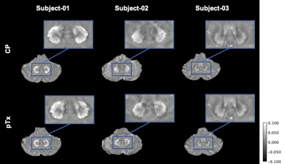 |
99 |
Optimized Quantitative Susceptibility Mapping of Deep Cerebellar
Nuclei using the phase of 3D-EPI Multi-Parametric Mapping at 7T
Mónica Ferreira1,
Rüdiger Stirnberg2,
Yannik Völzke2,
Daniel Löwen2,
Christian Langkammer3,
Simon Robinson3,4,
Stefan Ropele3,
Thomas Klockgether2,5,
Tony Stöcker2,6,
and Jennifer Faber2,5
1Clinical Research, DZNE Bonn, Bonn, Germany, 2DZNE Bonn, Bonn, Germany, 3Department of Neurology, Medical University of Graz, Graz, Austria, 4High Field MR Center, Department of Biomedical Imaging and Image-Guided Therapy, Medical University of Vienna, Vienna, Austria, 5Department of Neurology, University Hospital Bonn, Bonn, Germany, 6Department of Physics and Astronomy, University of Bonn, Bonn, Germany Keywords: Susceptibility, Quantitative Susceptibility mapping RF transmit field inhomogeneities at 7T usually degrade the image quality in regions such as the cerebellum and brainstem, which has so far delayed its use to study cerebellar ataxias with higher fields. We present an optimized pipeline that maximizes the QSM contrast of nuclei in the cerebellum based on multi-echo MPM data. We show that averaging the individual susceptibility maps increases the signal-to-noise ratio (SNR) and reduces the group standard deviation. We introduced a weighted averaging based on the individual acquisition SNRs. Final maps presented high contrast of the dentate nucleus and a high delineation of its denticulated silhouette. |
|
4186.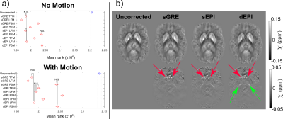 |
100 |
Dynamic Geometric Distortion Correction for Quantitative
Susceptibility Mapping Using a Multi-Echo 2D-EPI Sequence
Oliver C. Kiersnowski1,
Patrick Fuchs1,
Stephen J. Wastling2,3,
John S. Thornton2,3,
and Karin Shmueli1
1Department of Medical Physics and Biomedical Engineering, University College London, London, United Kingdom, 2Neuroradiological Academic Unit, UCL Queen Square Institute of Neurology, University College London, London, United Kingdom, 3Lysholm Department of Neuroradiology, National Hospital for Neurology and Neurosurgery, London, United Kingdom Keywords: Susceptibility, Quantitative Susceptibility mapping Echo planar imaging (EPI) suffers from geometric distortions, which can be corrected using reference acquisitions for single-echo EPI, or using field maps calculated from the multiple echoes in multi-echo EPI. The effect of distortion on EPI quantitative susceptibility mapping (QSM) has not been investigated. We compared static and dynamic geometric distortion correction for magnitude images and QSM using multi-echo 2D-EPI acquisitions with and without changes in head position. Multi-echo EPI corrected distortion in both magnitude and QSM images without the need for reference scans. Correcting the local field map in the QSM pipeline was optimal to reduce temporal variance. |
|
 Back to Meeting Home
 Back to the Program-at-a-Glance
The International Society for Magnetic Resonance in Medicine is accredited by the Accreditation Council for Continuing Medical Education to provide continuing medical education for physicians.
 Back to Meeting Home
 Back to the Program-at-a-Glance
 View Presentation Video