Digital Poster
Diffusion: Methods & Developments
ISMRM & ISMRT Annual Meeting & Exhibition • 03-08 June 2023 • Toronto, ON, Canada

Digital Poster
Diffusion: Methods & Developments
| Computer # | |||
|---|---|---|---|
5007.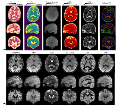 |
61 |
Optimized diffusion-prepared 3D-MRF for rapid high-resolution
whole-brain T1, T2, proton density, ADC and FA mapping
Xiaozhi Cao1,2,
Congyu Liao1,2,
Zihan Zhou3,
Zheng Zhong1,
Zhitao Li1,
Erpeng Dai1,
Siddharth Srinivasan Iyer1,4,
Airel Hannum1,5,
Mahmut Yurt1,2,
Sophie Schauman1,2,
Quan Chen1,
Nan Wang1,
Yifan Yan3,
Hongjian He3,
Stefan Skare6,
Jianhui Zhong7,
Adam Kerr2,
and Kawin Setsompop1,2
1Department of Radiology, Stanford university, Stanford, CA, United States, 2Department of Electrical Engineering, Stanford university, Stanford, CA, United States, 3Center for Brain Imaging Science and Technology, Department of Biomedical Engineering, Zhejiang University, Hangzhou, China, 4Department of Electrical Engineering and Computer Science, MIT, Cambridge, MA, United States, 5Department of Bioengineering, Stanford university, Stanford, CA, United States, 6Department of Clinical Neuroscience, Karolinska Institute, Solna, Sweden, 7Department of Imaging Sciences, University of Rochester, Rochester, NY, United States Keywords: Diffusion/other diffusion imaging techniques, Brain In this work, a diffusion preparation was implemented into the 3D spiral-projection MRF sequence to introduce additional diffusion weighting and enables whole-brain T1, T2, PD, ADC and FA mapping with 1-mm isotropic resolution within 10min. To maximize the diffusion signal and image-encoding efficiency, a diffusion-preparation without an amplitude stabilizer is employed, where robustness against phase variations is achieved using a combination of M1-compensated encoding, cardiac-gating, and an eddy-current compensating pre-pulse gradient. The MRF acquisition scheme and subspace reconstruction were also modified to enable effective data sharing across diffusion directions, which increase acceleration capability and improve mapping quality. |
|
5008.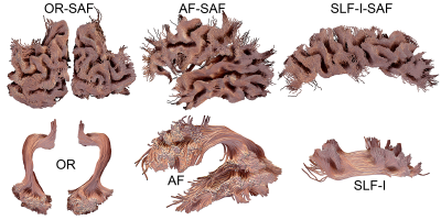 |
62 |
The link between superficial and deep white matter fibers
Maxime Chamberland1,
Dmitri Shastin2,
Derek K. Jones2,
David G. Norris1,
and Kurt G. Schilling3,4
1Donders Institute for Brain, Cognition and Behaviour, Radboud University, Nijmegen, Netherlands, 2Cardiff University Brain Research Imaging Centre (CUBRIC), Cardiff University, Cardiff, United Kingdom, 3Department of Radiology & Radiological Sciences, Vanderbilt University Medical Center, Nashville, TN, United States, 4Vanderbilt University Institute of Imaging Science, Vanderbilt University, Nashville, TN, United States Keywords: Tractography & Fibre Modelling, White Matter, U-fibers, Diffusion MRI, Tractometry, Visualization Here we visualized how superficial association fiber (SAF) systems relate to deep white matter bundles. We found that various shape features can be reliably extracted from those systems. The combination of superficial white matter tractography with traditional deep white matter bundles opens possibilities to study bundle-specific SAF in health and disease. |
|
5009.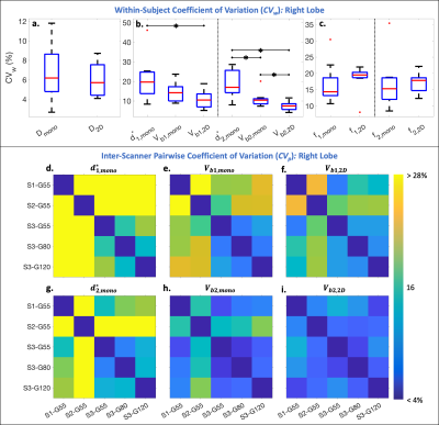 |
63 |
Multi-Scanner Reproducibility of IVIM Quantification in the
Liver using Pseudo-Diffusion and Physical IVIM Signal Models
Gregory Simchick1,2,
Timothy J Allen2,
and Diego Hernando1,2
1Radiology, University of Wisconsin-Madison, Madison, WI, United States, 2Medical Physics, University of Wisconsin-Madison, Madison, WI, United States Keywords: Diffusion/other diffusion imaging techniques, Data Acquisition Intravoxel incoherent motion (IVIM) quantification in the liver suffers from instability associated with separating multiple decaying signal components sampled along a single dimension (b-value). In this work, monopolar and 2D (b-value and first-order motion moment (M1)) noise-optimized IVIM-DWI acquisitions were acquired on three MR scanners with different MR gradient hardware. From each acquisition, IVIM estimates were obtained using pseudo-diffusion and physical (M1 dependent) IVIM signal models. Inter-scanner reproducibility and interlobar agreement were compared across acquisitions and models. 2D (b-M1) IVIM-DWI acquisitions combined with physical IVIM signal modeling improved reproducibility of IVIM quantification in comparison to monopolar acquisitions and pseudo-diffusion modeling. |
|
5010.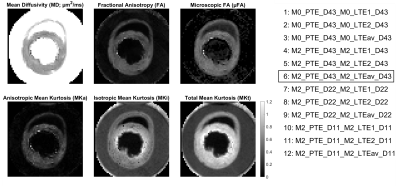 |
64 |
Time-dependence in Tensor-Valued Encoding in Ex Vivo Rat Heart
Richard J. Foster1,
Samo Lasič2,3,
Henrik Lundell2,
Filip Szczepankiewicz4,
Leah Khazin1,
Sven Plein1,
Erica Dall'Armellina1,
Nadira Y. Yuldasheva1,
Jürgen E. Schneider1,
and Irvin Teh1
1Leeds Institute of Cardiovascular and Metabolic Medicine, University of Leeds, Leeds, United Kingdom, 2Danish Research Centre for Magnetic Resonance, Centre for Functional and Diagnostic Imaging and Research, Copenhagen University Hospital Amager and Hvidovre, Copenhagen, Denmark, 3Random Walk Imaging, Lund, Sweden, 4Medical Radiation Physics, Clinical Sciences Lund, Lund University, Lund, Sweden Keywords: Diffusion/other diffusion imaging techniques, Heart, Time dependence, Myocardium, Microstructure, q-space, QTI Tensor-valued encoding is a promising technique for improving specificity in assessing the myocardial microstructure, but can be confounded by time-dependent diffusion (TDD). Here, the influence of TDD on q-space trajectory imaging (QTI) was examined in ex vivo rat heart, using 17 diffusion encoding waveforms with different frequency content and b-tensor shapes. We report apparent over/underestimation of QTI parameters when waveforms had different sensitivity to TDD, and demonstrate a means of frequency matching that reduced the apparent bias. Acquiring QTI at higher frequencies may provide greater sensitivity to intracellular structures, complementing QTI at lower frequencies. |
|
5011.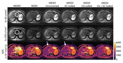 |
65 |
M1-optimized Liver DWI using Tetrahedral Diffusion Gradients on
Conventional Gradient Systems
Timothy J Allen1,
Srijyotsna Volety1,
Greg Simchick2,
and Diego Hernando1,2
1Medical Physics, University of Wisconsin-Madison, Madison, WI, United States, 2Radiology, University of Wisconsin-Madison, Madison, WI, United States Keywords: Diffusion/other diffusion imaging techniques, Motion Correction M1-optimized diffusion gradient waveforms allow for motion-robust liver DWI and ADC estimation. However, existing implementations have been limited to 3.0T scanners with high-performance gradient systems. This work introduces M1-optimized waveforms using a tetrahedral diffusion encoding to allow for high quality, motion-robust DWI on a 1.5T system with conventional gradient performance. Additionally, it addresses complications of this approach, namely increased concomitant gradients and eddy currents. Phantom experiments demonstrate a reduction in geometric distortion and bias of ADC maps generated with this approach compared to Stejskal–Tanner (monopolar) and non-tetrahedral gradient designs. Human subject imaging demonstrates increased motion-robustness. |
|
5012.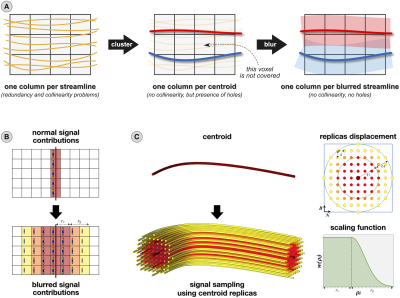 |
66 |
A novel streamline representation to reduce redundancy in
tractography
Ilaria Gabusi1,
Matteo Battocchio1,2,
Sara Bosticardo1,3,
Simona Schiavi1,4,
and Alessandro Daducci1
1Department of Computer Science, University of Verona, Verona, Italy, 2Department of Computer Science, University of Sherbrooke, Sherbrooke, QC, Canada, 3Department of Biomedical Engineering, University of Basel, Basel, Switzerland, 4Department of Neuroscience, Rehabilitation, Ophthalmology, Genetics, Maternal and Child Health (DINOGMI), University of Genoa, Genoa, Italy Keywords: Tractography & Fibre Modelling, Tractography & Fibre Modelling Diffusion MRI tractography allows one to characterize brain connectivity in vivo, and it is common practice to reconstruct millions of streamlines and filter them a posteriori. However, redundancy among streamlines leads to collinearity in the linear operators used by existing filtering algorithms. To solve this problem, we propose a novel streamline representation which uses a combination of clustering and spatial blur to reduce redundancy. This representation is as accurate as state-of-the-art filtering methods and more robust to noise/perturbations in the input, but requires only ≈5% of the input streamlines thus decreasing both storage requirements and computational complexity. |
|
5013.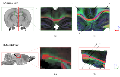 |
67 |
Novel light-sheet scattering microscopy for voxel-wise
validation of 3D-fibre orientations in murine white matter
Mario Corral-Bolaños1,2,
Tram Nguyen1,
Hans Martin Kjer1,
Marco Pizzolato1,2,
Casper Bo Gravesen Salinas3,
Johanna Perens3,
Jeppe Revall Frisvad1,
Julien Colombelli4,
and Tim Bjørn Dyrby1,2
1Department of Applied Mathematics and Computer Science, Technical University of Denmark, Kongens Lyngby, Denmark, 2Danish Research Centre for Magnetic Resonance (DRCMR), Centre for Functional and Diagnostic Imaging and Research, Copenhagen University Hospital, Amager & Hvidovre, Copenhagen, Denmark, 3Gubra, Hørsholm, Denmark, 4Institute for Research in Biomedicine (IRB Barcelona), The Barcelona Institute for Science and Technology (BIST), Barcelona, Spain Keywords: Validation, Tractography & Fibre Modelling, 3D-Fibre orientation This work develops a novel 3D validation technique that reveals the white matter orientation with micron resolution in cleared murine brains. Light-sheet elastic scattering microscopy (LSSM) can be used to exploit the scattering signal exhibited by white matter fibres, which is dependent on their orientation. The rotation and imaging of the sample yield a scattering profile, which corresponds to that of infinitely long cylinders. Our work combines two orthogonal acquisitions to voxel-wise reconstruct 3D-fibre orientations in large brain volumes. LSSM could be the definitive validation method for diffusion MRI orientation methods. |
|
5014.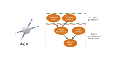 |
68 |
Hierarchical modelling of crossing fibres in the white matter
Hossein Rafipoor1,
Frederik Lange1,
Michiel Cottaar1,
and Saad Jbabdi1
1Wellcome Centre for Integrative Neuroimaging, FMRIB, Nuffield Department of Clinical Neurosciences, University of Oxford, Oxford, United Kingdom Keywords: Tractography & Fibre Modelling, White Matter, Fixel based analysis Fibre orientation distribution function (fODF) estimation is usually applied independently for each subject. This can lead to inconsistent fODF estimation across subjects complicating tract-level (i.e., fixel-based) analysis. Here we propose a hierarchical model to extract consistent tract-specific metrics across subjects that permit group comparisons and the development of more reliable biomarkers for disease and biological processes. |
|
5015.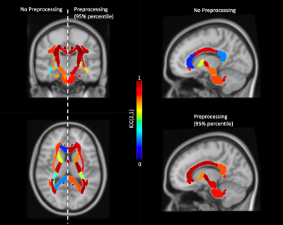 |
69 |
Assessing the variability of brain diffusion MRI preprocessing
pipelines using a Region-of-Interest analysis
Jelle Veraart1,
Stefan Winzeck2,3,
Álvaro Planchuelo-Gómez4,5,
Björn Fricke6,
Evgenios N. Kornaropoulos7,8,
Harri Merisaari9,10,
Tomasz Pieciak5,
Yukai Zou11,12,
and Maxime Descoteaux13
1Center for Biomedical Imaging, Dept. Radiology, NYU Grossman School of Medicine, New York, NY, United States, 2BioMedIA Group, Department of Computing, Imperial College London, London, United Kingdom, 3Division of Anaesthesia, Department of Medicine, University of Cambridge, Cambridge, United Kingdom, 4CUBRIC, Cardiff University, Cardiff, United Kingdom, 5ETSI Telecomunicación, Universidad de Valladolid, Valladolid, Spain, 6Department of Systems Neuroscience, University Medical Center Hamburg-Eppendorf, Hamburg, Germany, 7CRMBM, Aix-Marseille University, Marseille, France, 8Diagnostic Radiology, Lund University, Lund, Sweden, 9Department of Radiology, University of Turku, Turku, Finland, 10TBMC, University of Turku, Turku, Finland, 11Medical Physics Department, University Hospital Southampton NHS Foundation Trust, Southampton, United Kingdom, 12Clinical Neurosciences, Clinical and Experimental Sciences, University of Southampton, Southampton, United Kingdom, 13Sherbrooke Connectivity Imaging Lab (SCIL), Computer Science department, Université de Sherbrooke,, Sherbrooke, QC, Canada Keywords: Diffusion/other diffusion imaging techniques, Data Processing The lack of a standardized preprocessing pipeline is a significant source of variability that might lower the reproducibility of studies, especially across sites and with incomplete description of the preprocessing workflows. We evaluate the downstream impact of variability in preprocessing workflow by quantifying the reproducibility and variability of region-of-interest (ROI) analyses. While many pipelines achieve excellent reproducibility in most ROI, we observed a large variability in performance of preprocessing workflows to the extent that some pipelines are detrimental to the data quality and reproducibility. |
|
5016.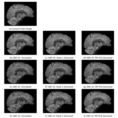 |
70 |
Voxel-level denoising of diffusion MRI using a rank-1
decomposition
Siebe Leysen1,2,
Stefan Sunaert2,3,
Frederik Maes1,2,
and Daan Christiaens1,2
1Department of Electrical Engineering, ESAT/PSI, KU Leuven, Leuven, Belgium, 2Medical Imaging Research Center, UZ Leuven, Leuven, Belgium, 3Translational MRI, Department of Imaging and Pathology, KU Leuven, Leuven, Belgium Keywords: Diffusion/other diffusion imaging techniques, Signal Representations, Denoising A voxel-wise rank-1 decomposition in spherical harmonics of the dMRI signal allows for a denoising method with minimal assumptions on signal and noise distributions. A voxel-wise denoising strategy is compared to MP-PCA on in vivo data and simulated data. The rank-1 decomposition and MP-PCA show visually similar results on the in vivo data, indicating that a voxel-level denoising approach has potential. However, results on the simulations differ. This may have various explanations; further investigation is necessary to strengthen the credibility of a rank-1 decomposition as a denoising approach. |
|
5017.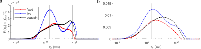 |
71 |
Multiexponential analysis of diffusion exchange times reveals a
distinct exchange process associated with metabolic activity
Teddy Xuke Cai1,2,
Nathan Hu Williamson1,
Rea Ravin1,3,
and Peter Joel Basser1
1Eunice Kennedy Shriver National Institute of Child Health and Human Development, National Institutes of Health, Bethesda, MD, United States, 2Wellcome Centre for Integrative Neuroimaging, FMRIB, Nuffield Department of Clinical Neurosciences, University of Oxford, Oxford, United Kingdom, 3Celoptics, Inc., Rockville, MD, United States Keywords: Diffusion/other diffusion imaging techniques, Data Analysis, Exchange Contrary to prevailing views, recent work suggests that steady-state water exchange between the intra- and extracellular space is driven, in part, by active metabolic processes. To support these findings, we investigate whether exchange exhibits multiexponential behavior consistent with distinct exchange processes. We find a bimodal distribution of exchange times in live neural tissue, with only the faster peak being reduced upon the introduction of a sodium-potassium pump inhibitor, thus supporting the existence of active exchange. Furthermore, we describe a time-efficient method of isolating exchange and fitting multiexponential exchange times using diffusion exchange spectroscopy, paving the way for future studies. |
|
5018.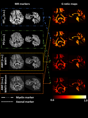 |
72 |
Improving in vivo MR g-ratio mapping via calibration of linearly
transformed MR markers from in situ to ex vivo
Jan Malte Oeschger1,
Francisco Javier Fritz1,
Mohammad Ashtarayeh2,
Maria Morozova3,4,
Tobias Streubel1,
Henriette Rusch4,
Markus Morawski4,5,
Nikolaus Weiskopf5,6,
and Siawoosh Mohammadi1,5
1Department of Systems Neurosciences, University Medical Center Hamburg-Eppendorf, Hamburg, Germany, 2Department of Systems Neurosciences,, University Medical Center Hamburg-Eppendorf, Hamburg, Germany, 3Department of Neurophysics,, Max Planck Institute for Human Cognitive and Brain Sciences, Leipzig, Germany, 4Paul Flechsig Institute of Brain Research, University of Leipzig, Leipzig, Germany, 5Department of Neurophysics, Max Planck Institute for Human Cognitive and Brain Sciences, Leipzig, Germany, 6Faculty of Physics and Earth Sciences, Felix Bloch Institute for Solid State Physics, Leipzig, Germany Keywords: Diffusion/other diffusion imaging techniques, Diffusion/other diffusion imaging techniques, G-ratio In vivo MR g-ratio mapping relies on a calibrated method that relates myelin and axon MR markers to volume fractions. To estimate the calibration parameter, fixed ex vivo tissue measured with MRI and histology is used. The so-determined calibration parameter is then applied in vivo, neglecting the difference in the MR markers due to fixation. Here, we proposed a new calibration method accounting for this difference. The new calibration reduced the variability across MR g-ratios based on different myelin and axon markers, indicating that it can improve the accuracy of MR g-ratios. |
|
5019.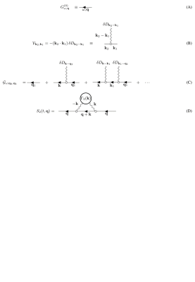 |
73 |
Diffusion diffraction in disordered systems
Sune Jespersen1,2 and
Dmitry S. Novikov3
1Department of Clinical Medicine, Aarhus University, Aarhus, Denmark, 2Department of Physics and Astronomy, Aarhus University, Aarhus, Denmark, 3Radiology, New York University School of Medicine, New York, NY, United States Keywords: Microstructure, Diffusion/other diffusion imaging techniques, diffraction, disordered systems, exchange We identify a new regime for the diffusion signal in non-confining disordered systems with locally varying diffusivity. It occurs at long times and large diffusion wavevectors, reminiscent of diffusion diffraction in closed pores. Remarkably, while for free diffusion the signal is exponentially strongly dephased, scattering off heterogeneities results in a much weaker, power-law decrease of the signal, thereby offering an enhanced sensitivity to the medium’s structure. In particular, we show how correlation functions of the medium’s microstructure to arbitrary order can be measured. We compare our theoretical predictions for the second order correlation function to numerical simulations with good agreement. |
|
5020.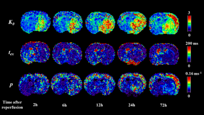 |
74 |
Estimating transcytolemmal water exchange from the Kärger model
using a Bayesian method in a rat model of ischemic stroke
Ruicheng Ba1,
Yuhui Ma2,
Kuiyuan Liu1,
Tianshu Zheng1,
Haotian Li1,
Chen Li2,
Xiaoli Wang2,
and Dan Wu1
1Key Laboratory for Biomedical Engineering of Ministry of Education, Department of Biomedical Engineering, College of Biomedical Engineering & Instrument Science, Zhejiang University, Hanzhou, China, 2Department of Medical Imaging, Weifang Medical University, Shandong, China Keywords: Diffusion/other diffusion imaging techniques, Brain Transcytolemal water exchange can be estimated using diffusion-time-dependent diffusion kurtosis imaging acquired at long diffusion times. However, dMRI signals acquired at long diffusion times using STEAM sequences are typically noisy, and fitting of the nonlinear kurtosis model and the Kärger model accumulates fitting errors. Here, we proposed a Bayesian method for estimating transcytolemal exchange time from the Kärger model and compared accuracy and robustness with conventional least square fitting method in both simulated data and rat brain data in a model of transient middle cerebral artery occlusion. Results indicated improved fitting accuracy and robustness against noise using the Bayesian approach. |
|
5021.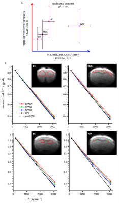 |
75 |
Micro-anisotropy and time-dependent diffusion in the mouse brain
in vivo with spherical tensor encoding and the spectral
principal axis system
Samo Lasic1,
Nathalie Just1,
Matthew Budde2,
and Henrik Lundell1
1Danish Research Centre for Magnetic Resonance, Centre for Functional and Diagnostic Imaging and Research, Copenhagen University Hospital Amager and Hvidovre, Copenhagen, Denmark, 2Department of Neurosurgery, Neurobiology, and Anatomy, Medical College of Wisconsin, Milwaukee, , USA, WI, United States Keywords: Microstructure, Diffusion/other diffusion imaging techniques, Tensor-valued encoding, time-dependent diffusion Accounting for time-dependent diffusion (TDD) in tensor-valued encoding is required for unbiased assessment of microscopic anisotropy (µA). We have previously introduced the spectral principle axis system (SPAS) to find linear tensor encoding (LTE) projections of spherical tensor encoding (STE) with maximum spread of sensitivity to TDD. This can be used to simultaneously achieve unbiased µA and TDD contrasts within a single protocol. Here we present results from in vivo experiments in a mouse brain. The two independent contrasts (µA- TDD) indicate consistent variations in different brain regions. |
|
5022.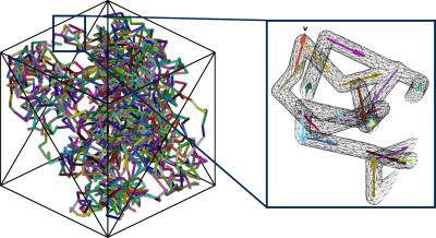 |
76 |
Toolkit for capillary pseudo-diffusivity Monte Carlo simulations
Elizabeth Powell1,
Geoff JM Parker1,2,3,
and Marco Palombo4,5
1Centre for Medical Image Computing, Medical Physics and Biomedical Engineering, University College London, London, United Kingdom, 2Queen Square MS Centre, Institute of Neurology, University College London, London, United Kingdom, 3Bioxydyn Limited, Manchester, United Kingdom, 4Cardiff University Brain Research Imaging Centre, School of Psychology, Cardiff University, Cardiff, United Kingdom, 5School of Computer Science and Informatics, Cardiff University, Cardiff, United Kingdom Keywords: Diffusion/other diffusion imaging techniques, Simulations, Pseudo-diffusivity The pseudo-diffusivity effect - arising from blood flow in capillaries - is used to characterise the diffusion-weighted MRI signal at low b-values (b<200 s/mm2) in several biophysical models. We propose here a generative model for producing vascular meshes with tunable properties - such as vessel segment radius and length, and vessel volume fraction – that are compatible with common Monte Carlo diffusion simulation frameworks. Monte Carlo simulations of pseudo-diffusivity are performed, under varying conditions, by adapting the Camino simulation toolkit to include a plug flow component to spin dynamics inside the vascular mesh; the resulting signals are validated against analytical solutions. |
|
5023.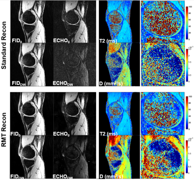 |
77 |
SNR-Enhanced T2 and Diffusion-Weighted 3D Dual-Echo Steady-State
MRI Using Random Matrix Theory-Based Denoising Reconstruction
Zhaohuan Zhang1,
Shu-Fu Shih1,
and Holden H. Wu1
1Department of Radiology, University of California, Los Angeles, Los Angeles, CA, United States Keywords: Diffusion/other diffusion imaging techniques, Diffusion/other diffusion imaging techniques Three-dimensional (3D) dual-echo steady-state (DESS) MRI can produce multi-contrast (MC)images with T2 and diffusion weighting (DW) for simultaneous T2 and D mapping in knee and prostate. A primary challenge of MC DESS is the low signal-to-noise ratio (SNR) of DW DESS leads to less reliable diffusion mapping. In this work, we investigated a new random matrix theory-based denoising reconstruction to improve MC T2W/DW DESS by taking advantages of the inherent redundancy in noise statistics across multiple coil channel and contrasts dimensions. Our in-vivo knee and prostate experiments showed promising improvements in SNR and quality of T2 and D mapping. |
|
5024.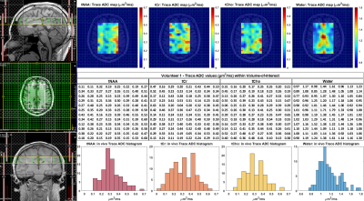 |
78 |
Single Shot Diffusion Trace Spectroscopic Imaging using Radial
Echo Planar Trajectories
Andres Saucedo1,2 and
M. Albert Thomas1,2
1Radiological Sciences, David Geffen School of Medicine, University of California, Los Angeles, Los Angeles, CA, United States, 2Physics and Biology in Medicine Interdepartmental Graduate Program, David Geffen School of Medicine, University of California, Los Angeles, Los Angeles, CA, United States Keywords: Diffusion/other diffusion imaging techniques, Spectroscopy, Spectroscopic Imaging We present the first demonstration of single shot diffusion trace spectroscopic imaging using radial echo planar k-space trajectories on a clinical 3T scanner. Conventional DW-MRS requires three separate acquisitions to compute the trace apparent diffusion coefficient (ADC), whereas the single shot technique generates a trace-weighted signal in one measurement, although this has so far only been applied in NMR and to a limited extent in DW-MRI. Our preliminary results indicate good agreement with expected trace ADC values both in phantom and in healthy brain, showing a promising approach for determining the orientation-independent trace ADC value with non-Cartesian diffusion-weighted spectroscopic imaging. |
|
5025.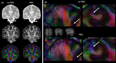 |
79 |
Prospective motion correction improves gSlider accelerated
diffusion imaging
Kerrin J Pine1,
Luke J Edwards1,
Marianna Schmidt1,2,
Juliane Damm1,
Fuyixue Wang3,
Susie Huang3,
Evgeniya Kirilina1,
and Nikolaus Weiskopf1,4
1Department of Neurophysics, Max Planck Institute for Human Cognitive and Brain Sciences, Leipzig, Germany, 2Max Planck School of Cognition, Leipzig, Germany, 3A.A. Martinos Center for Biomedical Imaging, Department of Radiology, Massachusetts General Hospital, Charlestown, MA, United States, 4Felix Bloch Institute for Solid State Physics, Faculty of Physics and Earth Sciences, Leipzig University, Leipzig, Germany Keywords: Diffusion/other diffusion imaging techniques, Motion Correction, DTI High resolution diffusion weighted imaging (DWI) is required to map cortical fibres and short association fibres in superficial white matter. The recently developed gSlider sequence allows high-resolution whole-brain DWI but is prone to motion-induced artefacts due to the long volume acquisition. We combined gSlider with prospective motion correction (PMC) using optical tracking to acquire high angular, high spatial resolution DWI. In three healthy participants, we demonstrated that PMC led to a reduction of motion artifacts, an increase of temporal SNR of around 15% and better estimates of fibre characteristics in the cortex and superficial white matter. |
|
 Back to Meeting Home
 Back to the Program-at-a-Glance
The International Society for Magnetic Resonance in Medicine is accredited by the Accreditation Council for Continuing Medical Education to provide continuing medical education for physicians.
 Back to Meeting Home
 Back to the Program-at-a-Glance
 View Presentation Video